Systems and methods for localizing vascular architecture, and evaluation and monitoring of functional behavior of same

a technology of vascular architecture and localization, applied in the field of systems and methods for localizing vascular architecture, and evaluation and monitoring of functional behavior of same, can solve the problems of crude use of infrared systems, limited sensitivity and speed of existing infrared systems, and inability to achieve significant value, etc., to achieve enhanced contrast between vascular architecture and surrounding tissue within the area being scanned

Inactive Publication Date: 2006-04-13
ADVANCED BIOPHOTONICS
View PDF8 Cites 30 Cited by
  • Summary
  • Abstract
  • Description
  • Claims
  • Application Information

AI Technical Summary

Benefits of technology

[0010] In another embodiment, the present invention provides a method for evaluating vascular architecture of a tissue area on a patient. The method includes initially maintaining, at substantially normal body temperature, the tissue area having the vascular architecture of interest on the patient. Next, photon flux emitted from the tissue area may be detected. In one embodiment, a stream of individual frames of data from detected

Problems solved by technology

The early technology used, unfortunately, had neither the sensitivity, resolution nor speed to be of substantial value.
The existing infrared systems, however, are limited in sensitivity and speed.
As such, the use of these systems for identifying, for instance, vessel architecture, can be crude and can require the use of contrast enhancement techniques, such as cooling the area of interest or the use of contrast agents.
Moreover, existing medical imaging systems are limited to collecting information about the tissue physiology in a single band (i.e., wavelength spectrum) of emission.
With certain surgical procedures, such as reconstructive surgery where free tissue transfer may be involved, there can arise problems that prevent a successful outcome of such a procedure.
These systems, however, do not typically employ infrared imaging.
However, such

Method used


Image

  • Systems and methods for localizing vascular architecture, and evaluation and monitoring of functional behavior of same
  • Systems and methods for localizing vascular architecture, and evaluation and monitoring of functional behavior of same
  • Systems and methods for localizing vascular architecture, and evaluation and monitoring of functional behavior of same

Examples


Embodiment Construction

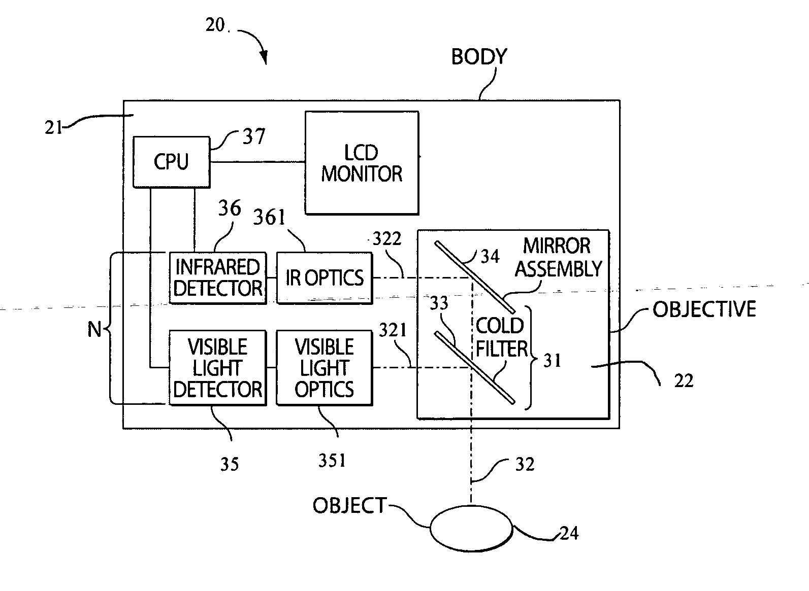
[0022] The present invention, in one embodiment, is directed to a system and method for localizing vascular architecture, and for the evaluation and monitoring of the functional behavior of the vascular architecture for pre-operative, post-operative and diagnostic purposes. The system employs a detection network of at least one detector, single or multiple bands, that is capable of collecting photons of various wavelengths for dynamic imaging of a tissue area of interest. The dynamic imaging system of the present invention may also permit a user to view multiple bands of electromagnetic radiation concurrently as individual images or as a merged or superimposed image.

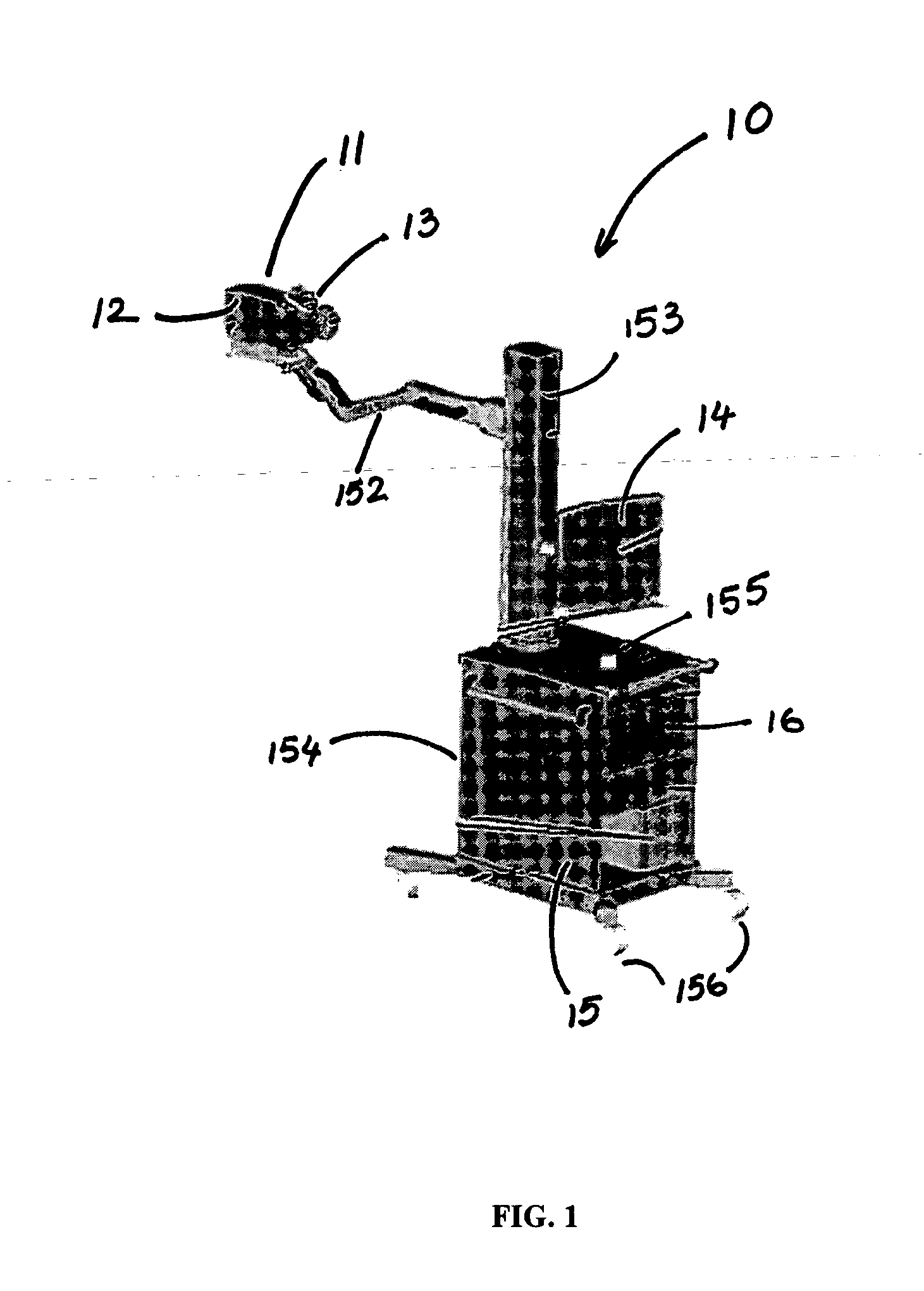
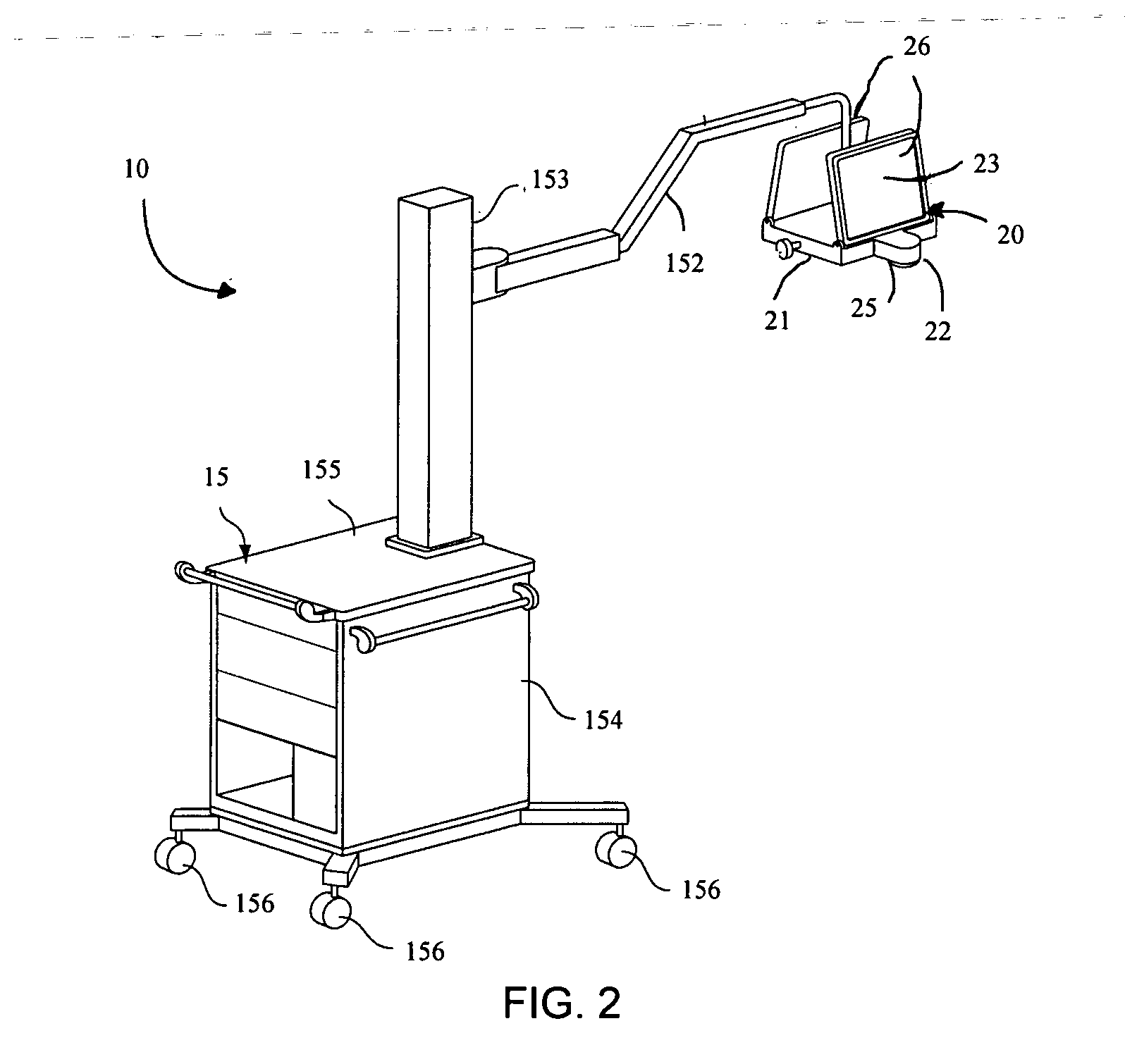
[0023] With reference now to FIG. 1, there is illustrated a dynamic imaging system 10 of the present invention. The system 10 includes, in one embodiment, a scanner 11 positioned on a mobile cart 15 for ease of use. The scanner 11 includes a body portion 12, within which detection components of the scanner 11 may be pos...


PUM


Abstract

A system is provided utilizing dynamic imaging protocol to localize vascular architecture, and to evaluate and monitor functional behavior of the vascular architecture for pre-operative, post-operative and diagnostic purposes. The system includes, among other things, a scanner having within its objective portion an assembly for capturing a photon beam emitted from an object being monitored. The system is also provided with a detection network designed to convert, into electronic signals, data correlated from the beam. A processor can be provided for generating discrete image data from the electronic signals for subsequent display as an image. The system can also include a display for viewing the image data. The system can further include a ruler for positioning on the object being monitored to permit subsequent translation of the image viewed in the display onto the object. Methods for evaluating, monitoring, and localizing the vascular architecture are also provided.

Description

RELATED U.S. APPLICATIONS [0001] The present application claims priority to U.S. application Ser. No. 60 / 585,806, filed Jul. 6, 2004, which application is hereby incorporated herein by reference.TECHNICAL FIELD [0002] The present invention relates to a system and method using dynamic infrared imaging for localizing vascular architecture, and for evaluating and monitoring functional behavior of the vascular architecture for pre-operative, post-operative, and diagnostic purposes. BACKGROUND ART [0003] The concept of infrared imaging for biomedical applications has been explored for some time. The early technology used, unfortunately, had neither the sensitivity, resolution nor speed to be of substantial value. Infrared imaging has now advanced to where it is being used for a range of applications in medicine, and has multiple advantages over conventional medical imaging techniques, including, low cost, no ionizing radiation and minimal need for contrasting agents. The existing infrare...

Claims


Application Information

Patent Timeline
IPC IPC(8): A61B5/05
CPCA61B5/0059A61B5/413A61B5/489A61B5/7257
Inventor FAUCI, MARK A.SALVITTI, MATTHEW A.PAWLOWSKI, MAREK
Owner ADVANCED BIOPHOTONICS
Why Eureka
Try Eureka