Method of preparing a packaged antimicrobial medical device

Active Publication Date: 2006-05-04
ETHICON INC
View PDF64 Cites 35 Cited by
  • Summary
  • Abstract
  • Description
  • Claims
  • Application Information

AI Technical Summary

Benefits of technology

[0007] The present invention relates to packaged antimicrobial medical devices and methods for preparing such packaged medical devices. In accordance with embodiments of the present invention, an antimicrobial agent is disposed on the surfaces of the medical device. The medical device is positioned within a package or within a packaging component such as a containment compartment within a package, and upon being subjected to sufficient conditions, a portion of the antimicrobial agent transfers from the medical device to the package and/or the containment compartment. The transfer of the antimicrobial agent is in an amount sufficient to inhibit bacterial growth on and about the medical device, the package and/or the containment compartment.
[0008] An embodiment of the packaged antimicrobial medical device includes at least one package having an inner surface with an antimicrobial agent disposed thereon, the antimicrobial agent being selected from halogenated hydroxyl ethers, acyloxydiphenyl ethers, and combinations thereof, in an amount sufficient to substantially inhibit bacterial colonization on the package; and at least one medical device positioned within the package, the medical device having one or more surfaces having an antimicrobial agent disposed thereon, the antimicrobial agent being selected from halogenated hydroxyl ethers, acyloxydiphenyl ethers, and combinations thereof, in an amount sufficient to substantially inhibit bacterial colonization on the medical device.
[0009] Another embodiment of the packaged antimicrobial medical device includes a package having an inner surface and a containment compartment for securing the medical device and that resides within the package. In this embodiment, at least one surface of the containment compartment includes an antimicrobial agent disposed thereon, present in an amount sufficient to substantially inhibit bacterial colonization on the containment compartment. In an alternate embodiment, the inner surface of the package and at least one surface of the containment compartment include an antimicrobial agent disposed thereon, present in an amount sufficient to substantially inhibit bacterial colonization on the package and the containment compartment. The packaged medical device also includes at least one medical device positioned within the containmen

Problems solved by technology

Such bacterial colonization on the medical device may lead to infection and trauma to th

Method used


Image

  • Method of preparing a packaged antimicrobial medical device
  • Method of preparing a packaged antimicrobial medical device
  • Method of preparing a packaged antimicrobial medical device

Examples


Example

EXAMPLE 1

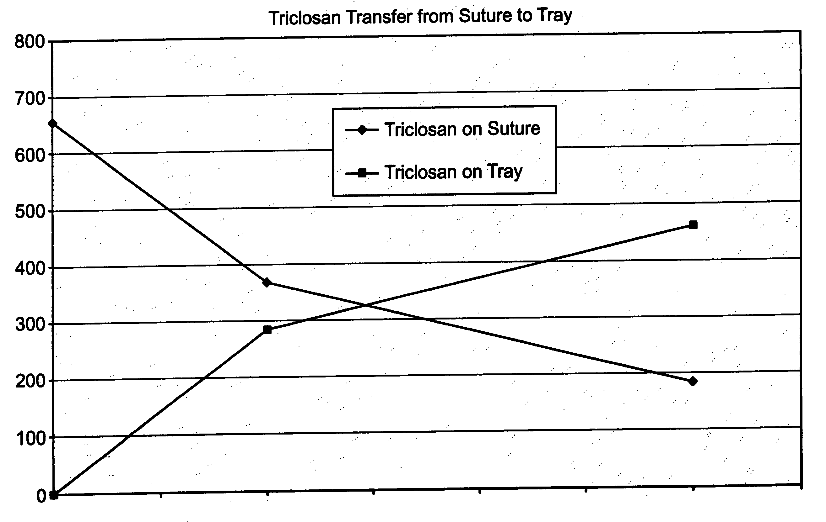
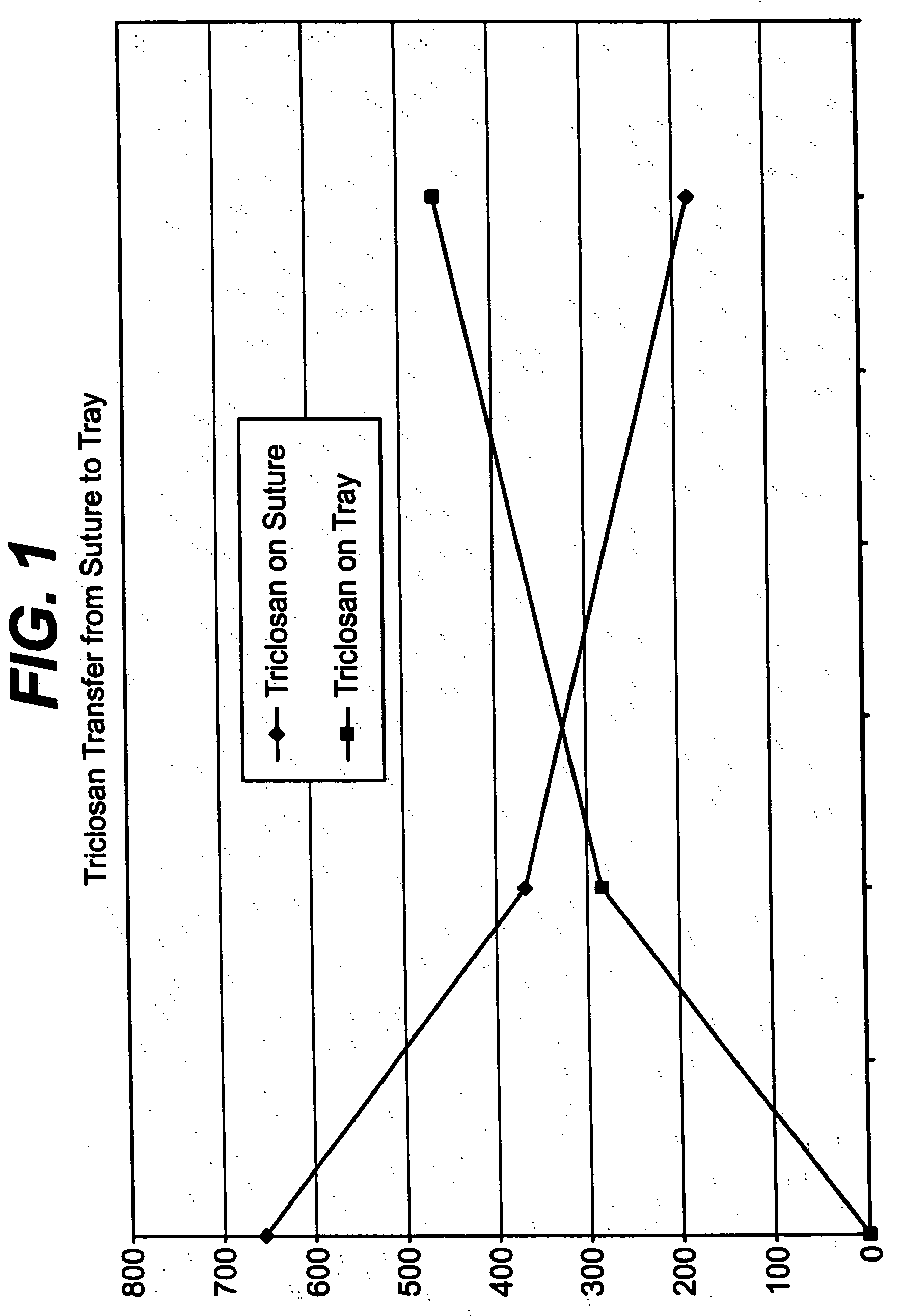
[0045] A series of USP standard size 5-0 coated polyglactin 910 sutures were coated with a 2% triclosan coating composition so that each suture contained about a total of 23.2 μg triclosan before sterilization. The coated sutures each were placed in a package as described herein above including a containment component, i.e., a tray, for holding the suture and a paper component for covering the suture in the tray. The suture in the containment component and packaging were sterilized as described herein above. After sterilization, it was determined that that suture contained about 5.5 μg triclosan, the tray about 0.2 μg triclosan, the paper component about 2.3 μg triclosan, and the package heat seal coating about 1.5 μg triclosan. Triclosan not recovered after sterilization was about 13.7 μg triclosan. FIG. 1 indicates triclosan transfer from the antimicrobial suture to the tray of the package as a function of time at 55° C.

[0046] After sterilization, the paper component an...

Example

EXAMPLE 2

[0050] This example is a 24-hour aqueous immersion assay. The purpose of this assay was to determine the effect of aqueous exposure on the antimicrobial properties of suture material for a range of suture diameters. Sterile sutures in USP sizes 2-0, 3-0, 4-0, and 5-0, with and without a 1% triclosan coating applied thereto, were aseptically cut into 5-cm pieces. One half of the cut pieces were stored in a sterile Petri dish and kept under a dry nitrogen atmosphere for 24 hours (dry suture). One half of the cut pieces were aseptically transferred to sterile 0.85% saline and incubated at 37° C. for 24 hours (wet sutures).

[0051] The dry and wet sutures were then aseptically placed in individual sterile Petri dishes and challenged with 100 microliters of inoculum containing 105 colony-forming units (CFU) of Staphylococcus aureus or Staphylococcus epidermidis. Ten replicates of each suture size were used for each organism and for both the dry and wet sample groups. TSA was pou...

Example

EXAMPLE 4

[0055] This example is directed to a 7-day aqueous immersion assay. The purpose of this assay was to determine if the antimicrobial effect of triclosan treatment would endure for 7 days in a buffered aqueous environment.

[0056] Sterile USP size 2-0 coated polyglactin 910 suture coated with a 1%, 2%, and 3% triclosan coating solution, respectively, and ethylene oxide sterilized USP size 2-0 coated polyglactin suture were aseptically cut into 5-cm pieces. Samples were tested on each of 7 days in triplicate.

[0057] On day 1, 3 pieces of each suture material were placed into individual sterile Petri dishes and inoculated with 0.1 mL of challenge organism containing approximately 104 CFU. TSA was poured into each dish and allowed to solidify. All remaining pieces of suture material were placed into 100 mL of sterile phosphate buffered 0.85% saline (PBS). Every 24 hours for the next 6 days, 3 pieces of each suture material were removed from the PBS, inoculated, and pour plated i...


PUM

PropertyMeasurementUnit
Inhibitionaaaaaaaaaa

Abstract

A method of making a packaged antimicrobial suture comprising the steps of providing a containment compartment that is substantially free of an antimicrobial agent; positioning a suture within the containment compartment, said suture comprising one or more surfaces having an antimicrobial agent disposed thereon, said antimicrobial agent being selected from the group consisting of halogenated hydroxyl ethers, acyloxydiphenyl ethers, and combinations thereof; placing the containment compartment having the suture in an outer package; and subjecting the outer package, the containment compartment and the suture to time, temperature and pressure conditions sufficient to vapor transfer an effective amount of the antimicrobial agent from the suture to the containment compartment, while retaining an effective amount of said antimicrobial agent on the suture, thereby substantially inhibiting bacterial colonization on the suture and the containment compartment.

Description

CROSS-REFERENCE TO PRIOR APPLICATIONS [0001] This application claims the benefit of U.S. Ser. No. 10 / 808,669 filed on Mar. 25, 2004 and Ser. No. 10 / 367,497 filed on Feb. 15, 2003. U.S. Ser. No. 10 / 808,669 is a continuation-in-part of U.S. Ser. No. 10 / 603,317 filed on Jun. 25, 2003, which is a continuation-in-part of U.S. Ser. No. 10 / 367,497 filed on Feb. 15, 2003, which claimed the benefit of U.S. Provisional Application No. 60 / 416,114 filed on Oct. 4, 2002.FIELD OF THE INVENTION [0002] The present invention relates to a packaged antimicrobial medical device and its methods of making. BACKGROUND OF THE INVENTION [0003] Each year, patients undergo a vast number of surgical procedures in the United States. Current data shows about twenty-seven million procedures are performed per year. Post-operative or surgical site infections (“SSIs”) occur in approximately two to three percent of all cases. This amounts to more than 675,000 SSIs each year. [0004] The occurrence of SSIs is often ass...

Claims


Application Information

Patent Timeline
IPC IPC(8): A61B19/02A61B17/06A61L17/00
CPCA61B17/06114A61B17/06166A61B19/02A61L2300/606A61L17/005A61L2300/202A61L2300/404A61B2017/00889A61B50/00
Inventor SCALZO, HOWARDFISCHER, JEROME A.ROTHENBURGER, STEPHENCERWIN, ROBERTMCDIVITT, JAMES R.
Owner ETHICON INC
Why Eureka
Try Eureka