Interventional devices for chronic total occlusion recanalization under MRI guidance

a technology of total occlusion recanalization and interventional devices, which is applied in the field of catheters, can solve the problems of limiting characteristics of conventional x-ray imaging, inability to visualize soft tissue by x-ray imaging, and inability to provide full and complete visualization of vascular geometry by conventional x-ray imaging, so as to improve mr guidance, enhance visualization, and enhance the effect of mr image visibility

Inactive Publication Date: 2006-05-18
THE JOHN HOPKINS UNIV SCHOOL OF MEDICINE
View PDF4 Cites 29 Cited by
  • Summary
  • Abstract
  • Description
  • Claims
  • Application Information

AI Technical Summary

Benefits of technology

[0011] Accordingly, one advantage of the present invention is improved MR guidance by providing MR images in which the position of the catheter is more clearly distinguishable in relation to the surrounding anatomy. For example, the present invention provides guide catheters that are visible in MR images along the length of the catheter, and whereby the distal end of the catheter has enhanced visibility in MR images. This is important in vascular procedures such as chronic total occlusion recanalization, in which enhanced visualization helps prevent inadvertent perforation of the blood vessel wall.
[0012] Another advantage of the present invention is improved MR guidance by providing MR images in which a distal section of the catheter tip is clearly distinguishable in the surrounding anatomy.
[0013] Still another advantage of the present invention is improved MR guidance by providing MR images in which at least a substantial portion of the catheter, including the tip and the shaft of the catheter are clearly distinguishable within the MR image.
[0014] In accordance with a first aspect of the present invention, the aforementioned and other advantages are achieved through a guide catheter, which comprises a loop antenna disposed at the distal end of the guide catheter, and a loopless antenna disposed on the guide catheter.

Problems solved by technology

However, there are a number of limiting characteristics associated with conventional X-ray imaging.
Also, soft tissue visualization by x-ray imaging is not possible.
First, conventional X-ray does not provide a full and complete visualization of the vascular geometry.
X-ray does not provide an image of the occluded portion of a blood vessel since the contrasting agent injected into the vasculature does not penetrate the occluded segment of the blood vessel.
As such, the practitioner does not know the geometry of the occluded portion of the blood vessel.
Another limiting feature associated with conventional X-Ray is its inability to provide cross-sectional images of the vasculature.
Still another less desirable feature is the exposure of the patient to potentially harmful X-Ray radiation.
Thus excellent soft tissue contrast and multiplaner imaging capability of MRI will enable superior anatomical imaging, however, conventional commercially available interventional devices cannot be visualized in an MRI environment and may not be safe to use in an MRI environment for safety concerns (e.g. RF heating, ferromagnetic issues).

Method used


Image

  • Interventional devices for chronic total occlusion recanalization under MRI guidance
  • Interventional devices for chronic total occlusion recanalization under MRI guidance
  • Interventional devices for chronic total occlusion recanalization under MRI guidance

Examples


Embodiment Construction

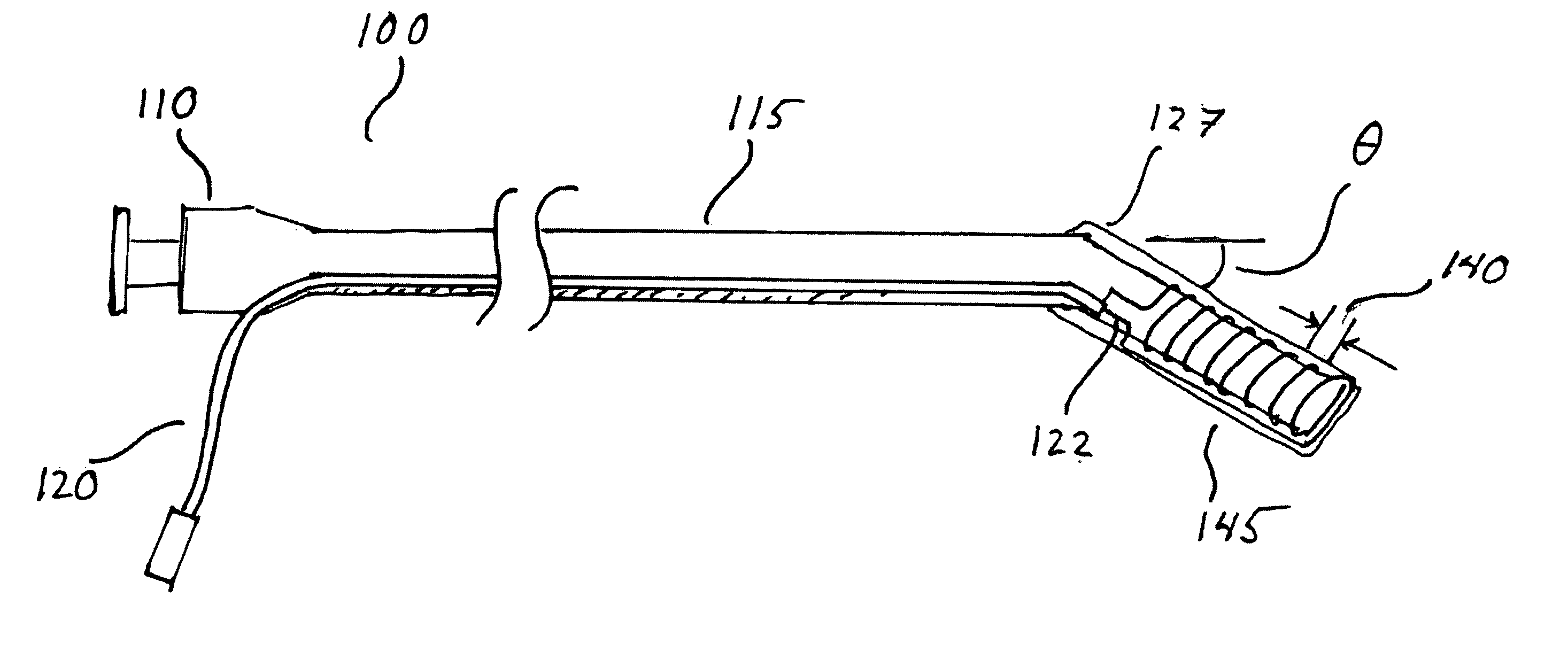
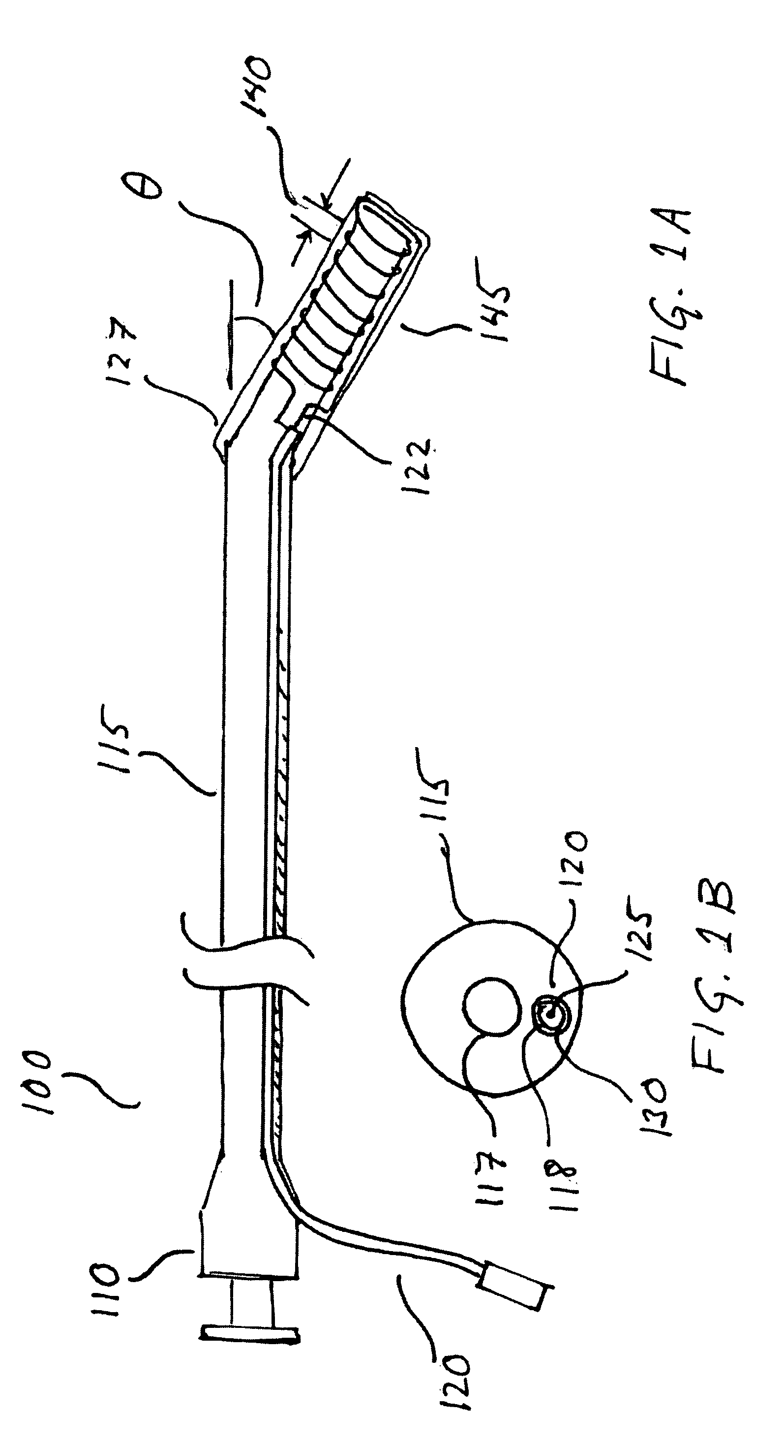
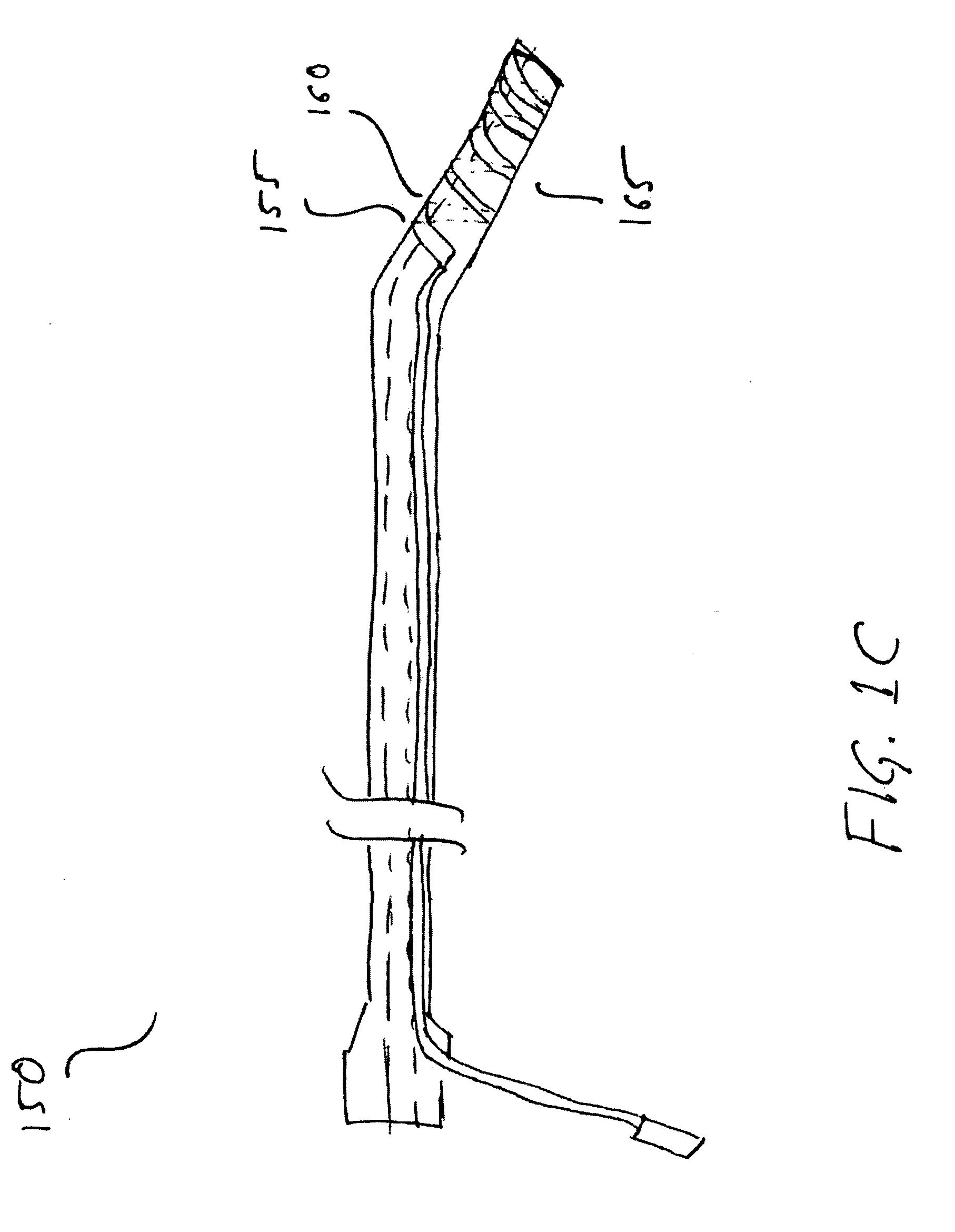
[0037] The present invention involves the use of an inductor loop coil in conjunction with a guide catheter such that the inductor loop coil (hereinafter “coil”) acts as an antenna that is matched and tuned to the Larmor frequency of MRI (0.25 Tesla-11 Tesla). This antenna receives RF signal from the surrounding tissue generated in response to external RF energy applied by the MRI system, which the MRI system subsequently detects and displays in MR images.

[0038]FIG. 1A illustrates an exemplary single loop coil guide catheter 100 according to the present invention. Single loop coil guide catheter 100 includes a multi-lumen polymeric flexible tubing 115, which may be braided, non braided, metallic or non-metallic; a hub 110; a microcoaxial cable 120; and a loop coil 145 formed of a loop wire 122.

[0039] As used herein, “microcoaxial cable” refers to a cable having an inner conductor and a shield, wherein the cable has a diameter that makes it suitable for minimally invasive medical u...


PUM


Abstract

Disclosed is a guide catheter that includes one or more RF antennas to enhance the visibility of the guide catheter in MR imagery. One embodiment of the guide catheter includes a loop coil at the distal end of the guide catheter and a loopless antenna between the distal end and the proximal end. By combining a loop coil and a loopless antenna on the catheter, the shaft of the catheter may be visible in MR imagery while the distal end may appear in the MR imagery more brightly than the shaft.

Description

[0001] This application claims the benefit of U.S. Provisional Patent Application No. 60 / 572,038 filed on May 18, 2004, which is hereby incorporated by reference for all purposes as if fully set forth herein.[0002] The research and development effort associated with the subject matter of this patent application was supported by the NIH Division of Intramural Research under Z01-HL005062-01 CVB and HL57483.BACKGROUND OF THE INVENTION [0003] 1. Field of the Invention [0004] The present invention generally relates to catheters, which are introduced into a biological duct, blood vessel, hollow organ, body cavity, or the like, during a medical procedure. More particularly, the present invention relates to catheters that employ one or more RF antennas to improve the visibility of the catheter and the surrounding tissue for various diagnostic and / or therapeutic purposes in an MRI environment. [0005] 2. Discussion of the Related Art [0006] Catheters have long been used for the purpose of pro...

Claims


Application Information

Patent Timeline
Patent Type & Authority Applications(United States)
IPC IPC(8): A61B5/05A61B19/00A61F2/44A61M25/00G01R33/28
CPCA61B19/54A61B2019/5458A61M25/00A61M2025/1079G01R33/286G01R33/287G01R33/34084A61B90/39A61B2090/3958
Inventor KARMARKAR, PARAG V.ATALAR, ERGINLEDERMAN, ROBERT J.RAVAL, AMISH N.
Owner THE JOHN HOPKINS UNIV SCHOOL OF MEDICINE
Why Eureka
Try Eureka