Specific epitope based immunological diagnosis of tuberculosis

a tuberculosis and immunological diagnosis technology, applied in the field of specific epitope based immunological diagnosis of tuberculosis, can solve the problems of increasing risk, reducing detection accuracy, and reducing detection accuracy, so as to improve detection accuracy and broaden the recognition

Active Publication Date: 2006-06-01
STATENS SERUM INST
View PDF1 Cites 42 Cited by
  • Summary
  • Abstract
  • Description
  • Claims
  • Application Information

AI Technical Summary

Benefits of technology

[0018] The invention is related to detecting infections caused by species of the tuberculosis complex (M. tuberculosis, M. bovis, M. africanum) and discriminate between TB infection and vaccination with the attenuated BCG strain or exposure to non-pathogenic environmental mycobacteria. The invention discloses specific diagnostic reagents, which can be used either in vivo or in vitro to detect a cellular response to M. tuberculosis infections. By using a cocktail or fusion protein of immunogenic epitopes from several different antigens we broaden the recognition, thereby making the test more sensitive. DETAILED DISCLOSURE OF THE INVENTION
[0052] Each polypeptide is encoded by a specific nucleic acid sequence. It will be understood that such sequences include analogues and variants hereof wherein such nucleic acid sequences have been modified by substitution, insertion, addition or deletion of one or more nucleic acid. Substitutions are preferably silent substitutions in the codon usage which will not lead to any change in the amino acid sequence, but may be introduced to enhance the expression of the protein.
[0064] Also, a linker or spacer can be introduced between the antigens being fused in order to enhance the immunogenicity of the fusion molecule. The linker could e.g. 1) introduce one or more protease cleavage sites which would lead to a cleavage of the fusion molecule in the macrophage, 2) introduce a sequence leading to polymerization of the fusion molecule, 3) incorporate a sequence facilitating transport of the fusion molecule across the cell membrane leading to MHC I presentation, or 4) induce a different folding of the protein leading to an altered folding of the fusion molecule and thereby a different processing resulting in presentation of another group of epitopes. The term “linker” includes such linkers.

Problems solved by technology

Tuberculosis (TB) is a major cause of morbidity and mortality throughout the world.
Immunocompetent individuals infected with M. tuberculosis in general have a lifetime risk of 10% for developing active TB, this risk increases many times if the individual is co infected with HIV.
This demands a certain bacterial load and access to the infection site, and is therefore not suitable in early diagnosis.
However it is far from ideal in detecting M. tuberculosis infection.
This broad cross-reactivity of PPD causes the poor specificity of the TST, leading to a situation where BCG vaccination and exposure to non-tuberculosis mycobacteria gives a test result similar to that seen in a M. tuberculosis infected individual.

Method used


Image

  • Specific epitope based immunological diagnosis of tuberculosis
  • Specific epitope based immunological diagnosis of tuberculosis
  • Specific epitope based immunological diagnosis of tuberculosis

Examples


example 1

Assay Conditions

[0097] PBMC were obtained from healthy BCG vaccinated donors with no history of contact to M. tuberculosis and from TB patients with microscopy- or culture-proven infection. Blood samples were drawn from TB patients 0-6 months after diagnosis. PBMC were freshly isolated by gradient centrifugation of heparinized blood on Lymphoprep (Nycomed, Oslo, Norway) and stored in liquid nitrogen until use. The cells were resuspended in complete RPMI 1640 medium (Gibco BRL, Life Technologies) supplemented with 1% penicillin / streptomycin (Gibco BRL, Life Technologies), 1% non-essential-amino acids (FLOW, ICN Biomedicals, CA, USA), and 10% heat-inactivated normal human AB serum (NHS). The viability and number of the cells were determined by Nigrosin staining.

[0098] PBMC cell cultures were established in triplicates with 1.25×10⁵ PBMCs in 100 μl in microtitre plates (Nunc, Roskilde, Denmark) and stimulated with 5 μg / ml PPD, peptide pools spanning the entire length of the four prot...

example 2

Selection of Immunogenic Antigens

[0102] We have screened a large proportion (more than 70) of the ORF's deleted from BCG and only a few (approximately 10) of them are immunoreactive. The ORF's were tested for immunoreactivity using either isolated PBMC from TB patients or using T-cell lines derived from TB patients.

[0103] In table 1 the results from 10 deleted ORF's are shown. These results illustrates the fact that not all RD proteins are immunoreactive when tested with sensitised lymphocytes, and none of these 10 example antigens is recognised by the T-cells.

TABLE 1Table 1. Examples of antigens belonging to the RD regions tested inT-cell lines derived from TB patients. Results are giving in pg / ml IFNgamma. Positive results are marked with bold.RD regionAntigenLine 1Line 2Line 3Line 4Line 5Non61140PHA33131750203323881127RD4Rv 0221310597RD4Rv 022316145RD10Rv 1256022911RD3Rv 1574202390RD3Rv 15802650220RD15Rv 197051000RD2Rv 198200326RD12Rv 2074041225RD5Rv 31191403716RD11Rv 3426192...

example 3

Definition of Specific Regions in the Selected Antigens

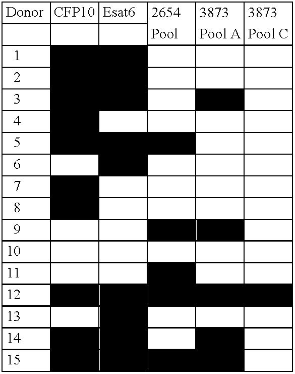
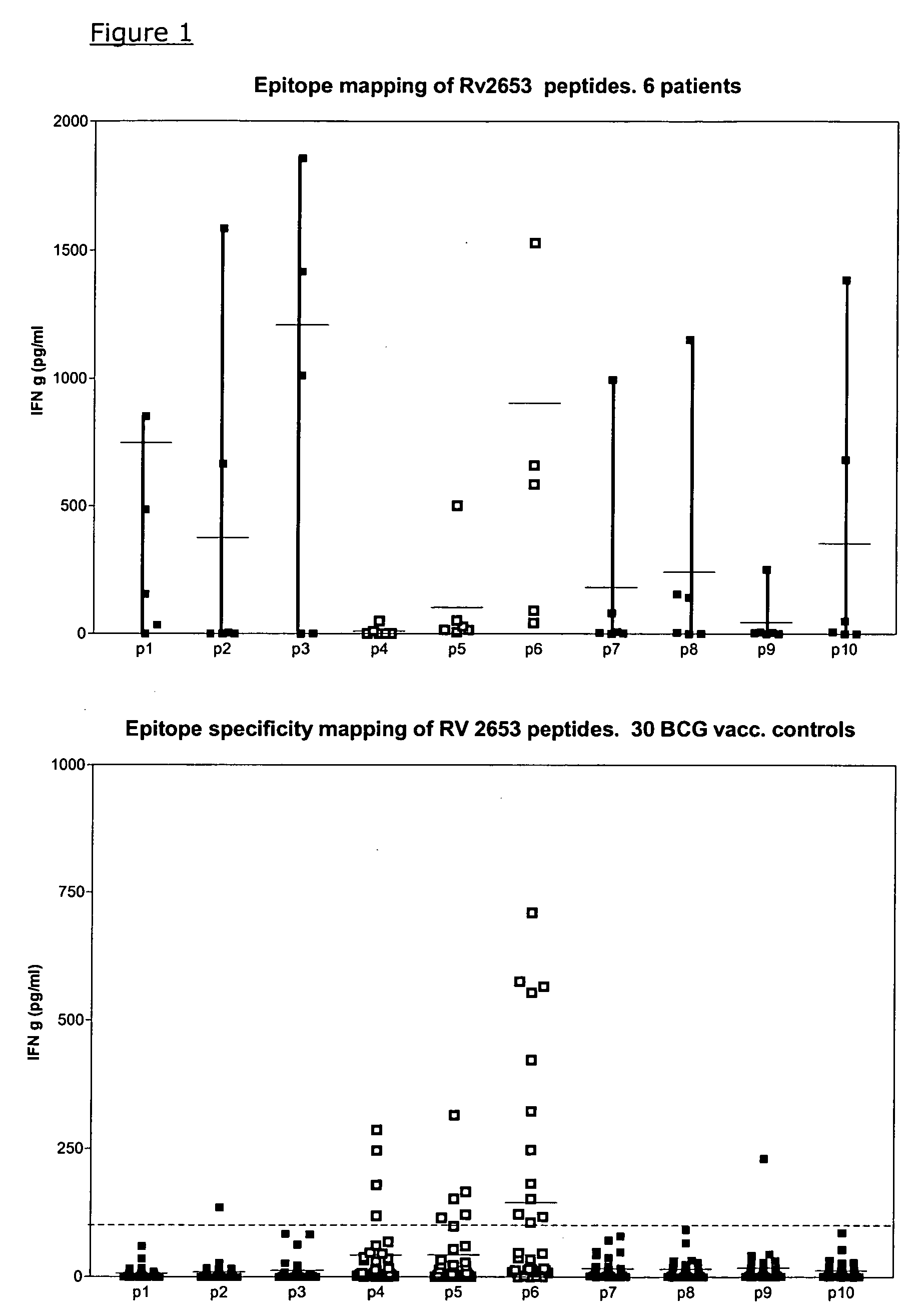
[0109] The antigens were initially tested for recognition by PBMC from TB patients and controls. Even though the four new selected proteins were chosen from the RD regions of the M. tub. genome, we most surprisingly found substantial recognition of two of the antigens by PBMC from BCG vaccinated healthy controls with no known contact to mycobacteria. All participants in the control panel were carefully asked for prior exposure to mycobacteria, occupational or otherwise, and all had only one known exposure: the BCG vaccination.

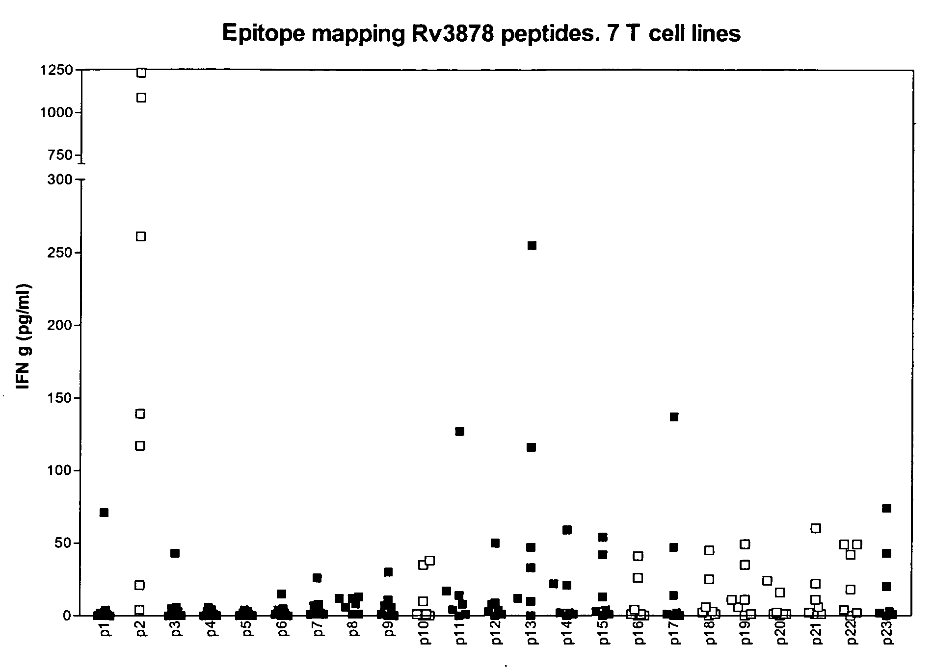
[0110] As seen in table 3 the immunological testing of recombinant Rv3873 (the full length protein), Rv 3878 peptide pools spanning the whole protein and Rv 2653 peptide pools spanning the whole protein gave rise to substantial (between 23% and 59%) recognition in terms of IFN-g release in samples of healthy BCG vaccinated persons.

[0111] To further investigate this phenomena of cross reactivity, syntheti...


PUM

PropertyMeasurementUnit
temperatureaaaaaaaaaa
concentrationaaaaaaaaaa

Abstract

The currently used method for immunological diagnosis of tuberculosis infection, the tuberculin skin test, is problematic for a number of reasons; it has low specificity in BCG vaccinated individuals, a high interobserver variance and requires skill to be read and interpreted. Furthermore it requires an extra visit to the clinic to have the test read. Both people vaccinated with BCG and those exposed to non-tuberculosis mycobacteria give a positive skin test result similar to that seen in a TB infected individual. This also applies for purified protein derivative (PPD) when used in a blood cell based test. The present invention discloses the development of an immunological TB diagnostic tool based on a combination of epitopes from proteins encoded by regions of the M. Tuberculosis (M. tub.) genome, that are not present in the BCG vaccine strain or in the most common non-tuberculosis mycobacteria. Four recently characterized proteins with this diagnostic potential were selected. Peptides from these proteins were tested one by one with peripheral blood mononuclear cells from microscopy or culture confirmed TB patients as well as from healthy BCG vaccinated controls. Some combinations of peptides showed a sensitivity level comparable to the level seen with the two wellknown M. tuberculosisspecific proteins ESAT 6 and CFP 10. An epitope combination with these peptides combined with ESAT 6 and CFP 10 gave a sensitivity of 93%, representing a raise in sensitivity of about 26-33% compared to using ESAT6 or CFP 10 alone. The results from a panel of TB patients, using a collection of the new specific epitopes clearly demonstrates, that addition of other specific epitopes to the already known specific antigens, increases the sensitivity of a diagnostic assay based on cell mediated immune response.

Description

REFERENCE TO RELATED APPLICATIONS [0001] The present invention is a continuation-in-part of International Patent Application PCT / DK2004 / 000314 filed May 6, 2004 and published as WO 2004 / 099771 on Nov. 18, 2004, which claims priority from Denmark Patent Appplication PA 2003 00699 filed May 8, 2003. Each of the above referenced applications, and each document cited in this text (“application cited documents”) and each document cited or referenced in each of the application cited documents, and any manufacturer's specifications or instructions for any products mentioned in this text and in any document incorporated into this text, are hereby incorporated herein by reference; and, technology in each of the documents incorporated herein by reference can be used in the practice of this invention. [0002] It is noted that in this disclosure, terms such as “comprises”, “comprised”, “comprising”, “contains”, “containing” and the like can have the meaning attributed to them in U.S. patent law;...

Claims


Application Information

Patent Timeline
Patent Type & Authority Applications(United States)
IPC IPC(8): C12Q1/68C12P19/34C07K14/35G01N33/50G01N33/569G01N33/68
CPCC07K14/35G01N33/505G01N33/5695G01N33/6812
Inventor ANDERSEN, PETERBROCK, INGERWELDINGH, KARIN
Owner STATENS SERUM INST
Why Eureka
Try Eureka