Anti-TNF chimeric antibody fragments

Inactive Publication Date: 2006-06-29
NEW YORK UNIVERSITY +1
8 Cites 20 Cited by

AI-Extracted Technical Summary

Problems solved by technology

Some of these mabs were used to map epitopes of human TNF and develop enzyme immunoassays (Fendly et al., infra; Hirai et al., infra; Moller et al., infra) and to assist in the purification of recombinant TNF (Bringman et al., infra) However, these studies do not provide a basis for producing TNF neutralizing antibodies that can be used for in vivo diagnostic or therapeutic uses in humans, due to immunogenicity, lack of specificity and/or pharmaceutical suitability.
To date, experience with anti-TNF...

Method used

[0085] Chimeric antibodies are molecules different portions of which are derived from different animal species, such as those having variable region derived from a murine mAb and a human immunoglobulin constant region, which are primarily used to reduce immunogenicity in application and to increase yields in production, for example, where murine mabs have higher yields from hybridomas but higher immunogenicity in humans, such that human/murine chimeric mAbs are used. Chimeric antibodies and methods for their production are known in the art (Cabilly et al, Proc. Natl. Acad. Sci. USA 81:3273-3277 (1984); Morrison et al., Proc. Natl. Acad. Sci. USA 81:6851-6855 (1984); Boulianne et al., Nature 312:643-646 (1984); Cabilly et al., European Patent Application 125023 (published Nov. 14, 1984); Neuberger et al., Nature 314:268-270 (1985); Taniguchi et al., European Patent Application 171496 (published Feb. 19, 1985); Morrison et al., European Patent Application 173494 (published Mar. 5, 1986); Neuberger et al., PCT Application WO 86/01533, (published Mar. 13, 1986); Kudo et al., European Patent Application 184187 (published Jun. 11, 1986); Morrison et al., European Patent Application 173494 (published Mar. 5, 1986); Sahagan et al., J. Immunol. 137:1066-1074 (1986); Robinson et al., International Patent Publication #PCT/US86/02269 (published 7 May 1987); Liu et al., Proc. Natl. Acad. Sci. USA 84:3439-3443 (1987); Sun et al., Proc. Natl. Acad. Sci. USA 84:214-218 (1987); Better et al., Science 240:1041-1043 (1988); and Harlow and Lane ANTIBODIES: A LABORATORY MANUAL Cold Spring Harbor Laboratory (1988)). These references are entirely incorporated herein by reference.
[0168] Once armed with the present disclosure, one skilled in the art would also be able to create derivatives of the immunoreceptor peptides of the invention. Such derivatives are intended to be within the scope of this invention. For example, amino acids in the immunoreceptor that constitute a protease recognition site can be modified to avoid protease cleavage and thus confer greater stability, such as KEX2 sites.
[0176] It was discovered using the molecules of the present invention that the p55/light chain fusion proteins and p55/heavy chain fusion proteins would assemble with each other and dimerize to form an antibody-like molecule that is tetravalent with respect to p55. The resulting tetravalent p55 molecules can confer more protection against, and have greater affinity for, TNFα or TNFβ than the bivalent p55 molecules. Despite the presumed close proximity of the two light chain p55 domains to the heavy chain p55 domains, they do not appear to stereocilia hinder or reduce the affinity for TNF.
[0178] In initial experiments, an irrelevant light chain was co-transfected with the p55-heavy chain construct and subsequent analyses showed that the two chains did assemble and that the resulting fusion protein protected WEHI cells from TNFα. However, it was considered likely that the variable region of the irrelevant light chain would stereocilia hinder interactions between the p55 subunits and TNFα. For this reason, a mouse-human chimeric antibody light chain gene was engineered by (1) deleting the variable region coding sequence, and (2) replacing the murine J coding sequence with human J coding sequence. Use of this truncated light chain, which was shown to assemble and disulfide bond with heavy chains, increased the efficiency of TNF inhibition by approximately 30-fold compared to use of a complete irrelevant light chain.
[0182] The molecules of the present invention have demonstrated the same degree of protection against TNF in a 5000-fold lower molar concentration than monomeric p55. (See Table 1.) It is believed that the presence of the CH, chain in the molecules of a major embodiment of the present invention can confer greater flexibility to the molecule and avoid stearic hindrance with the binding of the TNF receptor.
[0212] Many vector systems are available for the expression of cloned anti TNF peptides H and L chain genes in mammalian cells (see Glover, ed., DNA Cloning, Vol. II, pp143-238, IRL Press, 1985). Different approaches can be followed to obtain complete H2L2 antibodies. As discussed above, it is possible to co-express H and L chains in the same cells to achieve intracellular association and linkage of H and L chains into complete t...

Benefits of technology

[0043] The present invention also provides antigenic polypeptides of hTNF, corresponding to peptides containing neutralizing epitopes or portions of TNF that, when such epitopes on TNF are bound by anti-TNF antibodies or peptides, neutralize or inhibit the biological activity of TNF in vitro, in situ or in vivo.
[0044] The present invention also provides anti-TNF antibodies and peptides in the form of pharmaceutical and/or diagnostic compounds and/or compositions, useful for the diagnostic and/or therapeutic methods of the present invention for diagnosing and/or treating TNF-related pathologies.
[0045] Anti-TNF Abs or anti-TNF peptides of the present invention are provided for use in diagnostic methods for detecting TNF in patients or animals suspected of suffering from conditions associated with abnormal TNF production, including methods wherein high affinity anti-TNF antibodies or peptides are contacted with a biological sample from a patient and an antigen-antibody reaction detected. Also included in the present invention are kits for detecting TNF in a solution using anti-TNF antibodies or peptides, preferably in detectably labeled form.
[0046] The present invention is also directed to an anti-hTNF chimeric antibody comprising two light chains and two heavy chains, each of the...

Abstract

Anti-TNF antibodies and anti-TNF peptides, specific for tumor necrosis factor (TNF) are useful for in vivo diagnosis and therapy of a number of TNF-mediated pathologies and conditions, as well as polynucleotides coding for anti-TNF murine and chimeric antibodies, peptides, methods of making and using the antibody or peptides in immunoassays and immuno-therapeutic approaches are provided, where the anti-TNF peptide is selected from a soluble portion of TNF receptor, an anti-TNF antibody or structural analog thereof.

Application Domain

Technology Topic

PeptideChimeric antibody +9

Image

  • Anti-TNF chimeric antibody fragments
  • Anti-TNF chimeric antibody fragments
  • Anti-TNF chimeric antibody fragments

Examples

  • Experimental program(8)

Example

EXAMPLE I
Production a Mouse Anti-Human TNF mAb
[0290] To facilitate clinical study of TNF mAb a high-affinity potent inhibiting and/or neutralizing mouse anti-human TNF IgG1 mAb designated A2 was produced.
[0291] Female BALB/c mice, 10 weeks old, were obtained from the Jackson Laboratory (Bar Harbor, ME). Forty μg of purified E. coli-derived recombinant human TNF (rhTNF) emulsified with an equal volume of complete Freund's adjuvant (obtained from Difco Laboratories) in 0.4 ml was injected subcutaneously and intraperitoneally (i.p.) into a mouse. One week later, an injection of 5 μg of rhTNF in incomplete Freund's adjuvant was given i.p. followed by four consecutive i.p. injections of 10 μg of TNF without adjuvant. Eight weeks after the last injection, the mouse was boosted i.p. with 10 μg of TNF.
[0292] Four days later, the mouse was sacrificed, the spleen was obtained and a spleen cell suspension was prepared. Spleen cells were fused with cells of the nonsecreting hybridoma, Sp2/0 (ATCC CRL1581), at a 4:1 ratio of spleen cells to Sp2/0 cells, in the presence of 0.3 ml of 30% polyethylene glycol, PEG 1450. After incubation at 37° C. for 6 hours, the fused cells were distributed in 0.2 ml aliquots into 96-well plates at concentrations of 2×10⁴ SP2/0 cells per well. Feeder cells, in the form of 5×10⁴ normal BALB/c spleen cells, were added to each well.
[0293] The growth medium used consisted of RPMl -1640 medium, 10% heat-inactivated fetal bovine serum (FBS) (HYCLONE), 0.1 mM minimum essential medium (MEM) nonessential amino acids, 1 mM sodium pyruvate, 2 mM L-glutamine, 100 U/ml penicillin, 100 μg/ml streptomycin (GIBCO Laboratories) and, for selection, hypoxanthine-aminopterin-thymidine (HAT) (Boehringer Mannheim). A solid-phase radioimmunoassay (RIA) was employed for screening supernatants for the presence of mAbs specific for rhTNFα. This assay is described in Example II, below. The background binding in this assay was about 500 cpm. A supernatant was considered positive if it yielded binding of 2000 cpm or higher.
[0294] Of 322 supernatants screened, 25 were positive by RIA. Of these 25, the one with the highest binding (4800 cpm) was designated A2. Positive wells were subcloned at limiting dilution on mouse feeder cells. Upon further analysis of the supernatants in neutralization assays, A2 was found to be the only positive clone showing potent inhibiting and/or neutralizing activity. Thus, the hybridoma line A2 was selected. This line was maintained in RPMl-1640 medium with 10% FBS (GIBCO), 0.1 mM nonessential amino acids, 1 mM sodium pyruvate, 2 mM L-glutamine, 100 U/ml penicillin and 100 μg/ml streptomycin.
[0295] Alternatively, anti-TNF antibodies which inhibit TNF biological activity can be screened by binding to peptide including at least 5 amino acids of residues 87-108 or both residues 59-80 and 87-108 of TNF (of SEQ ID NO:1) or combinations of peptides contained therein, which are used in place of the rTNF protein, as described above.

Example

EXAMPLE II
Characterization of an Anti-TNF Antibody of the Present Invention
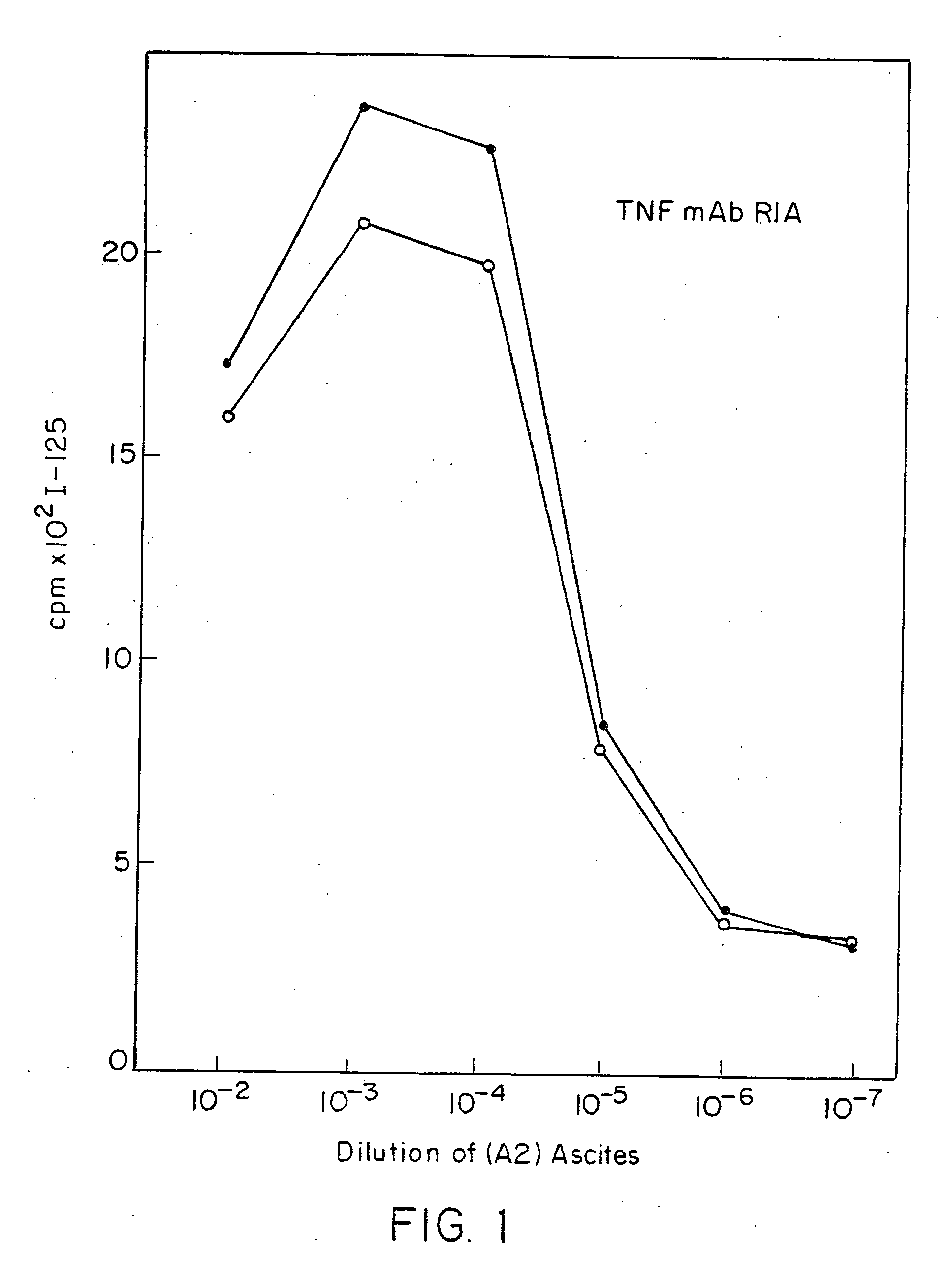
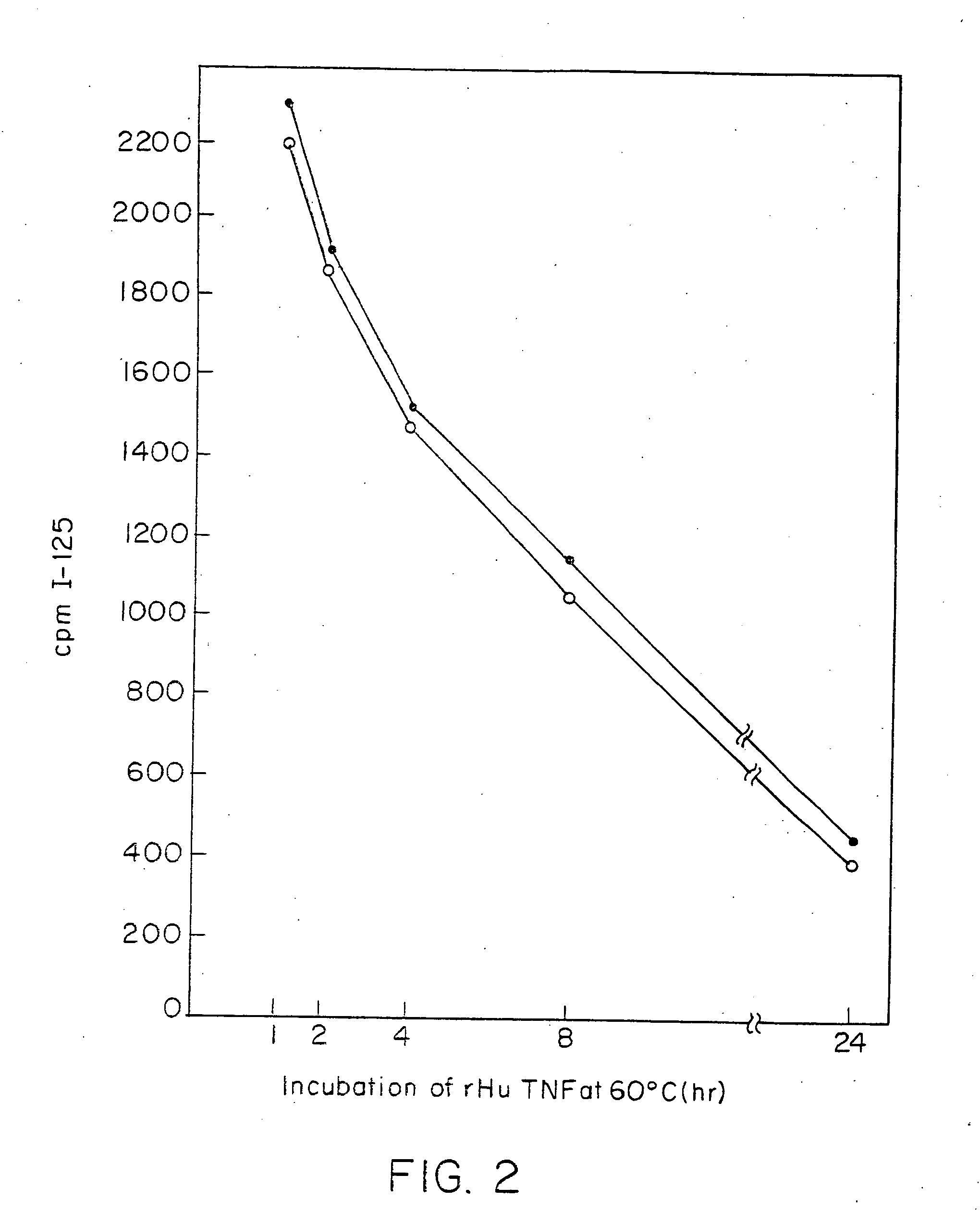
Radioimmunoassays
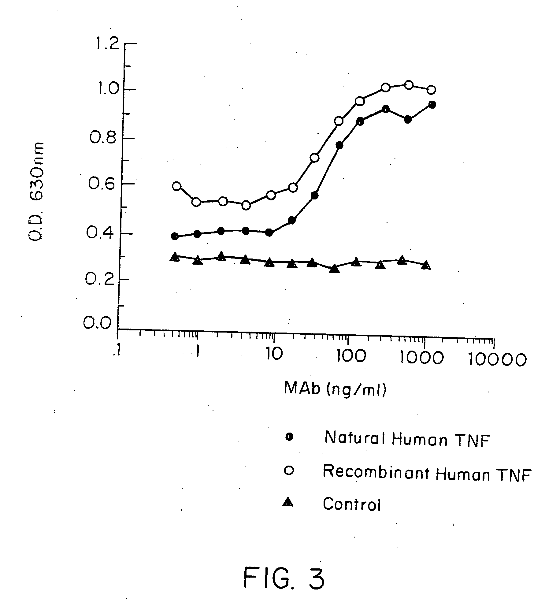
[0296]E. coli-derived rhTNF was diluted to 1 μg/ml in BCB buffer, pH 9.6, and 0.1 ml of the solution was added to each assay well. After incubation at 4° C. overnight, the wells were washed briefly with BCB, then sealed with 1% bovine incubated with 40 pg/ml of natural (GENZYME, Boston, Mass.) or recombinant (SUNTORY, Osaka, Japan) human TNFα with varying concentrations of mAb A2 in the presence of 20 μg/ml cycloheximide at 39° C. overnight. Controls included medium alone or medium +TNF in each well. Cell death was measured by staining with naphthol blue-black, and the results read spectrophotometrically at 630 nm. Absorbance at this wave length correlates with the number of live cells present.
[0297] It was found that A2 inhibited or neutralized the cytotoxic effect of both natural and rhTNF in a dose-dependent manner (FIG. 3).
[0298] In another experiment, the specificity of this inhibiting and/or neutralizing activity was tested. A673/6 cells were seeded at 3×10⁴ cells/well 20 hr before the TNF bioassay. Two-fold serial dilutions of rhTNF, E. coli-derived recombinant human lymphotoxin (TNFβ), and E. coli-derived recombinant murine TNF were prepared. The A2 hybridoma supernatant was added to an equal volume of the diluted TNF preparations, and the mixtures were incubated at room temperature for 30 min. Aliquots of 0.1 ml were transferred to the wells containing A673/6 cells, 20 μg/ml of cycloheximide was added, and the cells were incubated at 39° C. overnight. The cells were then fixed and stained for evaluation of cytotoxicity. The results indicate that mAb A2 specifically inhibited or neutralized the cytotoxicity of rhTNFα, whereas it had no effect on human lymphotoxin (TMFβ) (FIG. 4) or murine TNF (FIG. 5).
[0299] Experiments were next performed to analyze the cross-reactivity of mAb A2 with TNF derived from non-human primates. Monocytes isolated from B514 (baboon), J91 (cynomolgus) and RH383 (rhesus) blood by Ficoll gradient centrifugation and adherence, were incubated at 1×10⁵ cells/well in RPMl1640 medium with 5% FBS and 2 μg/ml of E. coli LPS for 3 or 16 hr at 37° C. to induce TNF production. Supernatants from duplicate wells were pooled and stored at 4° C. for less than 20 hr until the TNF bioassay was performed, as described above, using A673/6 cells. Two-fold dilutions of the culture supernatants were mixed with either medium or purified mAb A2 at a final concentration of 1 μg/ml, incubated at room temperature for 30 min and aliquots transferred to the indicator cells. The results showed that mAb A2 failed to significantly inhibit or neutralize the cytotoxic activity of TNF produced by baboon, cynomolgus and rhesus monkey monocytes.
[0300] A further experiment was conducted with chimpanzee TNF. Monocytes isolated from CH563 (chimpanzee) blood were incubated as described above to generate TNF-containing9 supernatants. The ability of 10 μg/ml of mAb A2 to inhibit or neutralize the bioactivity of these supernatants was assayed as above. Human TNF was used as a positive control. Results, shown in FIG. 6, indicate that mAb A2 had potent inhibiting and/or neutralizing activity for chimpanzee TNF, similar to that for human TNF (FIG. 7).
[0301] The inhibiting and/or neutralizing activity of mAb A2 was compared with three other murine mAbs specific for human TNF, termed TNF-1, TNF-2 and TNF-3, and a control mAb. Two-fold serial dilutions of purified mAbs were mixed with rhTNF (40 pg/ml), incubated at room temperature for 30 min, and aliquots tested for TNF bioactivity as above. It was found that mAbs TNF-1, TNF-2 and TNF-3 each had a similar moderate degree of inhibiting and/or neutralizing activity. In contrast, mAb A2 had much more potent inhibiting and/or neutralizing activity.

Example

EXAMPLE III
General Strategy for Cloning Antibody V and C Genes
[0302] The strategy for cloning the V regions for the H and L chain genes from the hybridoma A2, which secretes the anti-TNF antibody described above, was based upon the linkage in the genome between the V region and the corresponding J (joining) region for functionally rearranged (and expressed) Ig genes. J region DNA probes can be used to screen genomic libraries to isolate DNA linked to the J regions. Although DNA in the germline configuration (i.e., unrearranged) would also hybridize to J probes, this DNA would not be linked to a Ig V region sequence and can be identified by restriction enzyme analysis of the isolated clones.
[0303] The cloning utilized herein was to isolate V regions from rearranged H and L chain genes using JH and Jk probes. These clones were tested to see if their sequences were expressed in the A2 hybridoma by Northern analysis. Those clones that contained expressed sequence were cloned into expression vectors containing human C regions and transfected into mouse myeloma cells to determine if an antibody was produced. The antibody from producing cells was then tested for binding specificity and functionally compared to the A2 murine antibody.
EXAMPLE IV
Construction of a L Chain Genomic Library
[0304] To isolate the L chain V region gene from the A2 hybridoma, a size-selected genomic library was constructed using the phage lambda vector charon 27. High molecular weight DNA was isolated from A2 hybridoma cells and digested to completion with restriction endonuclease HindIII. The DNA was then fractionated on a 0.8% agarose gel and the DNA fragments of three different size ranges of approximately 3 kb, 4 kb and 6 kb were isolated from the gel by electroelution. The size ranges for library construction were chosen based upon the size of Hind III fragments that hybridized on a southern blot with the Jk probe. After phenol/chloroform extraction and ethanol precipitation, the DNA fragments from each size class were ligated with lambda charon 27 arms and packaged into phage particles in vitro using Gigapack Gold from Stratagene (LaJolla, Calif.).
[0305] These libraries were screened directly at a density of approximately 20,000 plaques per 150 mm petri dish using a 32P-labeled Jk probe. The mouse L chain Jk probe was a 2.7 kb HindIII fragment containing all five Jk segments. The probe was labeled with 32P by random priming using a kit obtained from Boehringer Mannheim. Free nucleotides were removed by centrifugation through a Sephadex G-50 column. The specific activities of the probe was approximately 109 cpm/μg.
[0306] Plaque hybridizations were carried out in 5×SSC, 50% formamide, 2× Denhardt's reagent, and 200 μg/ml denatured salmon sperm DNA at 42° C. for 18-20 hours. Final washes were in 0.5×SSC, 0.1% SDS at 65° C. Positive clones were identified after autoradiography.
EXAMPLE V
Construction of H Chain Genomic Library
[0307] To isolate the V region gene for the A2 H chain, a genomic library was constructed in the lambda gt10 vector system. High molecular weight DNA was digested to completion with restriction endonuclease EcoRI and fragments of approximately 7.5 kb were isolated after agarose gel electrophoresis. These fragments were ligated with lambda gt10 arms and packaged into phage particles in vitro using Gigapack Gold.
[0308] This library was screened at a density of 20, 000 plaques per 150 mm plate using a JH probe. The JH probe was a 2 kb BamHI/EcoRI fragment containing both J3 and J4 segments. The probe was labeled as in Example III and had a similar specific radioactivity. Hybridization and wash conditions were identical to those used in Example III.
EXAMPLE VI
Cloning of the TNF-Specific V Gene Regions
[0309] Several positive clones were isolated from the H and L chain libraries after screening approximately 106 plaques from each library using the JH and Jk probes, respectively. Following plaque purification, bacteriophage DNA was isolated for each positive clone, digested with either EcoRI (H chain clones) or HindIII (L chain clones) and fractionated on 1% agarose gels. The DNA was transferred to nitrocellulose and the blots were hybridized with the JH or the JK probe.
[0310] Several H chain clones were obtained that contained 7.5 kb EcoRI DNA encoding fragments of MAbs to the JH probe. For the light chain libraries, several clones from each of the three size-selected libraries were isolated that contained HindIII fragments that hybridize to the Jk probe. For the L chain, several independently derived HindIII fragments of 2.9 kb from the 2 kb library hybridized with a 1250 bp mRNA from A2, but not with SP2/0 mRNA (see Example VII). In addition, several HindIII fragments derived from the 4 kb library hybridized both to the A2 mRNA and the fusion partner mRNA. A 5.7 kb HindIII fragment from the 6 kb library did not hybridize to either RNA.
[0311] The observed lengths of hybridizing A2 mRNA were the correct sizes for H and L chain mRNA, respectively Because the RNA expression was restricted to the A2 hybridoma, it was assumed that the 7.5 kb H chain fragments and the 2.9 kb L chain fragments contained the correct V region sequences from A2. One example of each type was chosen for further study. The important functional test is the demonstration that these V regions sequences, when combined with appropriate C region sequences, are capable of directing the synthesis of an antibody with a specificity and affinity similar to that of the murine A2 antibody.
[0312] The 7.5 kb H chain fragment and the 2.9 kb L chain fragment were subcloned into plasmid vectors that allow expression of the chimeric mouse/human proteins in murine myeloma cells (see Examples VIII and IX). These plasmids were co-transfected into SP2/0 cells to ascertain if intact antibody molecules were secreted, and if so, if they were of the correct specificity and affinity. Control transfections were also performed pairing the putative anti-TNF H chain with an irrelevant, but expressed, L chain; the putative anti-TNF L chain was also paired with an irrelevant, but expressed, H chain. The results indicated that the 7.5 kb H chain fragment could be expressed, whereas the 2.9 kb L chain fragment could not. This was confirmed by DNA sequence analysis that suggested portions of the coding region were not in the proper amino acid reading frame when compared to other known L chain amino acid sequences.
[0313] Because the 2.9 kb HindIII fragment appeared not to contain a functional V gene, the 4.0 kb and 5.7 kb HindIII fragments isolated from L chain libraries were cloned into expression vectors and tested for expression of chimeric antibody after co-transfection with the 7.5 kb H chain. The 5.7 kb HindIII fragment was incapable of supporting antibody expression, whereas the 4.0 kb HindIII fragment did support antibody expression. The antibody resulting from the co-transfection of the 7.5 kb putative H chain V region and the 4.0 kb L chain V region was purified, tested in solid phase TNF binding assay, and found to be inactive. It was concluded that the V region contained on the 4.0 kb HindIII fragment was not the correct anti-TNF V regions, but was contributed to the hybridoma by the fusion partner. This was subsequently confirmed by sequence analysis of cDNA derived from the A2 hybridoma and from the fusion partner.
[0314] Other independently derived L chain clones containing 2.9 kb HindIII fragments that hybridized with A2 mRNA were characterized in more detail. Although the restriction maps were similar, the clones fell into two classes with respect tot the presence or absence of an AccI enzyme site. The original (non-functional) 2.9 kb fragment (designated clone 8.3) was missing an AccI site present in some other clones (represented by clone 4.3). The DNA sequence of clone 4.3 was extremely similar to clone 8.3, but contained a single amino acid reading frame with close homology to known L chains, unlike clone 8.3. The 2.9 kb HindIII fragment from clone 4.3 was subcloned into the L chain expression vector and co-transfected with the putative anti-TNF H chain into SP2/0 cells. An antibody was synthesized, purified and tested in the solid phase TNF binding assay. This antibody bound to TNF, and therefore, the clone 4.3 L chain V region was assumed to be the correct one.
[0315] The A2 murine hybridoma has been shown to contain at least four rearranged L chain V region genes. At least two of these are expressed as proteins: clone 4.3 (the correct anti-TNF L chain gene) and the gene contained in the 4.0 kb HindIII fragment (contributed by the fusion partner). The expression of two L chains implies that the resulting antibody secreted from the murine hybridoma is actually a mixture of antibodies, some using the correct L chain, some using the incorrect L chain, and some using one of each. The presence of two different L chains in the murine A2 antibody has been confirmed by SDS gel and N-terminal protein sequence analysis of the purified antibody. Because construction of the chimeric A2 antibody involves cloning the individual H and L chain genes and expressing them in a non-producing cell line, the resulting antibody will have only the correct L chain and therefore should be a more potent antibody (see Examples X, XI and XII).
EXAMPLE VII
Northern Analysis of Cloned DNA
[0316] Cloned DNA corresponding to the authentic H and L chain V regions from the A2 hybridoma would be expected to hybridize to A2 mRNA. Non-functional DNA rearrangements at either the H or L chain genetic loci should not be expressed.
[0317] Ten μg total cellular RNA was subjected to electrophoresis on 1% agarose/formaldehyde gels (Sambrook et al, infra) and transferred to nitrocellulose. Blots were hybridized with random primed DNA probes in 50% formamide, 2× Denhardt's solution, 5×SSC, and 200 μg/ml denatured salmon sperm DNA at 42° C. for 10 hours. Final wash conditions were 0.5×SSC, 0.1% SDS at 65° C.
[0318] The subcloned DNA fragments were labeled with 32P by random priming and hybridized to Northern blots containing total RNA derived from A2 cells or from cells of SP2/0, the fusion partner parent of A2. The 7.5 kb EcoRI H chain fragment hybridized with a 2 kb mRNA from A2, but not with SP2/0 mRNA. Similarly, the 2.9 kb L chain HindIII fragment (clone 4.3) hybridized with a 1250 bp mRNA from A2, but not with SP2/0 mRNA. The observed lengths of A2 mRNA hybridizing were the correct sizes for H and L chain mRNA, respectively, confirming that the V region sequences on these DNA fragments are expressed in A2 hybridoma cells.
EXAMPLE VIII
Construction of Expression Vectors
[0319] The putative L (clone 4.3) and H chain V genes described above were joined to human kappa and gamma1 constant region genes in expression vectors. The 7.5 kb EcoRI fragment corresponding to the putative VH region gene from A2 was cloned into an expression vector containing the human Cgamma1 gene and the Ecogpt gene to yield the plasmid designated pA2HG1apgpt (see FIG. 8).
[0320] The 2.9 kb putative VL fragment from clone 4.3 was cloned into a vector containing the human kappa Ck gene and the Ecogpt gene to allow selection in mammalian cells. The resulting plasmid was designated pA2HuKapgpt (See FIG. 8).
EXAMPLE IX
Expression of Chimeric Antibody Genes
[0321] To express the chimeric H and L chain genes, the expression plasmids were transfected into cells of the non-producing mouse myeloma cell line, SP2/0. Plasmid DNA to be transfected was purified by centrifuging to equilibrium in ethidium bromide/cesium chloride gradients twice. Plasmid DNA (10-50 μg) was added to 107 SP2/0 cells in medium containing Hank's salts, and the mixture was placed in a BIORAD electroporation apparatus. Electroporation was performed at 20 volts, following which the cells were plated in 96 well microtiter plates.
[0322] Mycophenolic acid selection was applied after 24 hours and drug resistant colonies were identified after 1-2 weeks. Resistant colonies were expanded to stable cell lines and tissue culture supernatant from these cell lines was tested for antibody using an ELISA assay with goat anti-human IgG Fc antibody and goat anti-human H+L conjugated with alkaline phosphatase (obtained from Jackson Laboratories).
[0323] The chimeric A2 antibody,was purified from tissue culture supernatant by Protein A-Sepharose chromatography. The supernatant was adjusted to 0.1 M Tris, 0.002 M EDTA, pH 8.0 and loaded on a Protein A-Sepharose column equilibrated in the same buffer. The IgG was eluted with 0.1 M citrate, pH 3.5, inhibited or neutralized with 1 M Tris, and dialyzed into phosphate buffered saline (PBS).
[0324] The purified chimeric antibody was evaluated for its binding and inhibiting and/or neutralizing activity.
EXAMPLE X
Specificity of an Anti-TNF Chimeric Antibody
[0325] Since the antigen binding domain of cA2 was derived from murine A2, these mAbs would be expected to compete for the same binding site on TNF. Fixed concentrations of chimeric A2 and murine mAb A2 were incubated with increasing concentrations of murine and chimeric A2 competitor, respectively, in a 96-well microtiter plate coated with rhTNF (Dainippon, Osaka, Japan). Alkaline-phosphatase conjugated anti-human immunoglobulin and anti-mouse immunoglobulin second antibodies were used to detect the level of binding of chimeric and murine A2, respectively. Cross-competition for TNF antigen was observed in this solid-phase ELISA format (FIG. 9). This finding is consistent with the expected identical epitope specificity of cA2 and murine A2.
[0326] The affinity constant for binding of mouse mAb A2 and cA2 to rhTNFα was determined by Scatchard analysis (see, for example, Scatchard, Ann. N.Y. Acad. Sci. 51:660 (1949)). The results are shown in FIG. 10. This analysis involved measuring the direct binding of 125I labelled cA2 to immobilized rhTNFα in a 96-well plate. The antibodies were each labelled to a specific activity of about 9.7 μi/μg by the iodogen method. An affinity constant (Ka) of 0.5×10⁹ liters/mole was calculated for the mouse mAb A2. Unexpectedly, the chimeric A2 antibody had a higher affinity, with a Ka of 1.8×10⁹ liters/mole. Thus, the chimeric anti-TNFα antibody of the present invention was shown to exhibit a significantly higher affinity of binding to human TNFα than did the parental murine A2 mAb. This finding was surprising, since murine and chimeric antibodies, fragments and regions would be expected to have affinities that are equal to or less than that of the parent mAb.
[0327] Such high affinity anti-TNF antibodies, having affinities of binding to TNFα of at least 1×10⁸ M−1, more preferably at least 1×10⁹ M−1 (expressed as Ka) are preferred for immunoassays which detect very low levels of TNF in biological fluids. In addition, anti-TNF antibodies having such high affinities are preferred for therapy of TNF-α-mediated conditions or pathology states.
[0328] The specificity of cA2 for TNF was confirmed by testing for cross-neutralization of human lymphotoxin (TNF-β). Lymphotoxin shares some sequence homology and certain biological activities, for example, tumor cell cytotoxicity, with TNF (Pennica, et al., Nature 312:724-729 (1984)). Cultured human A673 cells were incubated with increasing concentrations of human lymphotoxin (GENENTECH, San Francisco, Calif.) with or without 4 μg/ml chimeric A2 in the presence of 20 μg/ml cycloheximide at 39° C. overnight. Cell death was measured by vital staining with naphthol blue-black, as above. The results indicated that cA2 was ineffective at inhibiting and/or neutralizing human lymphotoxin, confirming the TNFα-specificity of the chimeric antibody.
[0329] The ability of A2 or cA2 to react with TNF from different animal species was also evaluated. As mentioned earlier, there are multiple epitopes on human TNF to which inhibiting and/or neutralizing mAbs will bind (Moller, et al., infra). Human TNF has bioactivity in a wide range of host animal species. However, certain inhibiting and/or neutralizing epitopes on human TNF are conserved amongst different animal species and others appear to be restricted to humans and chimpanzees.
[0330] Neutralization experiments utilized endotoxin-activated cell supernatants from freshly isolated human, chimpanzee, rhesus and cynomolgus monkey, baboon, pig, dog, rabbit, or rat monocytes as the TNF source. As discussed above, murine mAb A2 inhibited or neutralized activity of only human and chimpanzee TNF, and had no effect on TNF derived from other primates and lower animals. A2 also did not inhibit or neutralize the cytotoxic effect of recombinant mouse TNF.
[0331] Thus, the epitope recognized by A2 is one shared by human and chimpanzee TNFα. Chimeric A2 was also tested in this manner for cross-reactivity with monocyte-derived TNF from rat, rabbit, dog and pig, as well as with purified recombinant mouse TNFα, and natural and recombinant human TNFα. Chimeric A2 only inhibited or neutralized natural and recombinant human TNFα. Therefore, cA2 appears to share species specificity with murine A2.
EXAMPLE XI
In Vitro Activity and Neutralization Efficacy of a Chimeric Anti-TNF Antibody
[0332] Both the murine and chimeric anti-TNFα antibodies, A2 and cA2 were determined to have potent TNF-inhibiting and/or neutralizing activity. In the TNF cytotoxicity assay described above, murine A2, at a concentration of about 125 ng/ml completely inhibited or neutralized the biological activity of a 40 pg/ml challenge of rhTNFα. Two separate determinations of inhibiting and/or neutralizing potency, expressed as the 50% Inhibitory Dose (ID50) were determined to be 15.9±1.01 and 17.9±1.6 ng/ml (Mean±Std error). Thus the mAb A2 has an ID50 of about 17 ng/ml.
[0333] In this same experimental system, three other murine anti-TNFα antibodies (termed TNF-1, TNF-2 and TNF-3) of comparable binding affinity to TNF were found to have ID50 values of 1-2 orders of magnitude greater, and thus were significantly less potent in neutralization than A2.
[0334] The ability of cA2 to inhibit or neutralize human TNFα bioactivity in vitro was tested using the bioassay system described above. Cultured A673 cells were incubated with 40 pg/ml natural (Genzyme, Boston, Mass.) or recombinant (Suntory, Osaka, Japan) human TNF with or without antibody overnight as above, and cell death was measured by vital staining. As expected based upon the above results with the A2 mouse mAb, cA2 also inhibited or neutralized both natural and rhTNF in a dose-dependent manner in the cytotoxicity assay (FIG. 11). In this assay format, levels of cA2 as low as 125 ng/ml completely abolished the toxic activity of TNF. Upon repeated analysis, the cA2 exhibited greater TNF-inhibiting and/or neutralizing activity than did the parent murine A2 mAb. Such inhibiting and/or neutralizing potency, at antibody levels below 1 μg/ml, can easily be attained in the blood of a subject to whom the antibody is administered. Accordingly, such highly potent inhibiting and/or neutralizing anti-TNF antibodies, in particular the chimeric antibody, are preferred for therapeutic use in TNFα-mediated pathologies or conditions.
[0335] As mentioned above, TNF induces cellular secretion of IL-6. Furthermore, there is evidence that IL-6 is involved in the pathophysiology of sepsis, although the precise role of IL-6 in that syndrome is unclear (Fong, et al., J Exp Med 170:1627-1633 (1989); Starnes Jr., et al., J Immunol 145:4185-4191 (1990)). The ability of cA2 to inhibit or neutralize TNF-induced IL-6 secretion was evaluated using cultured human diploid FS-4 fibroblasts. The results in Table 2 show that cA2 was effective in blocking IL-6 secretion in cells that had been incubated overnight with TNF. TNF-induced IL-6 secretion was not inhibited in the absence of a mAb or in the presence of a control mAb specific for an irrelevant antigen. TABLE 2 IN VITRO NEUTRALIZATION OF TNF-INDUCED IL-6 SECRETION TNF Concentration (ng/ml) Antibody 0 0.3 1.5 7.5 None <0.20 1.36 2.00 2.56 Control mAb <0.20 1.60 1.96 2.16 cA2 <0.20 <0.20 <0.20 0.30
Values represent mean concentrations of IL-6 of duplicate wells, in ng/ml. RhTNF (Suntory, Osaka, Japan), with or without 4 μg/ml antibody, was added to cultures of FS-4 fibroblasts and after 18 h, the supernatant was assayed for IL-6 using the QUANTIKINE Human IL-6 Immunoassay (from R&D
# Systems, Minneapolis, MN).
Control mAb = chimeric mouse/human IgG1 anti-platelet mAb (7E3).
[0336] The ability of TNF to activate procoagulant and adhesion molecule activities of endothelial cells (EC) is thought to be an important component of pathology pathophysiology. In particular, this can be associated with the vascular damage, disseminated intravascular coagulation, and severe hypotension that is associated with the sepsis syndrome. Therefore, the ability of cA2 to block TNF-induced activation of cultured human umbilical vein endothelial cells (HUVEC) was evaluated. TNF stimulation of procoagulant activity was determined by exposing intact cultured HUVEC cells to TNF (with or without antibody) for 4 hours and analyzing a cell lysate in a human plasma clotting assay. The results in Table 3 show the expected upregulation by TNF of HUVEC procoagulant activity (reflected by a decreased clotting time). Chimeric antibody cA2 effectively inhibited or neutralized this TNF activity in a dose-dependent manner. TABLE 3 IN VITRO NEUTRALIZATION OF TNF-INDUCED PROCOAGULANT ACTIVITY TNF Concentration (ng/ml) Antibody μg/ml 250 25 0 None — 64 ± 4* 63 ± 1 133 ± 13 Control Ab 10.00 74 ± 6 N.D. 178 ± 55 cA2 10.00 114 ± 5 185 ± 61 141 ± 18 cA2 3.30 113 ± 2 147 ± 3 N.D. cA2 1.10 106 ± 1 145 ± 8 N.D. A2 0.37 73 ± 17 153 ± 4 N.D. cA2 0.12 64 ± 1 118 ± 13 N.D.
*Values represent mean plasma clotting time, in seconds (±S.D.). Clotting time was determined in normal human plasma after addition of the rhTNF (Dainippon, Osaka, Japan) with or without antibody-treated HUVEC lysate and Ca++ at 37° C.
N.D. = Not done.
Control Ab is a chimeric mouse/human IgG1 anti-CD4 antibody.
[0337] In addition to stimulating procoagulant activity, TNF also induces surface expression of endothelial cell adhesion molecules such as ELAM-1 and ICAM-1. The ability of cA2 to inhibit or neutralize this activity of TNF was measured using an ELAM-1 specific detection radioimmunoassay.
[0338] Cultured HUVEC were stimulated with 250 ng/ml rhTNF (Dainippon, Osaka, Japan) with or without antibody at 37° C. overnight in a 96-well plate format. Surface expression of ELAM-1 was determined by sequential addition of a mouse anti-human ELAM-1 mAb and 125I-labelled rabbit anti-mouse immunoglobulin second antibody directly to culture plates at 4° C.
[0339] As shown in FIG. 12, TNF induced the expression of ELAM-1 on the surface of cultured HUVEC cells, and this activity was again effectively blocked in a dose-related manner by cA2.
[0340] Finally, TNF is known to stimulate mitogenic activity in cultured fibroblasts. Chimeric A2 inhibited or neutralized TNF-induced mitogenesis of human diploid FS-4 fibroblasts cultures, confirming the potent inhibiting and/or neutralizing capability of cA2 against a broad spectrum of in vitro TNF biological activities.
EXAMPLE XII:
Determination of Amino Acid Sequences (Epitope) on Human TNF-α Recognized by cA2 mAb
[0341] Reagents The following reagents are readily available from commercial sources. FMOC-L-Ala-OPfp, FMOC-L-Cys(Trt)-OPfp, FMOC-L-Asp(OtBu)-OPfp, FMOC-L-Glu(OtBu)-OPfp, FMOC-L-Phe-OPfp, FMOC-Gly-OPfp, FMOC-L-His(Boc)-OPfp, FMOC-L-Ile-OPfp, FMOC-L-Lys(Boc)-OPfp, FMOC-L-Leu-OPfp, FMOC-L-Asn-OPfp, FMOC-L-Pro-OPfp, FMOC-L-Gln-OPfp, FMOC-L-Arg(Mtr)-OPfp, FMOC-L-Ser(tBu)-ODhbt, FMOC-L-Thr(tBu)-ODhbt, FMOC-L-Val-OPfp, FMOC-L-Trp-OPfp, FMOC-L-Try(tBu)-OPfp, and 1-hydroxybenotriazol (HOBT) were obtained from Cambridge Research Biochemicals. Piperidine and was obtained from Applied Biosystems, Inc. 1-Methyl-2-Pyrrolidinone (NMP) was obtained from EM Science; Methanol from JT Baker; Acetic Anhydride from Applied Biosystems, Inc., Trifluoroaccetic acid (TFA) from Applied Biosystems, Inc.; Diisopropylamine (DIEA), Triethylamine, Dithiothreitol (DTT) and Anisole from Aldrich and Hydrochloric Acid (HCl) from JT Baker.
[0342] Abbreviations: FMOC, 9-fluorenylmethoxycarbonyl; tBu t-butyl ether; OtB, t-butyl ester; Boc, t-butyloxycarbonyl; Mtr, 4-methoxy-2,3,6-trimethylbenzenesulfonyl; Trt, trityl; OPfp, pentafluorophenylester; ODhbt. oxo-benzotriazone ster;
[0343] A chimeric antibody of the present invention, designated cA2, was used to determine which portions of the TNF amino acid sequence were involved in inhibitory binding by the antibody by epitope mapping, whereby the amino acid sequences of TNF-α recognized by cA2 have been identified.
[0344] The complete primary sequence of human TNFα, according to Pennica et al, Nature 312:724-729 (1984) is shown in FIG. 13 (SEQ ID NO:1). Overlapping decapeptides beginning with every second amino acid and covering the entire amino acid sequence of human TNF-α were synthesized on polyethylene pins using the method of Gysen (Gysen et al., Peptides: Chemistry and Biological, Proceedings of the Twelfth American Peptide Symposium, p. 519-523, Ed, G. R. Marshall, Escom, Leiden, 1988). Sets of peptide pins bearing free N-terminal amino groups and acetylated N-terminal amino groups were individually prepared. Both sets of peptide pins were incubated in solutions containing the anti-TNF mAb cA2 to determine the amino acid sequences that make up the cA2 epitope on human TNF-α, as described below. FIG. 14A shows the results of binding to the overlapping decapeptides that comprise the entire sequence of human TNFα. The O.D. (optional density) correlates directly with the increased degree of cA2 binding. FIG. 14B shows the results of binding of cA2 to the same set of peptide pins in the presence of human TNFα. This competitive binding study delineates peptides which can show non-specific binding to cA2.
[0345] There are at least two non-contiguous peptide sequences of TNF-α recognized by cA2. Using the conventional protein numbering system wherein the N-terminal amino acid is number 1, the cA2 mAb recognizes an epitope composed at least in part of amino acids located within residues 87-108 or both residues 59-80 and 87-108 of TNF (SEQ ID NO:1). FIG. 15 presents these non-contiguous sequences within the TNF sequence. These sequences are also shown in a space filling model in FIG. 16B, along with a space filing model of the TNF monomer shown in FIG. 16A.
[0346] Unexpectedly, the mAb cA2 blocks the action of TNF-α without binding to the putative receptor binding locus, which can include one or more of, e.g., 11-13, 37-42, 49-57 or 155-157 of hTNFα (of SEQ ID NO:1). Preferred anti-TNF mabs are those that inhibit this binding of human TNF-α to its receptors by virtue of their ability to bind to one or more of these peptide sequences. These antibodies can block the activity of TNF by virtue of binding to the cA2 epitope, such binding demonstrated to inhibit TNF activity. The identification of those peptide sequences recognized by cA2 provides the information necessary to generate additional MAbs with binding characteristics and therapeutic utility that parallel the embodiments of this application.
[0347] Peptide Pin Synthesis. Using an epitope mapping kit purchased from Cambridge Research Biochemicals, Inc. (CRB), dodecapeptides corresponding to the entire sequence of human TNF-α were synthesized on polyethylene pins.
[0348] A synthesis schedule was generated using the CRB epitope mapping software. Prior to the first amino acid coupling, the pins were deprotected with a 20% piperidine in NMP solution for 30 minutes at room temperature. After deprotected, the pins were washed with NMP for five minutes at room temperature, followed by three methanol washes. Following the wash steps, the pins were allowed to air dry for at least 10 minutes.
[0349] The following procedure was performed for each coupling cycle:
[0350] 1) The amino acid derivatives and the HOBT were weighted out according to the weights required in the synthesis schedule.
[0351] 2) The HOBT was dissolved in the appropriate amount of NMP according to the synthesis schedule.
[0352] 3) The amino acid derivatives were dissolved in the recommended amount of HOBT solution and 150 microliters were pipeted into the appropriate wells as directed by the well position sheet of the synthesis schedule.
[0353] 4) The blocks containing the pins were placed into the wells, and the “sandwich” units stored in plastic bags in a 30° C. water bath for 18 hours.
[0354] 5) The pins were removed from the wells and washed once (for 5 minutes) with NMP, three times (for two minutes) with methanol and air dried for 10 minutes.
[0355] 6) The pins were deprotected as described above and the procedure repeated.
[0356] To acetylate the peptides on one block of pins, the peptide pins were washed, deprotected and treated with 150 microliters of a solution containing NMP; acetic anhydride:triethylamine (5:2:1) for 90 minutes at 30° C., followed by the washing procedure outlined above. The second set of peptide pins was deprotected by not acetylated to give free N-terminal amino groups.
[0357] The final deprotection of the peptides to remove the side chain protecting groups was done using a mixture of TFA:anisole:dithiothreitol, 95:2.5:2.5 (v/v/w) for four hours at ambient temperature. After deprotection, the pins were air dried for 10 minutes, followed by a 15 minute sonication in a solution of 0.% HCl in methanol/distilled water (1:1). The pins dried over night and were then ready for testing. ELISA Assay for cA2 Binding to TNF-α Peptide PINs
[0358] Reagents: Disruption Buffer: Sodium dihydrogen phosphate (31.2 g, Sigma cat # S-0751 or equivalent) and sodium dodecylsulfate (20.0 g, Sigma cat # L-3771 or equivalent) were dissolved in 2.0 L of milliQ water. The pH was adjusted to 7.2±0.1 with 50% w/w sodium hydroxide (VWR cat # VW6730-3 or equivalent).
[0359] Blocking Buffer: Sodium dihydrogen phosphate (0.39 g, Sigma cat #S-0751 or equivalent) disodium hydrogen phosphate (1.07 g, Baker cat # 3828-1 or equivalent) and sodium chloride (8.50 g, Baker cat # 3624-5 or equivalent were dissolved in 1.0 L of .milliQ water. The pH was adjusted to 7.2±0.1 with 50% w/w sodium hydroxide (VWR cat VW6730-3 or equivalent). Chicken egg albumin (10.0 g, Sigma cat #A-5503 or equivalent) and bovine serum albumin (10.0 g, Sigma, cat #A-3294 or equivalent) were dissolved at room temperature with gentle stirring. The solution was filtered, and to the solution was added Tween 20 (2. 0 ml, Sigma cat #P-13.79 or equivalent). The solution was stirred gently at room temperature for 30 min, filtered and stored at 40°.
[0360] PBS/Tween 20: A 10× concentrate was prepared by dissolving sodium dihydrogen phosphate (3.90 g, Sigma cat # S-0751 or equivalent), disodium hydrogen phosphate (10.70 g, Baker cat #3828-1 or equivalent) and sodium chloride (85.0 g, Baker cat #3624-5 or equivalent) in 1.0 L of milliQ water. The pH was adjusted to 7.2±0.1 with 50% w/w sodium hydroxide (VWR cat #VW 6730 or equivalent). To the solution was added Tween 20 (5.0 mL, Sigma cat #P-1379 or equivalent), and the mixture stirred gently. Just prior to use 100 mL of this solution was diluted to 1.0 L with milliQ water.
[0361] Substrate solution: Substrate buffer was prepared by dissolving citric acid (4.20 g, Malinckrodt cat #0627 or equivalent) and disodium hydrogen phosphate (7.10 g, Baker cat #3828-1 or equivalent) in 1.0 L of milliQ water. The pH was adjusted to 5.00 with 50% w/w sodium hydroxide (VWR cat #VW6730-3 or equivalent). Immediately prior to use an OPD substrate tablet (30 mg, Sigma cat #P-8412 or equivalent and 30% (v/v) hydrogen peroxide (40 μL, Sigma cat #P-1379 or equivalent) were added to the substrate buffer 25.0 mL). The solution was wrapped in foil and mixed thoroughly.
[0362] 4 NH2SO4; Sulfuric acid (53 mL, EM Science cat #SX1244-5 or equivalent) was slowly added to MILLI-Q water (447 mL) and cooled to room temperature prior to use.
[0363] Equipment: Molecular Devices Model nu-max plate reader or equivalent. Scientific Products Model R4140 Oscillating table shaker and equivalent. BRANSON Model 5200 ultra-sonic bath or equivalent. FINNPIPETTE Model 4172317 multichannel pipeter or equivalent. CORNING Model 25801 96 well disposable polystyrene Elisa Plates.
[0364] Prior to use and after each subsequent use the peptide pins were cleaned using the following procedure. Disruption buffer (2.0 L) was heated to 60° and placed in an ultra-sonic bath in a fume hood. To the disruption buffer was added dithiolthreitol (2.5 g, Sigma cat #D-0632 or equivalent). The peptide pins were sonicated in this medium for 30 min, washed thoroughly with milliQ waster, suspended in a boiling ethanol bath for 2 min, and air-dried.
[0365] Blocking buffer (200 μL) was added to a 96 well disposable polystyrene Elisa plate and the peptide pins suspended in the wells. The peptide pins and plate were incubated for 2 h at room temperature on an oscillating table shaker. The plates and peptide pins were washed with PBS/Tween 20 (four times). To each well was added a 20 μg/ml concentration of cA2 antibody (diluted with blocking buffer, 175 μL/well). TNF competition was done by incubation of TNFα (40 μg/ml) and cA2 (20 μg/ml) in BSA/ovalbumin/BBS for three hours at room temperature. The peptide pins were suspended in the plate and incubated at 4° overnight. The peptide pins and plate were washed with PBS/Tween 20 (four times). To each well was added anti-human goat antibody conjugated to horseradish peroxidase (diluted with blocking buffer to 1/2000, 175 μL/well, Jackson IMMUNORESEARCH Labs). The peptide pins were suspended in the plate, and incubated for 1 h at room temperature on a oscillating table shaker. The plates and peptide pins were washed with PBS/Tween 20 (four times). To each well added freshly prepared substrate solution (150 μL/well), the peptide pins were suspended in the plate and incubated for 1 h at room temperature on an oscillating table shaker. The peptide pins were removed and to each well is added 4N H2SO4 (50 μL). The plates were read in a Molecular Devices plate reader (490 nm, subtracting 650 nm as a blank), and the results are shown in FIGS. 14A and 14B, as described above.

PUM

PropertyMeasurementUnit
Van der Waals' coefficient b108.0L / mol
Temperature37.0°C
Volume1000.0L
tensileMPa
Particle sizePa
strength10

Description & Claims & Application Information

We can also present the details of the Description, Claims and Application information to help users get a comprehensive understanding of the technical details of the patent, such as background art, summary of invention, brief description of drawings, description of embodiments, and other original content. On the other hand, users can also determine the specific scope of protection of the technology through the list of claims; as well as understand the changes in the life cycle of the technology with the presentation of the patent timeline. Login to view more.

Similar technology patents

Peptides with capacity to bind to scurfin and applications

InactiveUS20100267623A1Inhibit biological activityUseful in treatmentAntibacterial agentsBiocidePeptideCrystallography
Owner:PROYECTO DE BIOMEDICINA CIMA

Nucleus factor-kB p50 subunit combination peptide, preparation and application thereof

InactiveCN1687128AInhibit biological activityInhibit expressionPeptide/protein ingredientsAntipyreticPeptideFactor ii
Owner:THE THIRD AFFILIATED HOSPITAL OF THIRD MILITARY MEDICAL UNIV OF PLA

Cystatin C as an antagonist of TGF-beta and methods related thereto

ActiveUS20050267021A1Inhibit biological activityDepsipeptidesPeptide preparation methodsCystatinBiology
Owner:NAT JEWISH HEALTH

Classification and recommendation of technical efficacy words

  • High affinity
  • Inhibit biological activity

Soluble tgf-b type III receptor fusion proteins

InactiveUS20070184052A1Increase activin signalHigh affinityAnimal cellsSugar derivativesOverproductionTgf β superfamily
Owner:THE GENERAL HOSPITAL CORP

Non-mammalian GnRH analogs and uses thereof in the immune system

InactiveUS20050043245A1High affinityGreat affinityPeptide/protein ingredientsLuteinising hormone-releasing hormoneGnRH AnalogChemistry
Owner:SILER KHODR THERESA

Nucleus factor-kB p50 subunit combination peptide, preparation and application thereof

InactiveCN1687128AInhibit biological activityInhibit expressionPeptide/protein ingredientsAntipyreticPeptideFactor ii
Owner:THE THIRD AFFILIATED HOSPITAL OF THIRD MILITARY MEDICAL UNIV OF PLA

Cystatin C as an antagonist of TGF-beta and methods related thereto

ActiveUS20050267021A1Inhibit biological activityDepsipeptidesPeptide preparation methodsCystatinBiology
Owner:NAT JEWISH HEALTH

Peptides with capacity to bind to scurfin and applications

InactiveUS20100267623A1Inhibit biological activityUseful in treatmentAntibacterial agentsBiocidePeptideCrystallography
Owner:PROYECTO DE BIOMEDICINA CIMA
Why Eureka
Try Eureka