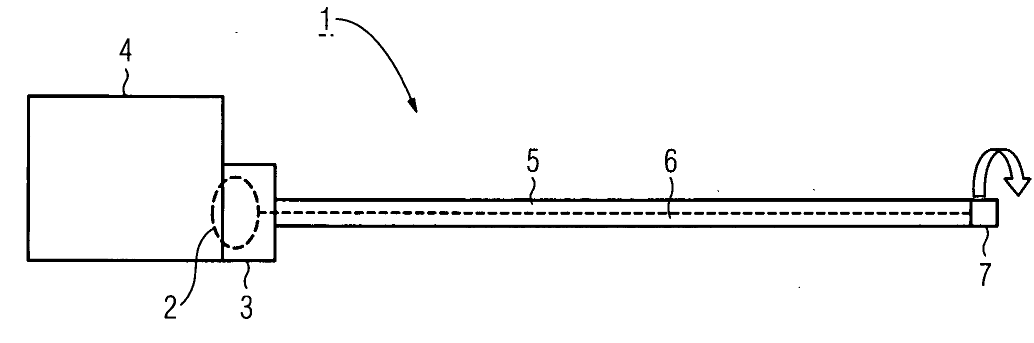
[0007] The intravenous pacemaker electrode according to the invention comprises an electrode cable and an electrode head linked thereto, which is provided in order to transmit electrical stimulation pulses. A duct and a line leading to the electrode head run in an electrode cable comprising an insulating sleeve. An
ultrasound catheter can be moved into the duct of the electrode cable, said ultrasound
catheter comprising a thread-like guide element and an ultrasound measurement element attached to its distal end. The guide element of the ultrasound catheter preferably serves both to advance the ultrasound measurement element in the electrode cable as well as to transmit electrical signals. A measurement element is understood as an ultrasound measurement element, said measurement element comprising both an ultrasound emitter and also an ultrasound
receiver. The combination of the pacemaker electrode with the ultrasound measurement element enables an
imaging diagnosis with a good resolution in the heart. This diagnosis is particularly advantageous during x-
ray illumination carried out simultaneously. The ultrasound catheter is not permanently linked to the remaining parts of the pacemaker electrode, but is however only inserted into the duct of the electrode cable if necessary.
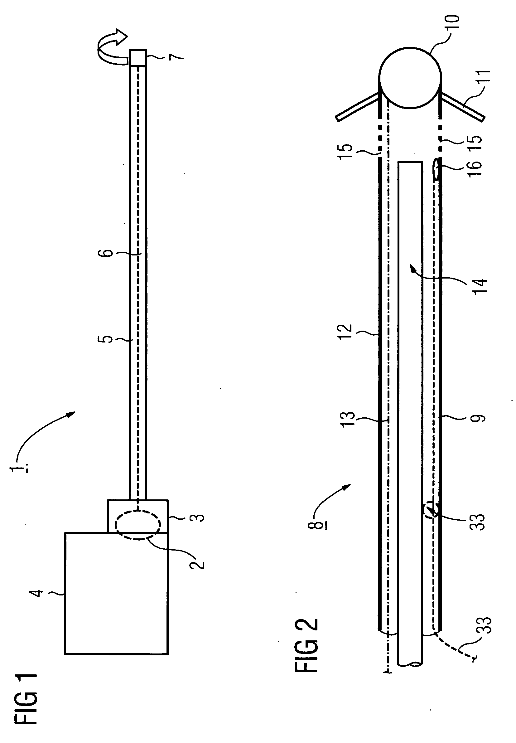
[0008] The duct is preferably closed to such an extent that the ultrasound catheter cannot come into contact with the patient's blood or
body tissue. Thus the ultrasound catheter can be used widely, even with different patients. The area of the electrode cable bordering the electrode head is preferably configured such that an ultrasound measurement is possible to a large extent uninfluenced by the material of the pacemaker electrode. For this purpose, an
axial distance between the distal end of the duct and the electrode head is advantageous, with the ultrasound catheter, in particular its ultrasound measurement element, being able to be moved through the duct towards the electrode head. At least one window which is transparent for the ultrasound, for instance a window ring, is arranged in the area of the insulating sleeve of the electrode cable bordering the electrode head.
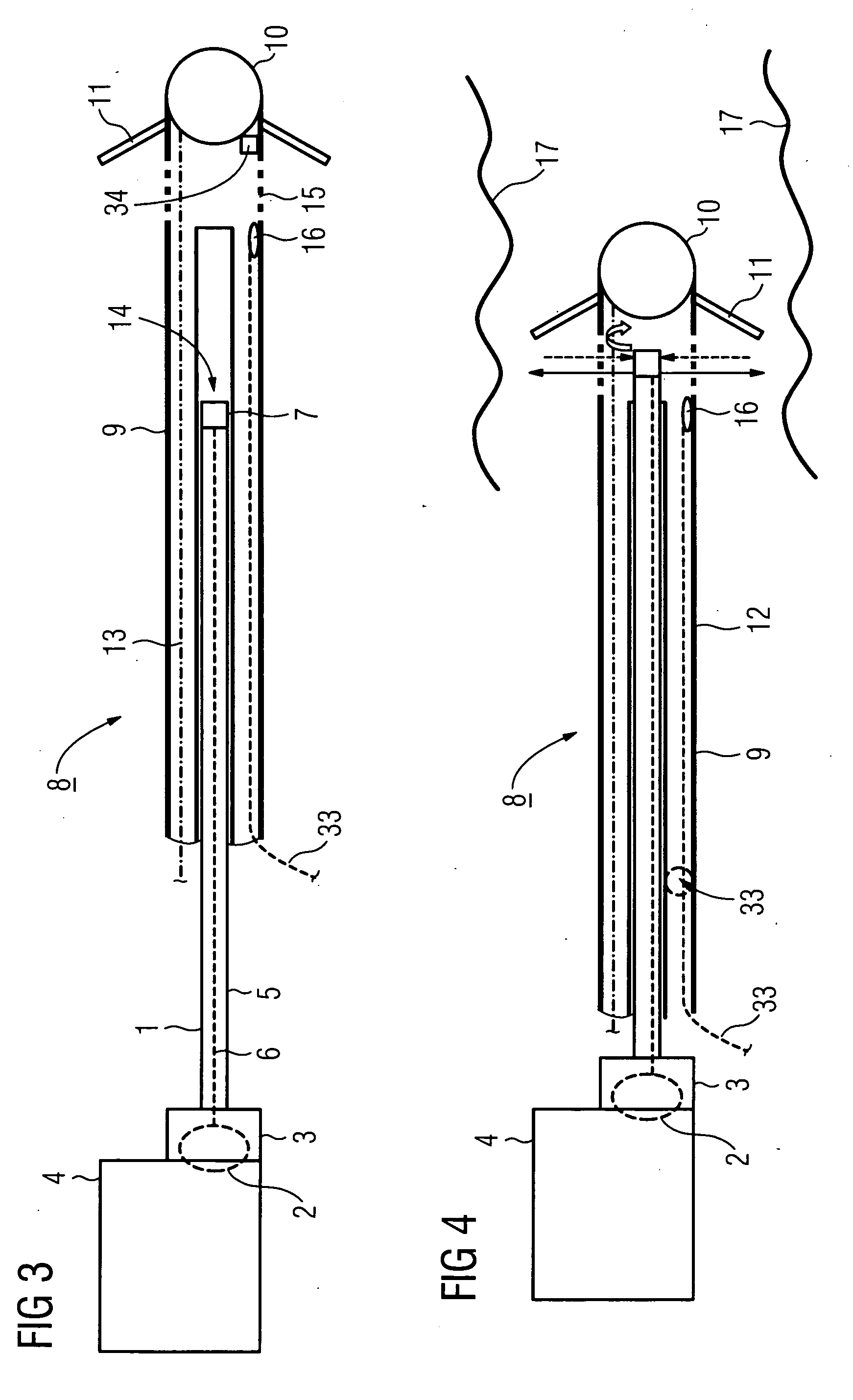
[0009] According to a preferred development, besides the duct for the ultrasound catheter, provision is made for a fluid duct suitable for conveying a contrast means towards the electrode head, and comprising an outlet opening in front of the electrode head. In contrast, provision can also be made for example to guide the contrast means through the same duct which is also suitable for inserting the ultrasound catheter. The use of a contrast means essentially broadens the diagnostic possibilities of the ultrasound examination. The outlet opening of the fluid duct preferably comprises a sealing device, which prevents body fluids from flowing into the electrode cable, in the manner of a non-return valve.
[0010] In addition to the duct for the ultrasound catheter and if necessary to the fluid duct for the contrast means, the electrode cable comprises an advantageous embodiment of a further guide duct provided for the
insertion of a guidewire. The guidewire can also be identical to one of the aforementioned ducts. Independent of the total number of ducts in the electrode cable, provision is made according to a preferred development for an exit opening for the ultrasound catheter in the region of the electrode head. This allows the ultrasound measurement element to be moved past the electrode head, provided that the exit opening is located in the electrode head, even out via the electrode head.
[0016] According to a second embodiment the diagnosis and treatment device additionally comprises a
telemetry module for intravenous pacemaker electrodes, in particular with the features of the claims, which is arranged in a pacemaker housing to which the electrode cable is connected and also comprises a
data link to the ultrasound measurement element. The
telemetry module allows the ultrasound measurement data to be read out even after the pacemaker has been implanted. The features of the two previously described exemplary embodiments of a diagnosis and treatment device are particularly advantageously combined. In this case the features of the first embodiment, in other words the computational consideration of the geometry of the pacemaker electrodes can be reduced within the implanted pacemaker or can be realized in an
extracorporeal evaluation unit.
[0017] The invention is particularly advantageous in that an
imaging diagnosis with good resolution is enabled in the heart by the combination of a pacemaker electrode with an ultrasound catheter which be arranged reversibly therein, whereby the risks involved with implantation are considerably reduced in comparison with an implantation exclusively undertaken using x-ray illumination.

