Treatment of cell proliferative disorders with chlorotoxin

a technology of chlorotoxin and proliferative disorders, applied in the field of cell physiology and oncology, can solve the problems of difficult to reach the pineal region, malignant, generally inability to remove tumors in this area, etc., and achieve the effect of inhibiting or arresting the growth of tumor cells and effectively inhibiting other types of cancer cells

Inactive Publication Date: 2006-07-27
MORPHOTEX INC
View PDF10 Cites 26 Cited by
  • Summary
  • Abstract
  • Description
  • Claims
  • Application Information

AI Technical Summary

Benefits of technology

[0012] The present inventors have found that low doses of chlorotoxin and/or derivatives thereof, on the order of about 10-1,000 times lower than previously disclosed for the targeted treatment of glioma and meningioma and can inhibit or arrest the growth of tumor cells when said low doses of chlorotoxin and/or derivatives thereof, on

Problems solved by technology

Tumors in this area generally cannot be removed.
Craniopharyngiomas develop in the region of the pituitary gland near the hypothalamus and are usually benign, however, they are sometimes considered malignant because they can press on or damage the hypothalamus and affect vital functions.
The pineal region is very difficult to reach, and these tumors often cannot be removed.
However, there remains controversy in placing all primitive neuroectodermal tumors into the same categories.
The only post-surgery adjuvant treatment which is known to work effectively on central nervous system tumors is radiation, and it can prolong survival.
Radiation treatment, however, has many undesirab

Method used


Image

  • Treatment of cell proliferative disorders with chlorotoxin
  • Treatment of cell proliferative disorders with chlorotoxin
  • Treatment of cell proliferative disorders with chlorotoxin

Examples


example 1

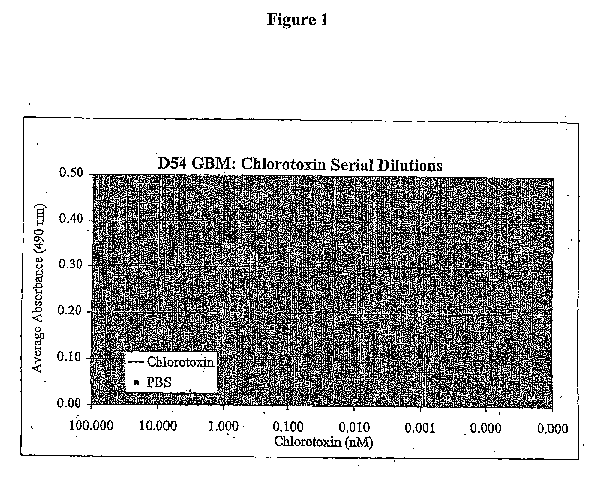
[0097] D54 glioblastoma cells were plated at a density of about 1000 cells / well in a 96-well flat bottom plate and incubated in 5% CO2 at 37° C. After twenty-four hours chlorotoxin was added at 1:4 limiting dilutions to a final concentration of 20, 5, 1.25, 0.313, 0.078, 0.0195, 0.0049, 0.0012, 0.00031 or 0.00008 nM. Control cells received vehicle only. Twenty-four hours after treatment, the effect of chlorotoxin was quantified using the MIT mitochondrial enzyme substrate with the Cell Counting Kit-8 (CCK-8) (Dojindo Inc.) according to the manufacturer's instructions. In brief, following the treatment period with chlorotoxin, cells were incubated with CCK-8 reagent. After incubation, plates were read on a microplate reader at a wavelength of 490 nm, with higher absorbance indicating greater cell viability. FIG. 1 shows that chlorotoxin incubation inhibited proliferation of the D54 cells at all concentrations tested down through 0.00120 nM as evidenced by the lower number of viable c...

example 2

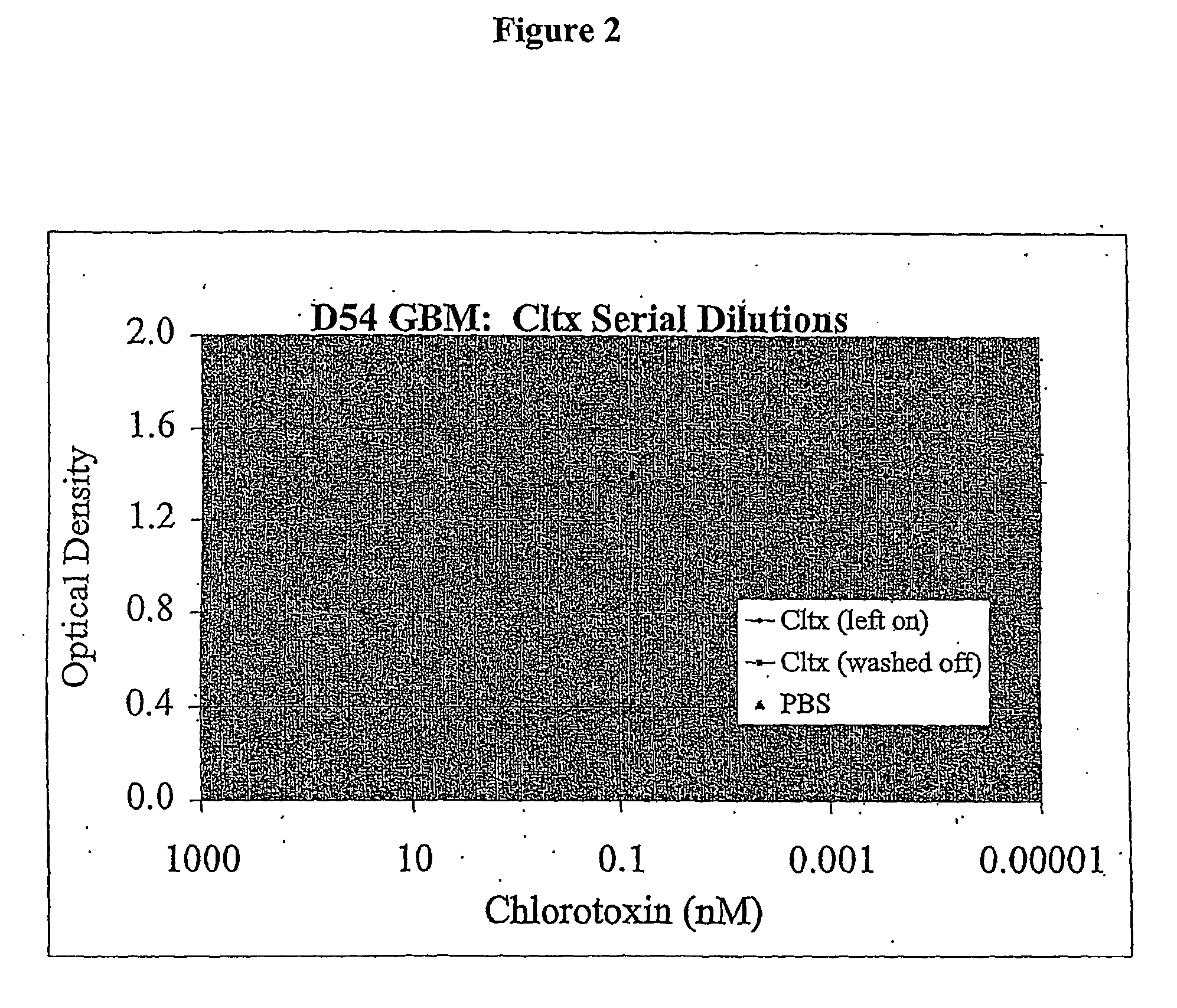
[0098] D54 glioblastoma cells were plated at a density of about 1000 cells / well in a 96-well flat bottom plate and incubated in 5% CO2 at 37° C. After twenty-four hours chlorotoxin was added at 1:4 limiting dilutions to a final concentration (in nM) of 20, 5, 1.25, 0.313, 0.078, 0.02, 0.0049, 0.0012, 0.0003, or 0.00008. Control cells received vehicle only. After twenty-four hours, half of the cells were washed free of chlorotoxin, the medium replaced with fresh medium. Cells in both conditions, chlorotoxin left on and chlorotoxin removed, were incubated for an additional four days. Following incubation, the effect of chlorotoxin was quantified using the MTT mitochondrial enzyme substrate with the CCK-8 as in Example 1. FIG. 2 shows that the long incubation time allowed the cells to overcome the effects of chlorotoxin with the additional days of proliferation and chlorotoxin did not appear to inhibit cell proliferation in this instance.

example 3

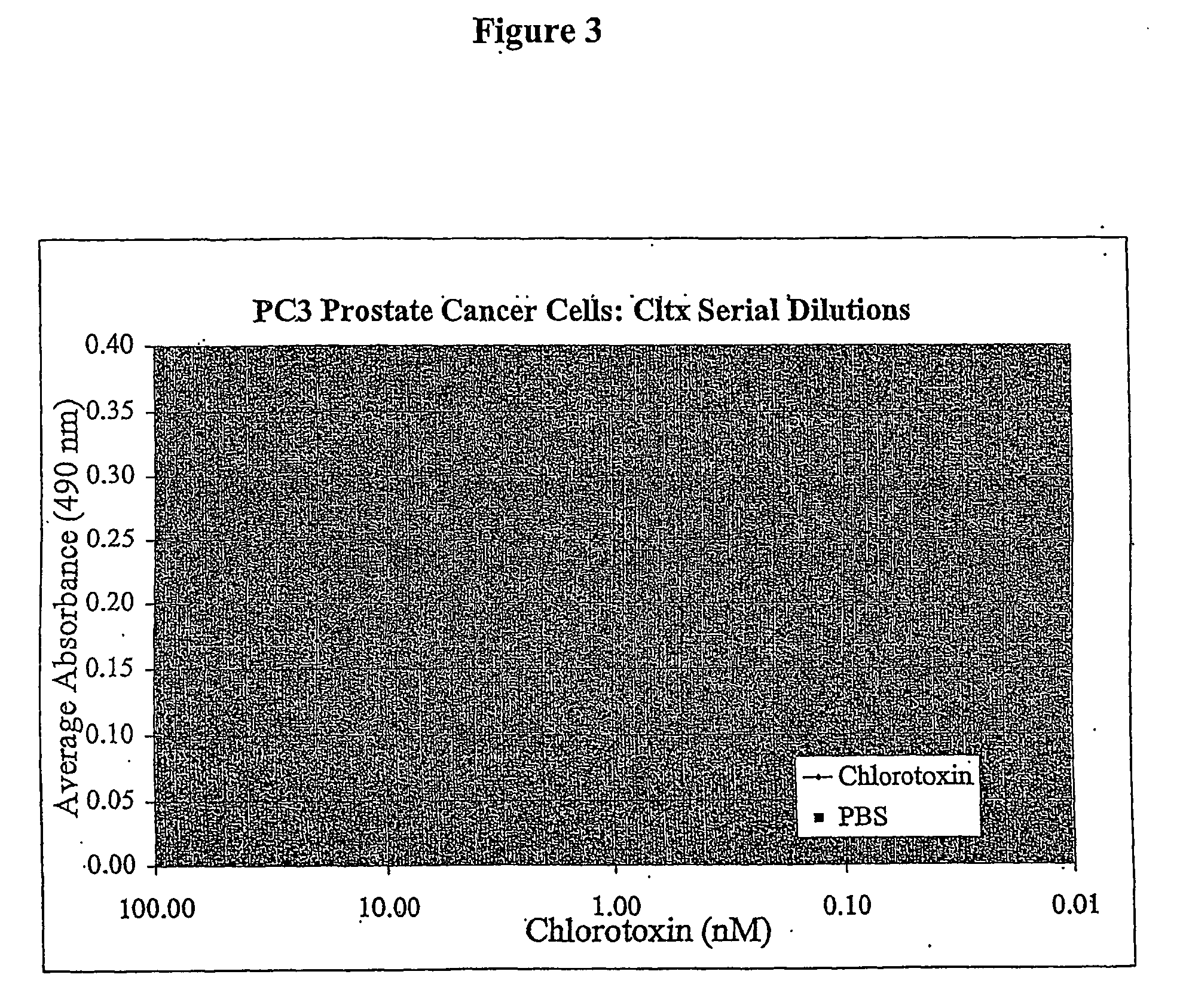
[0099] PC3 prostate cancer cells were plated at a density of about 1000 cells / well in a 96-well flat bottom plate and incubated in 5% CO2 at 37° C. After twenty-four hours chlorotoxin was added at 1:2 limiting dilutions to a final concentration (nM) of 20, 10, 5, 2.5, 1.25, 0.625, 0.313, 0.156, 0.078, and 0.039. Control cells received PBS vehicle only. Twenty-four hours after treatment, the effect of chlorotoxin was quantified using the MTT mitochondrial enzyme substrate with the CCK-8 as in Example 1. FIG. 3 shows that chlorotoxin incubation inhibited proliferation of the D54 cells at all concentrations tested as evidenced by the lower number of viable cells / well versus PBS control


PUM

PropertyMeasurementUnit
Dimensionless propertyaaaaaaaaaa

Abstract

Disclosed is a method of treating cell proliferative disorders, such as cancer, with low doses of chlorotoxin and/or derivatives, analogs or fragments thereof, which are effective to inhibit or arrest abnormal cell growth.

Description

RELATED APPLICATIONS [0001] This application claims the benefit of U.S. Provisional Application 60 / 406,033 (filed Aug. 27, 2002) and U.S. Provisional Application 60 / 384,171 (filed May 31, 2002) both of which are hereby incorporated by reference in their entirety.FIELD OF THE INVENTION [0002] The present invention relates generally to the fields of cell physiology and oncology. More specifically, the present invention relates to a novel method of treating cell proliferative disorders, such as cancers, with doses of chlorotoxin and / or derivatives thereof which are effective to inhibit or arrest abnormal cell growth. BACKGROUND OF THE INVENTION [0003] Tumors that originate in brain tissue are known as primary brain tumors as opposed to secondary brain tumors that develop when cancer metastasizes from other parts of the body to the brain. Primary brain tumors are classified by the type of tissue in which they begin. The most common brain tumors are gliomas, which begin in the glial (sup...

Claims


Application Information

Patent Timeline
IPC IPC(8): A61K38/08C07K7/06A61K31/00A61K38/00A61K45/00A61K38/17A61K45/06A61K51/08A61P35/00C07K14/435
CPCA61K31/00A61K31/175A61K31/4188A61K38/17A61K45/06C07K14/43522A61K2300/00A61K31/495A61P35/00A61P43/00
Inventor ALVAREZ, VERNONGONDA, MATTHEW
Owner MORPHOTEX INC
Why Eureka
Try Eureka