Methods and kits for diagnosing and monitoring hematopoietic cancers
a technology for hematopoietic cancer and kits, applied in the direction of instruments, biochemistry apparatus and processes, material analysis, etc., can solve the problems of hematopoietic cells, normal cells, clear lack of markers, and easy facilitation of markers
- Summary
- Abstract
- Description
- Claims
- Application Information
AI Technical Summary
Benefits of technology
Problems solved by technology
Method used
Image
Examples
example 1
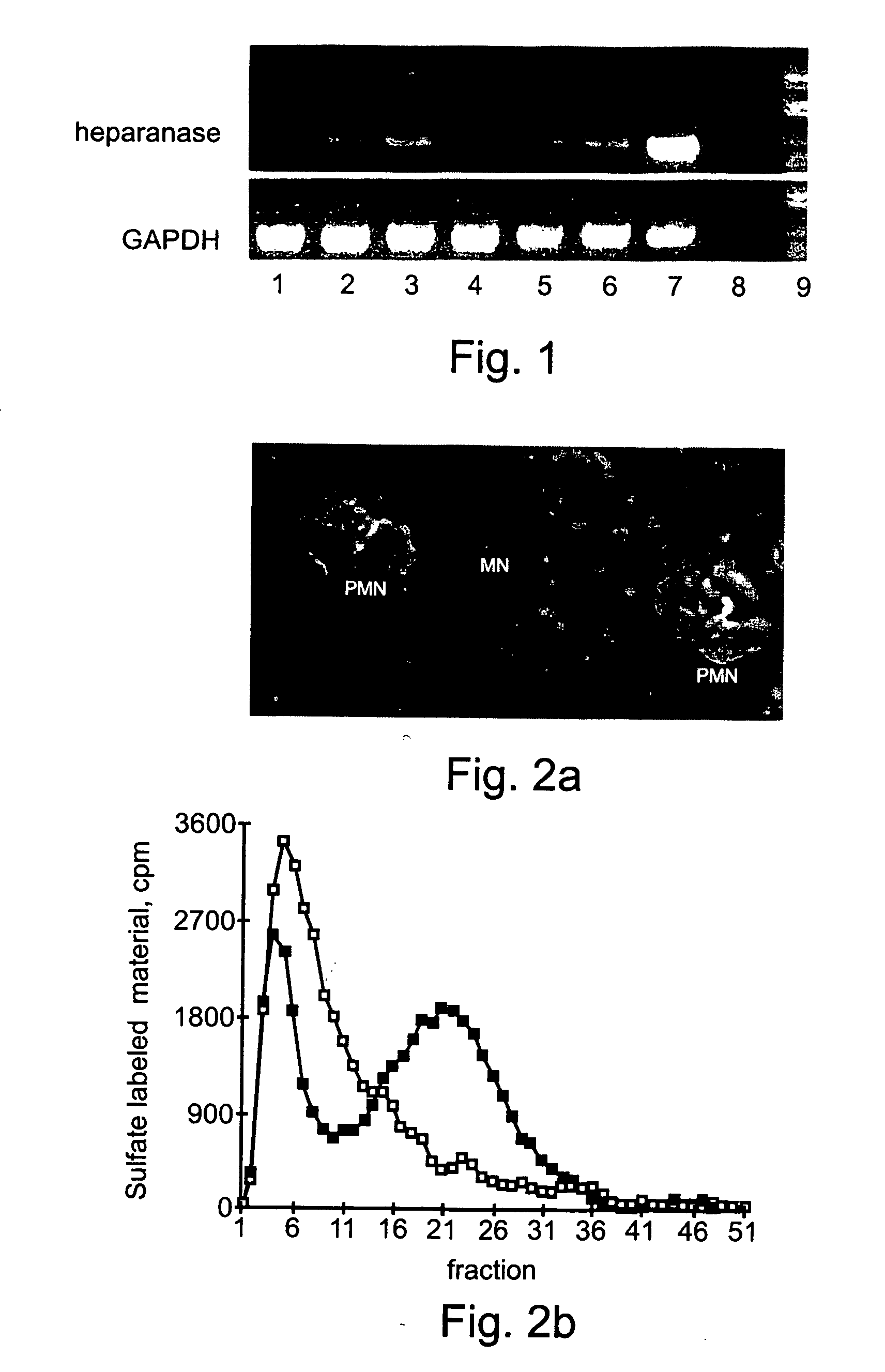
Heparanase Localization in Normal Peripheral Blood Granulocytes and Monocytes
Material and Experimental Methods
[0135] Obtaining Antibodies
[0136] The mouse anti-heparanase monoclonal antibody (mAb) 130, an IgG1 subclass, was generated as previously described (13, U.S. Pat. No. 6,177,545; U.S. patent application Ser. Nos. 09 / 704,772; 09 / 322,977; 09 / 186,200; 09 / 944,602; 09 / 759,207; and PCT Application Nos. US99 / 25451 and US99 / 09255). FITC-labeled anti-heparanase mAb 130 was prepared as follows; 2 mg of antibody were disloved in 1 ml of 0.1 M Sodium Carbonate, pH 9.0. 50 μl of 1 mg / ml fresh FITC solution in DMSO were added to 1 ml antibody solution, by 5 μl increments. The mixture was incubated for 8 hours at 4° C. in the dark. When OD495 nm / 280 nm in the range of 0.3 to 1.0, the reaction was stoped by adding NH4Cl to a final concentration of 50 mM and incubateed for 2 hours at 4° C. Xylene cylanol was then added to a final concentration of 0.1% and glycerol to a final concentration o...
example 2
Heparanase Localization in Myeloblasts of Leukemic Patient Samples
Material and Experimental Methods
[0151] Blood Samples, RNA Isolation, RT-PCR, Isolation of Peripheral Blood Monocytes and Immunocytochemistry
[0152] These procedures were conducted as per Example 1 above.
Experimental Results
[0153] Heparanase mRNA and Protein Expression in Leukemic Cells
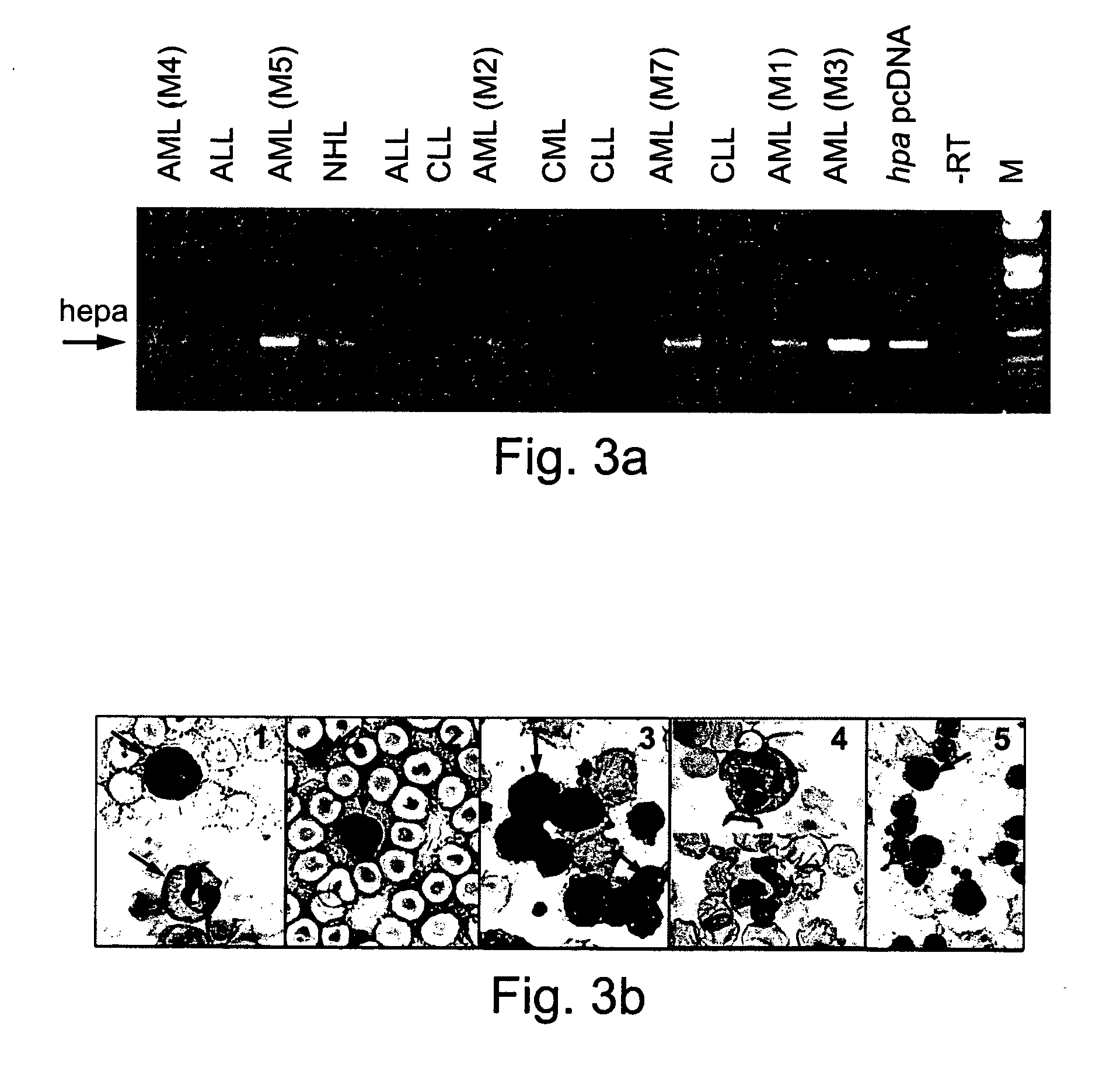
[0154] Aberrant expression of ECM-degrading metalloproteinases may characterize specific types of malignant hematopoietic cells (8). Heparanase expression was therefor evaluated both at the mRNA and protein levels in samples obtained from patients with a variety of hematological disorders, most predominantly with leukemias. RT-PCR amplification of mononuclear heparanase mRNA expression was evaluated in mononuclear of patient mRNA samples (FIG. 3A). Surprisingly, heparanase mRNA expression was detected primarily in AML samples. CLL samples failed to provide a positive signal for heparanase mRNA, regardless of CLL patient clinical st...
example 3
Functional Heparanase is Exclusive to AML Patient Leukocytes
Material and Experimental Methods
[0157] Blood Samples
[0158] Peripheral blood samples were obtained from AML stage 4 and CLL patients.
[0159] Isolation of Peripheral Blood Leukocytes,
[0160] Platelet depleted preparations of peripheral blood leukocytes were isolated by Ficoll-Hypaque density gradient centrifugation, prepared from AML stage M4 and CLL patients, as described in Example 1 above, and in (9-11, 14).
[0161] RNA Isolation, RT-PCR:
[0162] These procedures were conducted as detailed in Example 1, above.
[0163] Heparanase Activity
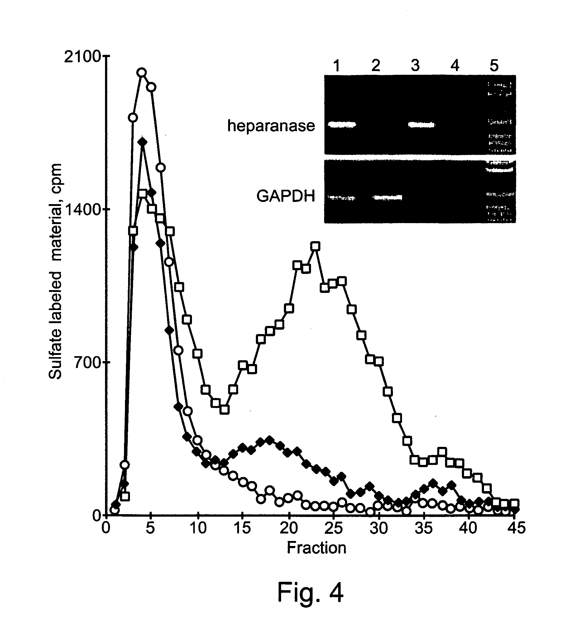
[0164] Dishes coated with metabolically labeled extracellular matrix (ECM) were prepared as previously described (9-11, 14). AML-M4 and CLL patient leukocytes were suspended (2.5×10⁶ / ml) in serum free RPMI medium and incubated (24 hours, 37° C., pH 6.6) with S-labeled ECM (with or without 2.5 μg / ml heparin). The incubation medium was centrifuged and the supernatant analyzed by gel filtrat...
PUM
| Property | Measurement | Unit |
|---|---|---|
| temperature | aaaaa | aaaaa |
| temperature | aaaaa | aaaaa |
| pH | aaaaa | aaaaa |
Abstract
Description
Claims
Application Information
 - R&D
- Intellectual Property
- Life Sciences
- Materials
- Tech Scout
- Unparalleled Data Quality
- Higher Quality Content
- 60% Fewer Hallucinations
Browse by: Latest US Patents, China's latest patents, Technical Efficacy Thesaurus, Application Domain, Technology Topic, Popular Technical Reports.
© 2025 PatSnap. All rights reserved.Legal|Privacy policy|Modern Slavery Act Transparency Statement|Sitemap|About US| Contact US: help@patsnap.com


