System and method for controlling neurological disorders with spatially separated detection and therapy locations

a spatial separation and neurological disorder technology, applied in the field of systems and methods for treating neurological disorders, can solve the problems of physical impairment, deterioration of other brain functions (including cognitive function), frequent limitation of sufferers, etc., and achieve the effect of longer advance notice and greater precision and reliability

Inactive Publication Date: 2006-10-05
NEUROPACE
View PDF7 Cites 25 Cited by
  • Summary
  • Abstract
  • Description
  • Claims
  • Application Information

AI Technical Summary

Benefits of technology

[0030] The application of stimulation signals remote from the epileptogenic region has several advantages. First, it is believed that such therapy will tend to avoid contributing to hypersynchronous activity in the epileptogenic region. Second, detection can be carried out at (or near) the same time stimulation is being performed, as it is less likely that the detection subsystems of the implantable neurostimulator will be affected by artifacts, specifically the stimulation signals transmitted through the brain tissue. Moreover, the ability to detect the effects of electrical stimulation on remote tissue in the patient's brain may contribute to advantages in detecting, identifying and treating seizures and other neurological events with greater precision and reliability, and with longer advance notice.

Problems solved by technology

Because epilepsy is characterized by seizures, its sufferers are frequently limited in the kinds of activities they may participate in.
Over time, epileptic seizures often become more frequent and more serious, and in particularly severe cases, are likely to lead to deterioration of other brain functions (including cognitive function) as well as physical impairments.
Unfortunately, those drugs typically have serious side effects, especially toxicity, and it is extremely important in most cases to maintain a precise therapeutic serum level to avoid breakthrough seizures (if the dosage is too low) or toxic effects (if the dosage is too high).
Besides being less than fully successful, these surgical approaches generally have a high risk of complications, and can often result in damage to eloquent (i.e., functionally important) brain regions and the consequent long-term impairment of various cognitive and other neurological functions.
Furthermore, for a variety of reasons, such surgical treatments are contraindicated in a substantial number of patients.
And unfortunately, even after radical brain surgery, many epilepsy patients are still not seizure-free.
However, currently approved and available electrical stimulation devices apply continuous electrical stimulation to neural tissue surrounding or near implanted electrodes, and do not perform any detection—they are not responsive to relevant neurological conditions.
Unfortunately, a much greater reduction in the incidence of seizures is needed to provide clinical benefit.
Unfortunately, continuous stimulation of deep brain structures for the treatment of epilepsy has not met with consistent success.
Recent research, however, indicates that the concept of a single epileptic focus does not necessarily accurately reflect the origins of partial epilepsy, at least in humans.
Although this approach is generally believed to achieve good results, for the most part, its computational expense renders it less than optimal for use in long-term implanted epilepsy monitor and treatment devices.
With current technology, the battery life in an implantable device computationally capable of performing the Dorfmeister method would be too short for it to be feasible.
Once more, the calculation of statistically relevant characteristics is not believed to be feasible in an implantable device.
To the extent responsive electrical stimulation is applied in response to a detection of epileptiform activity, artifacts of the stimulation received by the epileptiform activity detector may be significantly disruptive of the detection algorithms.
A potential solution to this problem is to blank the sensing amplifiers used to receive EEG signals during and for a period after the application of electrical stimulation, but this will lead to a loss of data during the blanking period.
It is believed to be advantageous to provide therapeutic electrical stimulation in a number of brain regions involved in a patient's epilepsy, but known approaches do not do this in any meaningful way.

Method used


Image

  • System and method for controlling neurological disorders with spatially separated detection and therapy locations
  • System and method for controlling neurological disorders with spatially separated detection and therapy locations
  • System and method for controlling neurological disorders with spatially separated detection and therapy locations

Examples


Embodiment Construction

[0043] The invention is described below, with reference to detailed illustrative embodiments. It will be apparent that a system according to the invention may be embodied in a wide variety of forms. Consequently, the specific structural and functional details disclosed herein are representative and do not limit the scope of the invention.

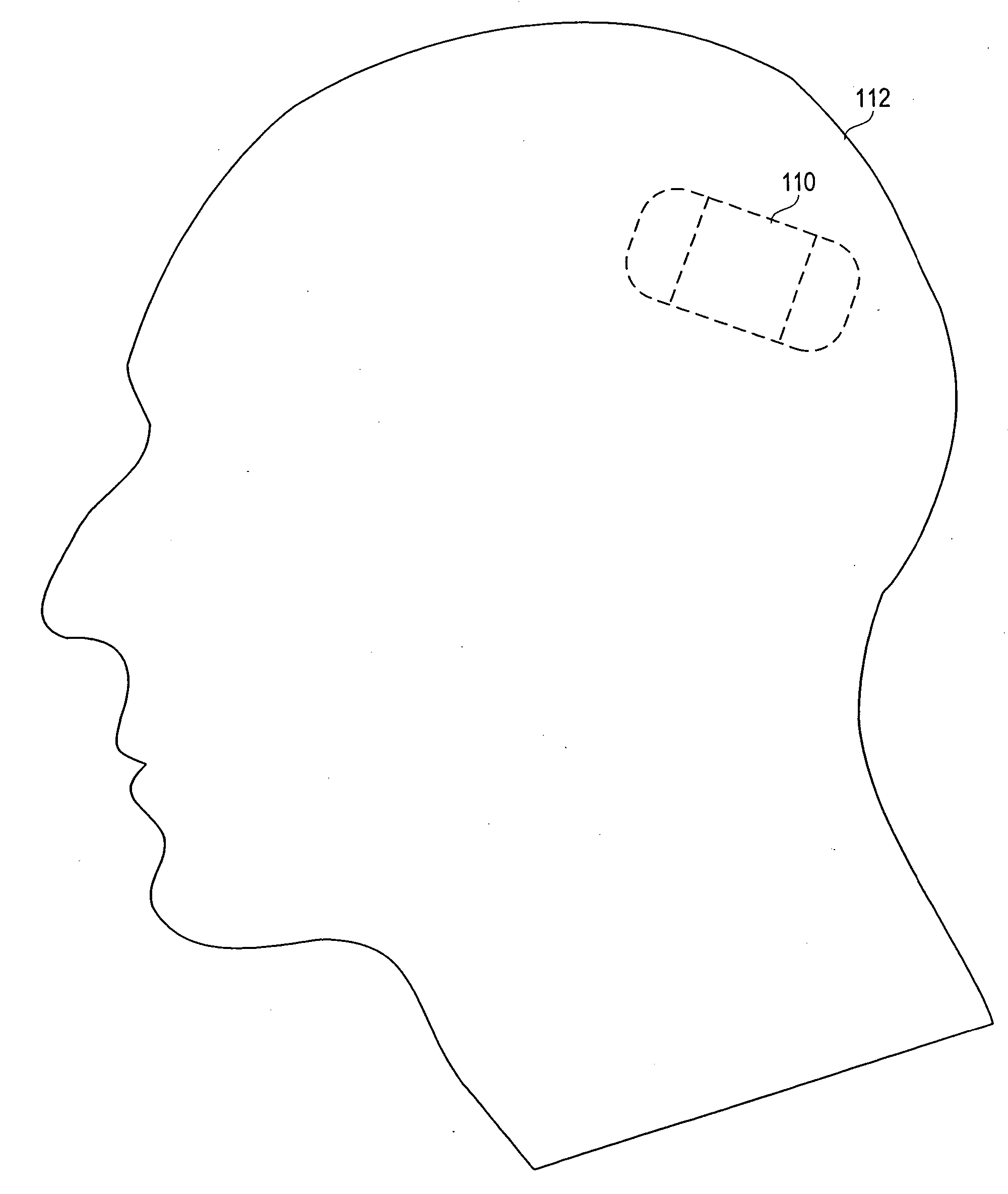
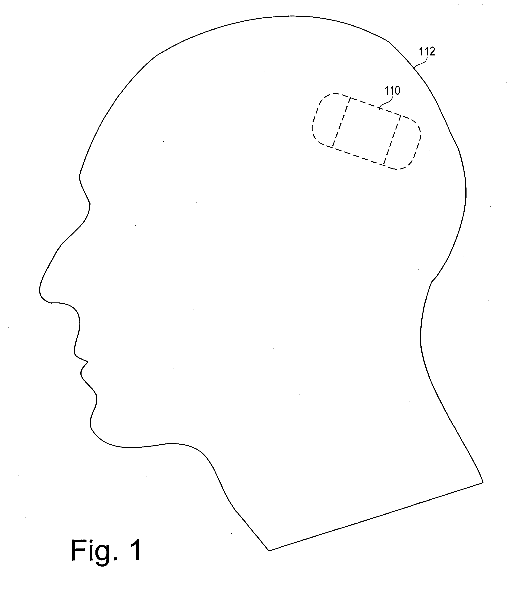
[0044]FIG. 1 depicts an intracranially implanted device 110 according to the invention, which in one embodiment is a small self-contained responsive neurostimulator. As the term is used herein, a responsive neurostimulator is a device capable of detecting ictal activity (or other neurological events) and providing electrical stimulation to neural tissue in response to that activity, where the electrical stimulation is specifically intended to terminate the ictal activity or treat the neurological event.

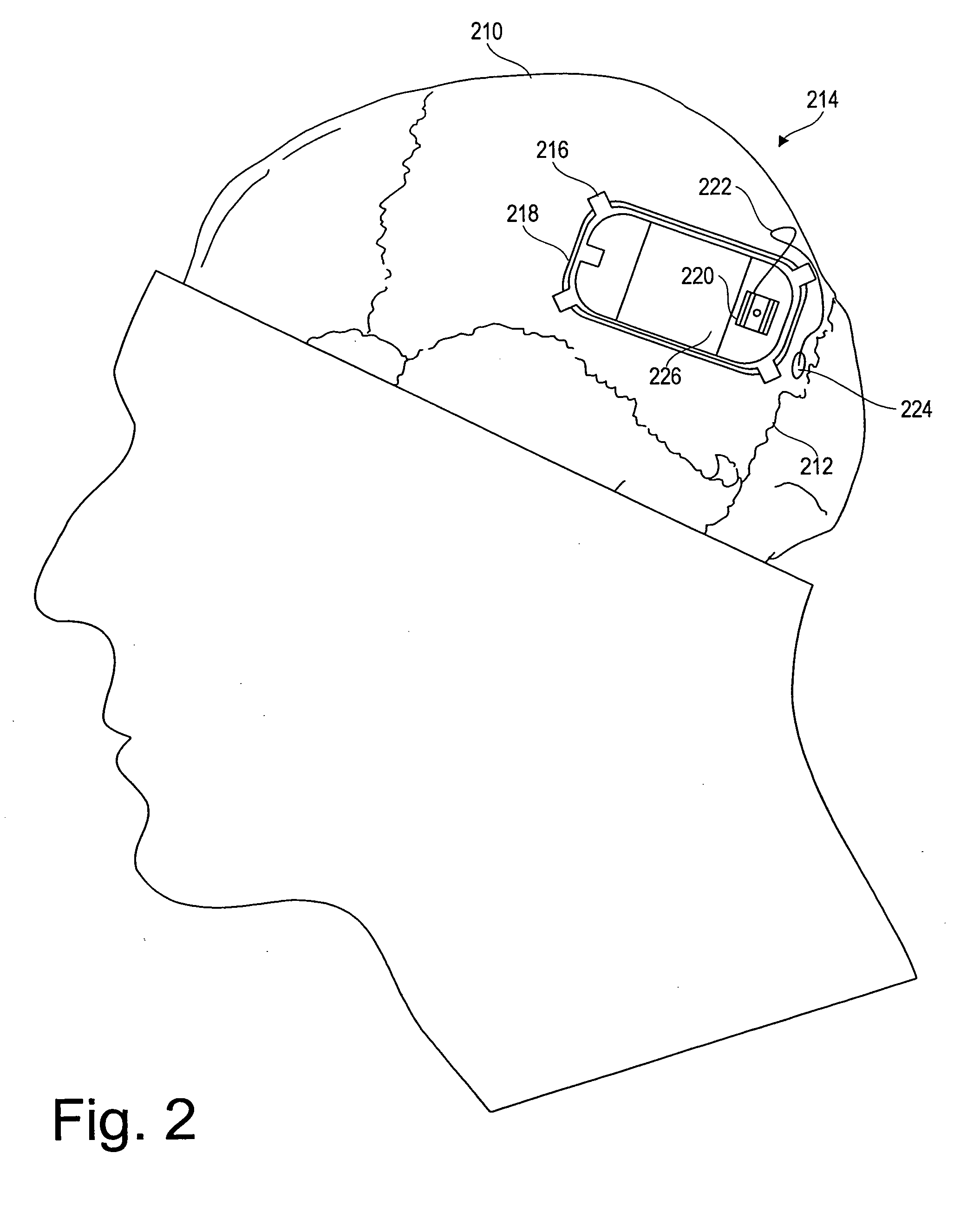
[0045] In the disclosed embodiment, the neurostimulator is implanted intracranially in a patient's parietal bone 210, in a location anterior to the...


PUM


Abstract

A system and method for controlling epilepsy and other neurological disorders by providing electrical stimulation to a patient's brain in response to detected neurological conditions. An implantable device includes a stimulation subsystem coupled to a stimulation electrode to provide responsive electrical brain stimulation in response to an event detected via an on-board processor's analysis of data received from a detection subsystem coupled to a detection electrode located in a different portion of the patient's brain.

Description

CROSS-REFERENCES TO RELATED APPLICATIONS [0001] This is a continuation of U.S. patent application Ser. No. 09 / 724,805, filed on Nov. 28, 2000 and issued as U.S. Pat. No. 6,597,954, which is a continuation-in-part of U.S. patent application Ser. No. 09 / 543,264, filed on Apr. 5, 2000; a continuation-in-part of U.S. patent application No. 09 / 373,676, filed on Aug. 13, 1999 and now issued as U.S. Pat. No. 6,230,049; and a continuation-in-part of U.S. patent application Ser. No. 09 / 628,977, filed on Aug. 2, 2000 and now issued as U.S. Pat. No. 6,360,122, which was a continuation of U.S. patent application Ser. No. 09 / 450,303, filed on Nov. 29, 1999 and now issued as U.S. Pat. No. 6,128,538, which was in turn a continuation of U.S. patent application Ser. No. 08 / 957,869, filed on Oct. 27, 1997 and now issued as U.S. Pat. No. 6,016,449.BACKGROUND OF THE INVENTION [0002] The invention relates to systems and methods for treating neurological disorders, and more particularly to a system and m...

Claims


Application Information

Patent Timeline
Patent Type & Authority Applications(United States)
IPC IPC(8): A61N1/00A61B5/00A61B5/0476A61M5/142A61N1/36
CPCA61B5/0002A61B5/0476A61M5/14276A61B5/7217A61N1/0534A61N1/36082A61N1/36064A61N1/0529A61B5/4094A61B5/369A61B5/372
Inventor PLESS, BENJAMIN D.FISCHELL, ROBERT E.FISCHELL, DAVID R.
Owner NEUROPACE
Why Eureka
Try Eureka