Microfluidic rare cell detection device

a rare cell detection and microfluidic technology, applied in the field of microfluidic devices, can solve the problems of lack of sensitivity of current detection methods to reproducibly detect disseminated cancer cells, and the scheme suffers from significant limitations, and achieves the effect of facilitating the detection of labeled cells

Inactive Publication Date: 2006-11-02
PERKINELMER HEALTH SCIENCES INC
View PDF29 Cites 78 Cited by
  • Summary
  • Abstract
  • Description
  • Claims
  • Application Information

AI Technical Summary

Problems solved by technology

The challenge for practical transfer of the diversity of molecular information being generated in the characterization of cancer cells into routine clinical practice is the development of reproducible, integrated and automated methods for their measurement.
Unfortunately, current detection methods lack adequate sensitivity to reproducibly detect disseminated cancer cells, which can be as few as 1-10 cells per 10 ml of blood.
However, this scheme suffers from a significant limitation—that the detectors may only detect and direct one cell at a time.
Unfortunately, due to the technically complex methodology and result interpretation involved in current flow cytometry methods, such analyses are generally performed by reference laboratories.

Method used


Image

  • Microfluidic rare cell detection device
  • Microfluidic rare cell detection device
  • Microfluidic rare cell detection device

Examples


example 1

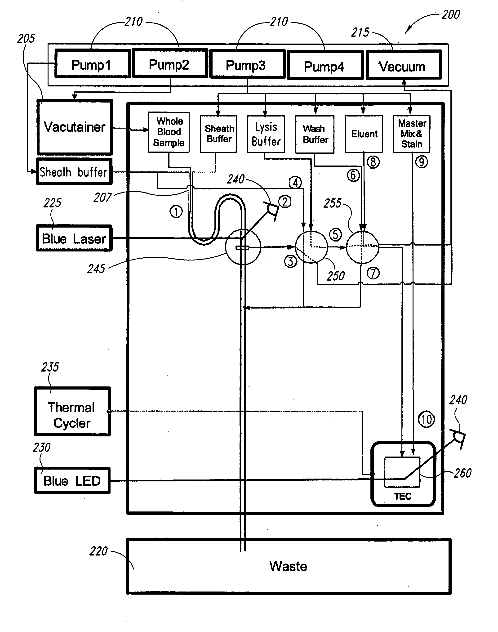
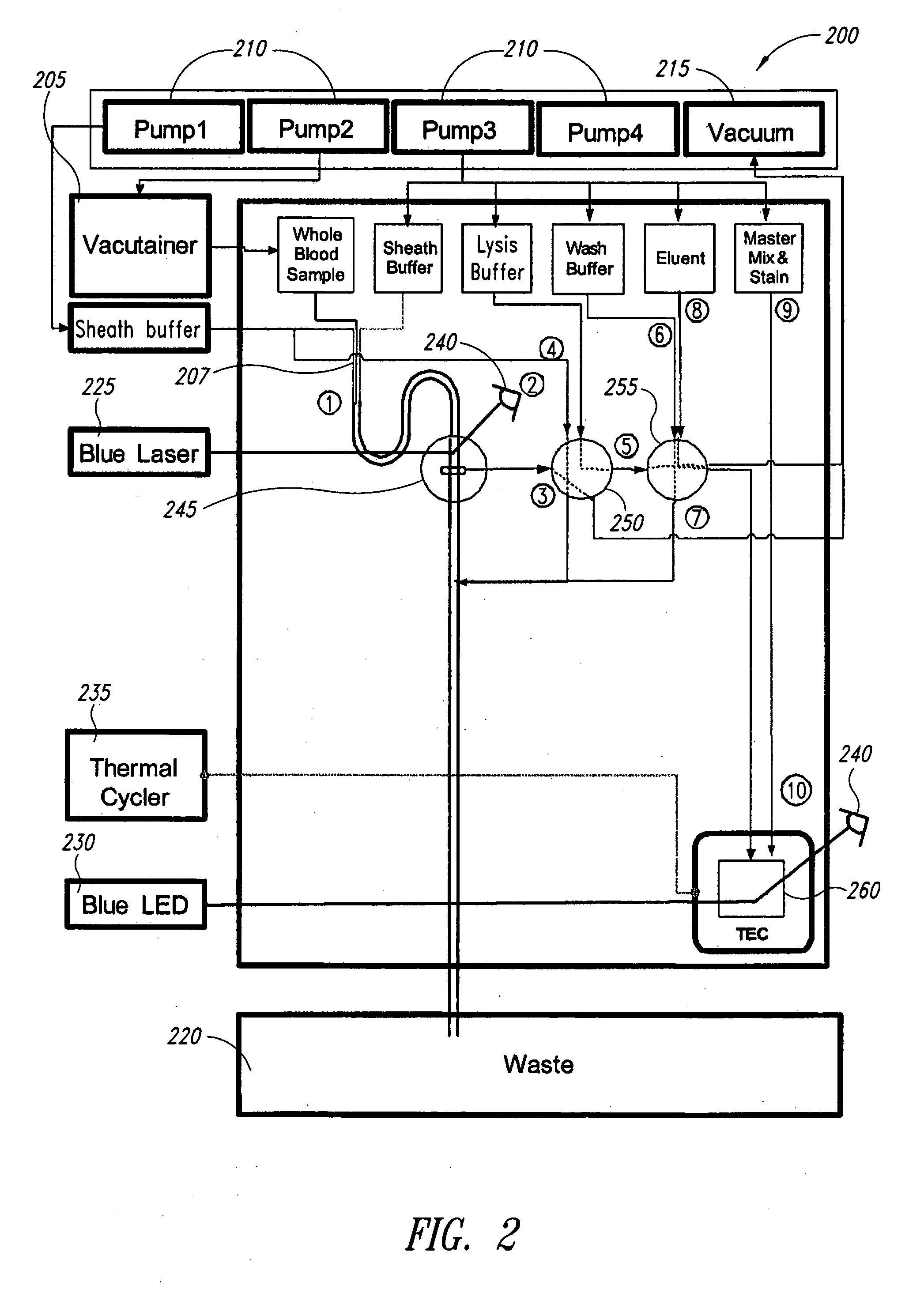
[0069] In the following example, the sample and antibody solutions were moved through the channels of the microfluidic devices by a microFlow™ system, which comprises a controller, pumps (250 μL and 2,300 μL capacity pumps), and a manifold. The microFlow™ system is a commercially available ultra-low-pulse pump system (Micronics, Inc.) with air, vacuum, forward and reverse pumping capabilities controlled by PC based software. In a microfluidic device, fluids can be transported by either air or Fluorinert™ FC-70 (Hampton Research HR2-797). Fluorinert™ FC-70 has a viscosity similar to water, with approximately 75% greater density, and is not miscible with aqueous solutions. In the following examples, Fluorinert™ FC-70 was used to prevent dilution of the sample and antibody solutions during processing.

Lab Card Design

[0070] A microfluidic device having the sub-circuits illustrated in FIGS. 3A-3C and 5A-5E was used for the following cell and / or bead counting and sorting experiments. Th...

example 2

Lab Card and Microfluidic Circuitry

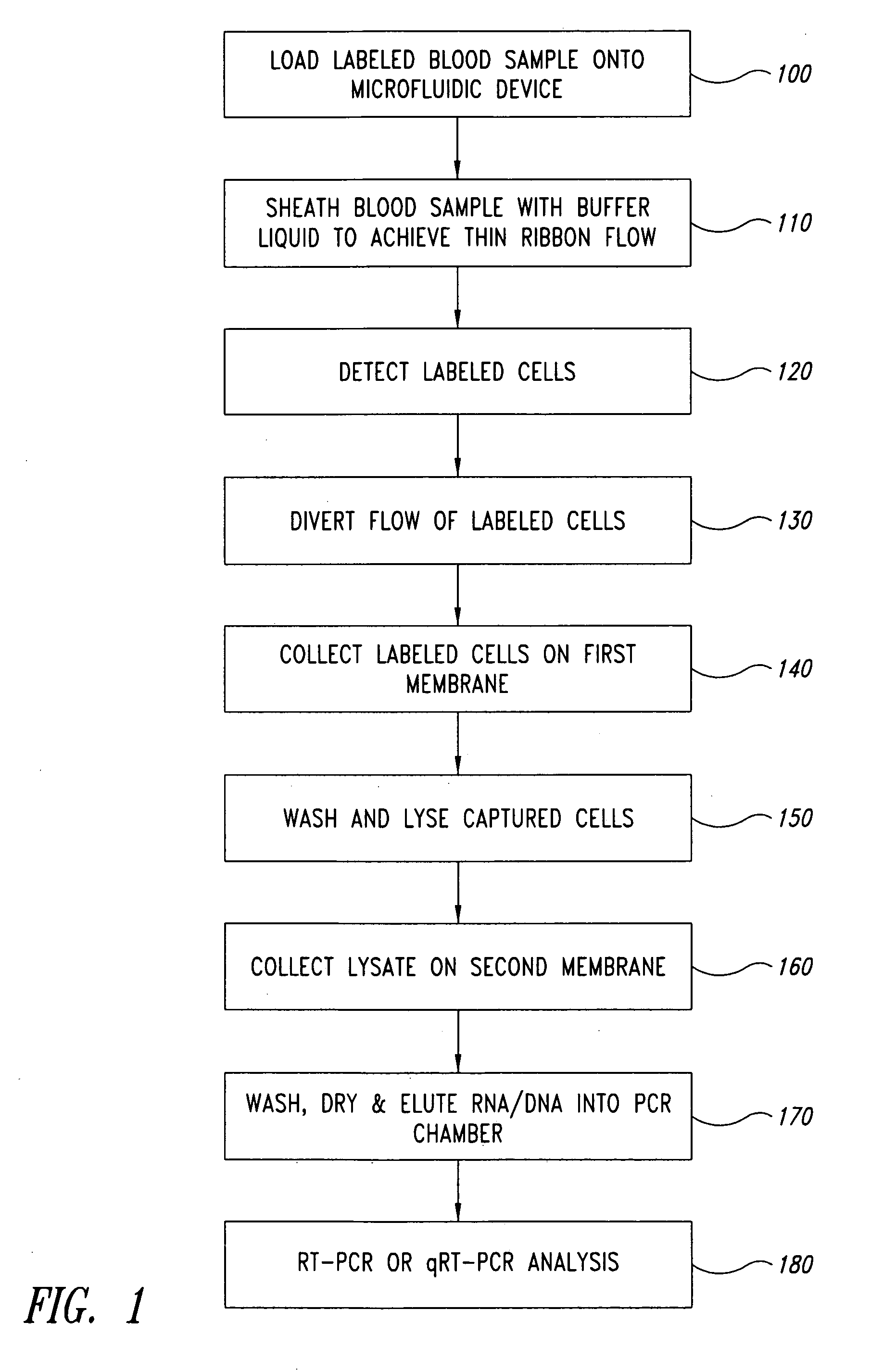
[0082] A microfluidic device having the sub-circuits illustrated in FIGS. 7A-7F was used to evaluate automated liquid handling steps for RNA extraction. The device comprised a 700 μL wash solution chamber, a 150 μL elution solution chamber, a 250 μL lysate / binding solution chamber, and a silica membrane assembly. The silica membrane assembly comprised two circular glass fiber filter type D membranes (GF / D, 8 mm diameter discs, Whatman). The fluidic circuitry used on-card valving to control fluid paths and a simple vacuum to deliver and draw solutions through the GF / D membranes Additionally, a small volume pump (˜150 μL) was used to deliver the elution solution. After loading the device with appropriate solutions, the device automated the following steps: (1) white blood cells in lysate solution were pulled across the silica membrane by vacuum; (2) nucleic acid from the cells were bound to the membrane under the lysis conditions used; (3) a wash s...


PUM

PropertyMeasurementUnit
sizeaaaaaaaaaa
speedsaaaaaaaaaa
widthaaaaaaaaaa

Abstract

The present invention relates to microfluidic devices and methods for detecting rare cells. The disclosed microfluidic devices and methods integrate and automate sample preparation, cell labeling, cell sorting and enrichment, and DNA / RNA analysis of sorted cells.

Description

CROSS-REFERENCE TO RELATED APPLICATION [0001] This application claims the benefit of U.S. Provisional Patent Application No. 60 / 643,833, filed Jan. 13, 2005, which application is hereby incorporated by reference in its entirety.BACKGROUND OF THE INVENTION [0002] 1. Field of the Invention [0003] The present invention relates generally to microfluidic devices, and, more particularly, to microfluidic devices and methods for detecting rare cells. [0004] 2. Description of the Related Art [0005] The biological changes that are now known to be associated with cancer cells encompass the full continuum from mutated or duplicated genomic sequences to shifts in gene expression patterns, as well as altered proteins. The challenge for practical transfer of the diversity of molecular information being generated in the characterization of cancer cells into routine clinical practice is the development of reproducible, integrated and automated methods for their measurement. One technical hurdle is d...

Claims


Application Information

Patent Timeline
Patent Type & Authority Applications(United States)
IPC IPC(8): C12M1/34B01L7/00
CPCB01L3/50273G01N2015/149B01L3/502753B01L3/502761B01L3/502776B01L7/52B01L2200/0636B01L2200/0647B01L2200/10B01L2300/0816B01L2300/0864B01L2300/0867B01L2300/087B01L2300/0874B01L2300/0887B01L2400/0487B01L2400/0633F16K99/0001F16K99/0007F16K99/0034F16K99/0046F16K2099/008F16K2099/0084G01N1/34G01N15/1459G01N15/1463G01N15/1484G01N2015/0069G01N2015/1409G01N2015/1486B01L3/502738
Inventor LANCASTER, CHRISTY A.BATTRELL, C. FREDERICKCAPODANNO, JASONGERDES, JOHNKOKORIS, MARKNABAVI, MELUDMORDUE, STEPHENMCRUER, ROBERTCLEMMENS, JOHNBREIDFORD, WAYNE L.
Owner PERKINELMER HEALTH SCIENCES INC
Why Eureka
Try Eureka