Low temperature deparaffinization
a low temperature deparaffinization and low temperature technology, applied in the field of low temperature deparaffinization, can solve the problem that the heat of immiscible liquid tends to have a harsh effect on the biological sampl
- Summary
- Abstract
- Description
- Claims
- Application Information
AI Technical Summary
Benefits of technology
Problems solved by technology
Method used
Image
Examples
example 1
Special Stains
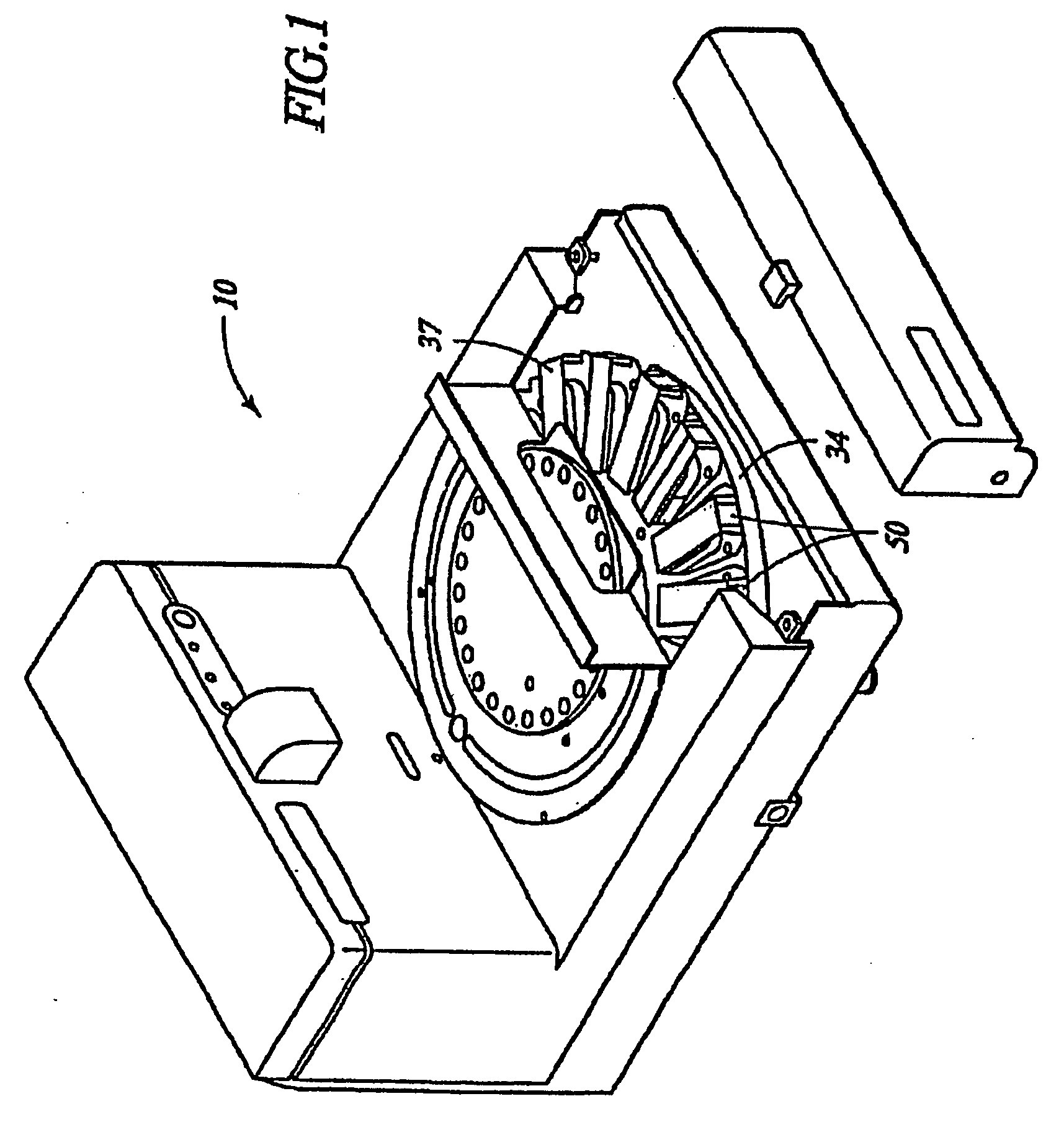
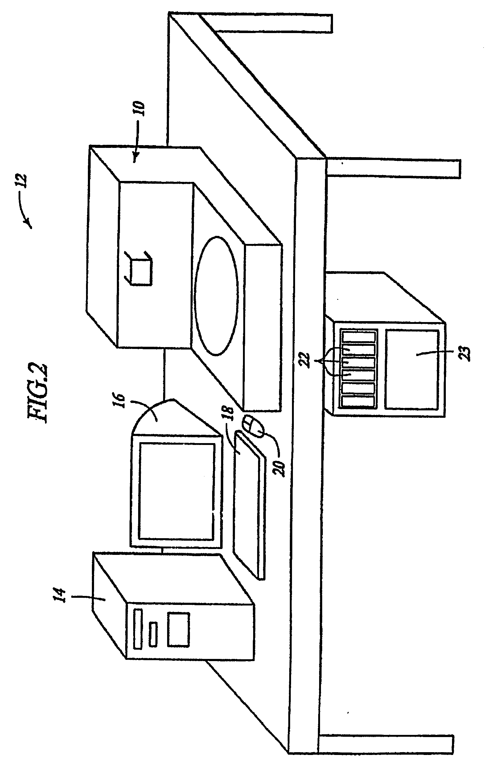
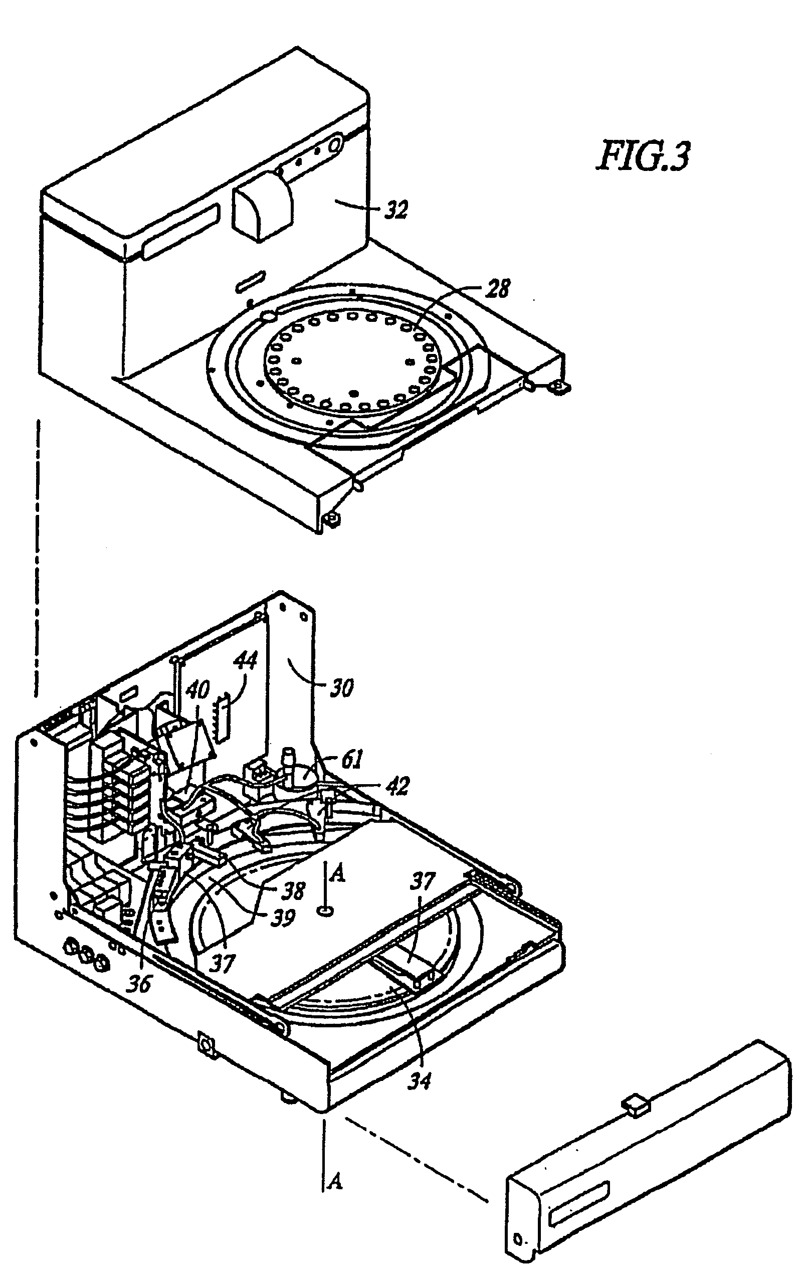
[0052] Various routinely prepared clinical paraffin tissue blocks were cut (5 μm) and placed onto glass slides. Air-dried slides were subjected to: Method 1) manual deparaffinization with xylene (control group) or Method 2) deparaffinization according to the methods of this invention for 12 minutes at 41° C. with NORPAR® 15 (test group) using NexES® Special Stains System manufactured by Ventana Medical Systems, Inc. (Tucson, Ariz.). The NORPAR® 15 containing solublized paraffin embedding material was then washed from the tissue samples using a tris-based washing buffer.
[0053] After the embedding material was removed from the biological samples by Method 1 or Method 2, the biological samples were processed for various automated special stain applications. These procedures were performed on a variety of cells stained with a variety of special stains. The special stain tests conducted are reported in Table 1 below. In all cases, the cells that were contacted with the l...
example 2
[0055] Formalin-fixed, paraffin-embedded mouse kidney samples were cut (5 μm) and placed onto glass slides. Air-dried slides were subjected to: Method 1) manual deparaffinization with xylene (control group 1); Method 2) automated heat deparaffinization with an aqueous solution (control group 2); or Method 3) deparaffinization at room temperature (37° C.) for 10 minutes (test group) with NORPAR® 15 on the Discovery® automated in situ hybridization apparatus manufactured by Ventana Medical Systems, Inc. (Tucson, Ariz). Following deparaffinization, all slides were processed for VEGF mRNA ISH using standard techniques.
[0056] The VEGF ISH results on mRNA deparaffinized by each of the methods above are set forth in the cross sections shown in FIGS. 6A-6C where 6A is the result for section deparaffinized manually with xylene of Method 1; 6B is the result for cells deparaffinized by heating the paraffin embedded tissues to a temperature above the melting point o...
example 3
[0058] Routinely processed clinical tissue samples were cut (5 μm) and placed onto glass slides. Air-dried slides were subjected to: Method 1) manual deparaffinization with xylene (control group 1); Method 2) automated heat deparaffinization (control group 2); or Method 3) deparaffinization with NORPAR 15 for 10 minutes at room temperature (37° C.) (test group) using the Discovery® automated ISH apparatus manufactured by Ventana Medical Systems, Inc. Following deparaffinization, all slides were processed by ISH for the presence of biological marker CD20.
[0059] The CD20 results of this example are reported in FIGS. 7A-7C in which 7A are the results of CD20 on tissue sections deparaffinized manually with xylene (Method 1), 7B are the results of CD20 detection on a tissue section deparaffinized using heat and an immiscible liquid (Method 2), and FIG. 7C are the results of CD20 detection on a tissue sample deparaffinized by a method of this invention (Method 3). T...
PUM
 Abstract
Description
Claims
Application Information
 - R&D
- Intellectual Property
- Life Sciences
- Materials
- Tech Scout
- Unparalleled Data Quality
- Higher Quality Content
- 60% Fewer Hallucinations
Browse by: Latest US Patents, China's latest patents, Technical Efficacy Thesaurus, Application Domain, Technology Topic, Popular Technical Reports.
© 2025 PatSnap. All rights reserved.Legal|Privacy policy|Modern Slavery Act Transparency Statement|Sitemap|About US| Contact US: help@patsnap.com


