
Methods and apparatus for endovascular heart valve replacement comprising tissue grasping elements

a heart valve and tissue grasping technology, applied in the field of endovascular heart valve replacement methods and apparatuses, can solve the problems of heart failure, adverse reactions to anesthesia medications, bleeding, stroke, etc., and achieve the effect of preventing blood backflow and allowing blood flow

Active Publication Date: 2006-11-09
BOSTON SCI SCIMED INC
View PDF99 Cites 538 Cited by
  • Summary
  • Abstract
  • Description
  • Claims
  • Application Information

AI Technical Summary

Benefits of technology

The solution enables minimally invasive heart valve replacement with reduced risks of mispositioning and migration, allowing for immediate functionality of the replacement valve and minimizing complications like coronary ostia blockage, while eliminating the need for life support during implantation.

Problems solved by technology

Valve replacement surgery is a highly invasive operation with significant concomitant risk.
Risks include bleeding, infection, stroke, heart attack, arrhythmia, renal failure, adverse reactions to the anesthesia medications, as well as sudden death.
However, the current devices suffer from several drawbacks.
First, many of the devices available today can become mispositioned with respect to the native valve.
This misposition may arise for a number of reasons, such as: the valve slipping after placement, improper initial positioning arising from the difficulties associated with visualizing the relative positions of the native and prosthetic valve, the difficulty in transmitting tactile feedback to the user through the delivery tool.
This is a critical drawback because improper positioning too far up towards the aorta risks blocking the coronary ostia of the patient.
Furthermore, a misplaced stent / valve in the other direction (away from the aorta, closer to the ventricle) will impinge on the mitral apparatus and eventually wear through the leaflet as the leaflet continuously rubs against the edge of the stent / valve.
Moreover, some stent / valve devices simply crush the native valve leaflets against the heart wall and do not grasp or engage the leaflets in a manner that would provide positive registration of the device relative to the native position of the valve.
This increases an immediate risk of blocking the coronary ostia, as well as a longer-term risk of migration of the device post-implantation.
Another drawback of the devices known today is that during implantation they may still require the patient to be on life support as the valve does not function for a portion of the procedure.
This further complicates the implantation procedure.

Method used


Image

  • Methods and apparatus for endovascular heart valve replacement comprising tissue grasping elements
  • Methods and apparatus for endovascular heart valve replacement comprising tissue grasping elements
  • Methods and apparatus for endovascular heart valve replacement comprising tissue grasping elements

Examples


Embodiment Construction

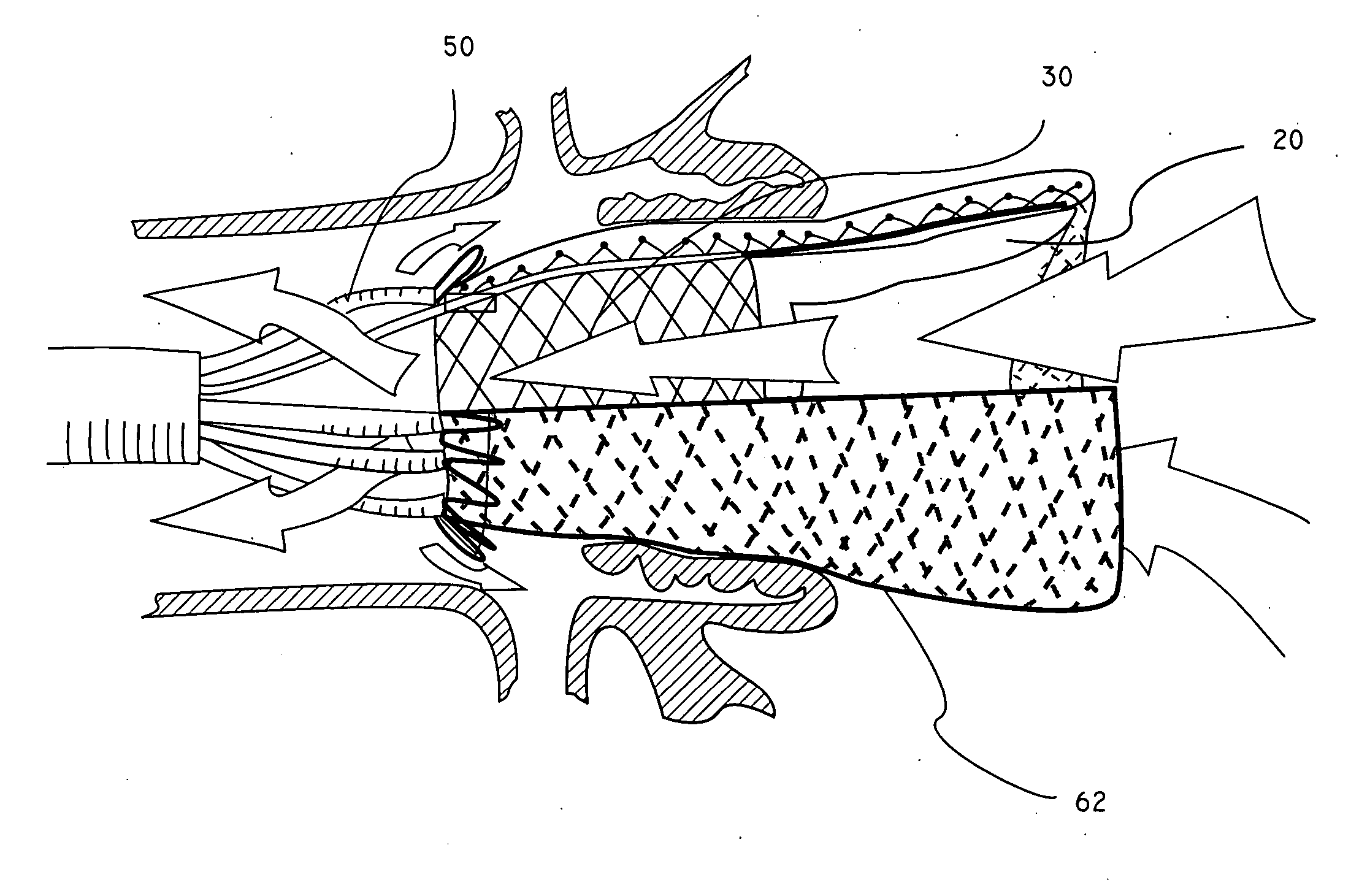
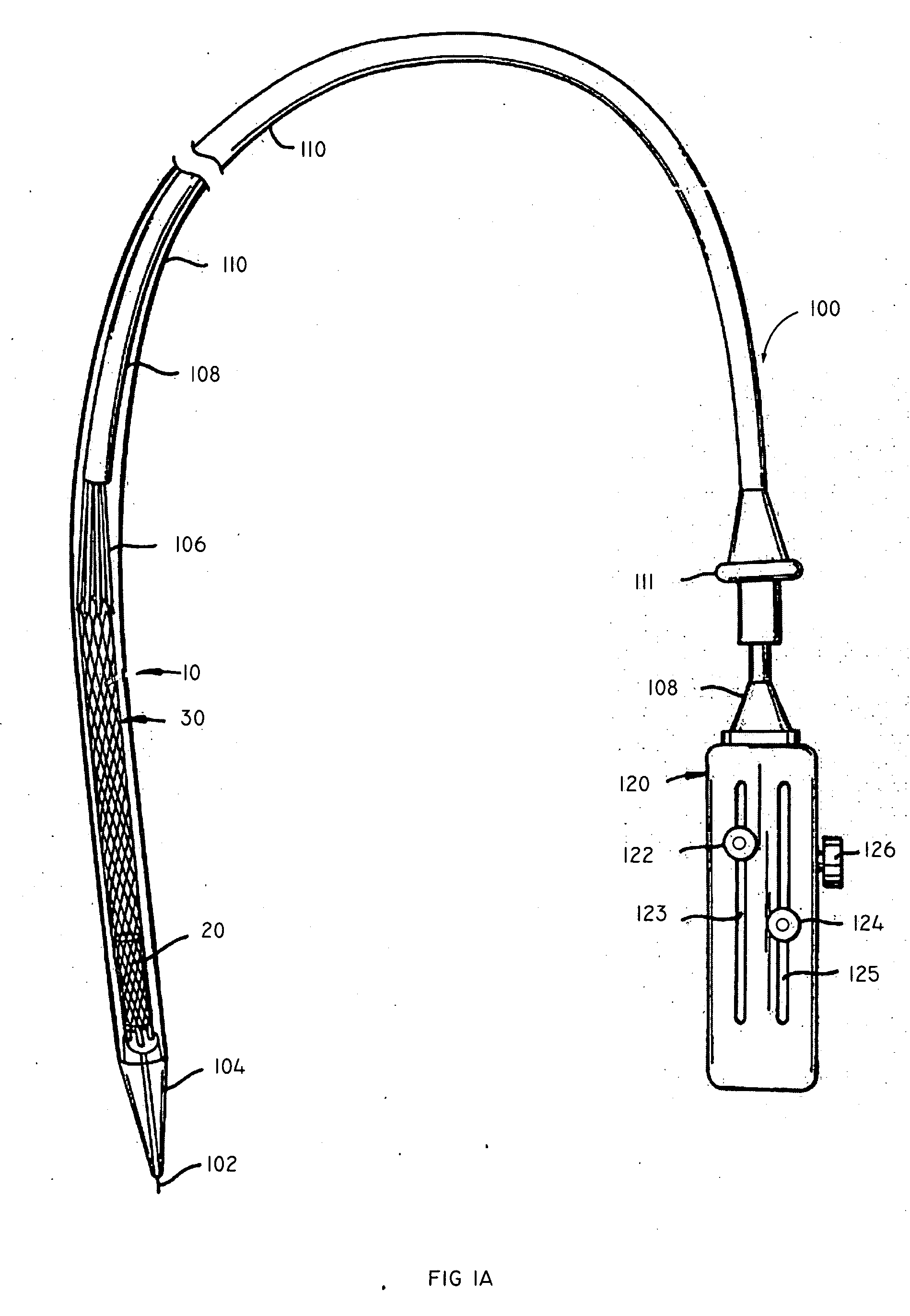
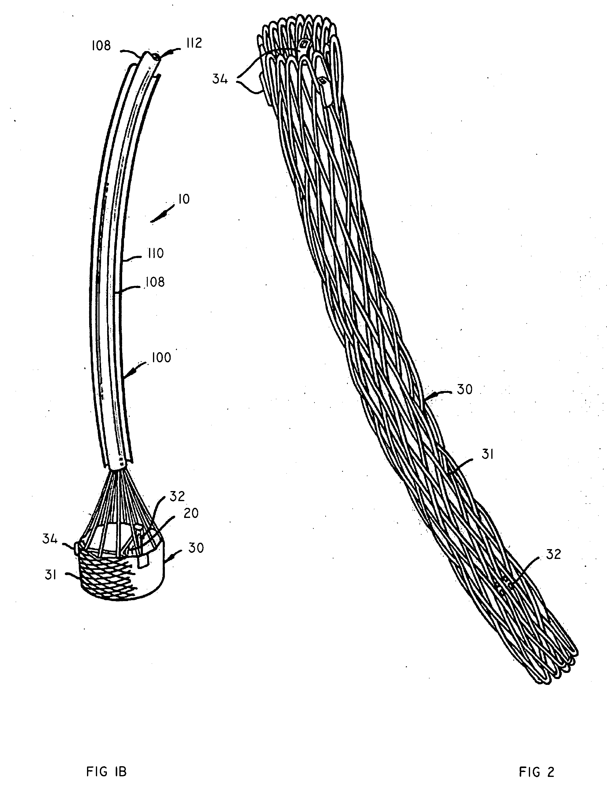
[0048] The present invention relates to an apparatus and methods for endovascularly delivering and deploying an aortic prosthesis within a patient's native heart valve, referred to hereinafter as “replacing” a patient's heart valve. The delivery system includes a sheath assembly, a multi-lumen shaft, and a guide wire for placing the apparatus endovascularly within a patient and a user control allowing manipulation of the aortic prosthesis. The apparatus includes an anchor and a replacement valve. The anchor and the replacement valve are adapted for percutaneous delivery and deployment within a patient's heart valve.

[0049] In some embodiments, the apparatus includes engagement elements and / or a seal inverting element situated along a proximal region of the anchor. The engagement elements are adapted to engage the native leaflets of the patient's heart, or more preferably the proximal edge and / or the commissural attachments of the native leaflets. The engagement elements need not ext...


PUM


Abstract

A method for endovascularly replacing a patient's heart valve including the following steps: endovascularly delivering an anchor and a replacement valve supported within the anchor to a vicinity of the heart valve in a collapsed delivery configuration, the anchor having grasping elements adapted to grasp tissue in a vicinity of the heart valve; expanding the anchor, thereby rotating the grasping elements; and grasping the tissue with the rotating grasping elements.

Description

CROSS-REFERENCE [0001] This application is a continuation-in-part application of application Ser. No. 10 / 972,287, filed Oct. 21, 2004, which is a continuation-in-part of application Ser. No. 10 / 746,240, filed Dec. 23, 2003, both of which are incorporated herein by reference in their entireties and to which applications we claim priority under 35 USC §120.BACKGROUND OF THE INVENTION [0002] The present invention relates to methods and apparatus for endovascularly replacing a heart valve. More particularly, the present invention relates to methods and apparatus for endovascularly replacing a heart valve with a replacement valve using an expandable anchor and tissue grasping elements. [0003] Heart valve surgery is used to repair or replace diseased heart valves. Valve surgery is an open-heart procedure conducted under general anesthesia. An incision is made through the patient's sternum (sternotomy), and the patient's heart is stopped while blood flow is rerouted through a heart-lung by...

Claims


Application Information

Patent Timeline
Patent Type & Authority Applications(United States)
IPC IPC(8): A61F2/24A61F2/90
CPCA61F2/2418A61F2/2439A61F2/848A61F2/90A61F2230/0078A61F2/2412A61F2220/0058A61F2230/005A61F2230/0054A61F2220/0016A61F2210/0014A61F2220/0033A61F2250/0069A61F2/2427A61F2/2433A61F2/88
Inventor SALAHIEH, AMRHILDEBRAND, DANIELSAUL, TOM
Owner BOSTON SCI SCIMED INC
Learn More