Quinazolinone compositions for regulation of gene expression related to pathological processes
- Summary
- Abstract
- Description
- Claims
- Application Information
AI Technical Summary
Benefits of technology
Problems solved by technology
Method used
Image
Examples
example 1
Effect of Halofuginone on TAA-Induced Liver Fibrosis
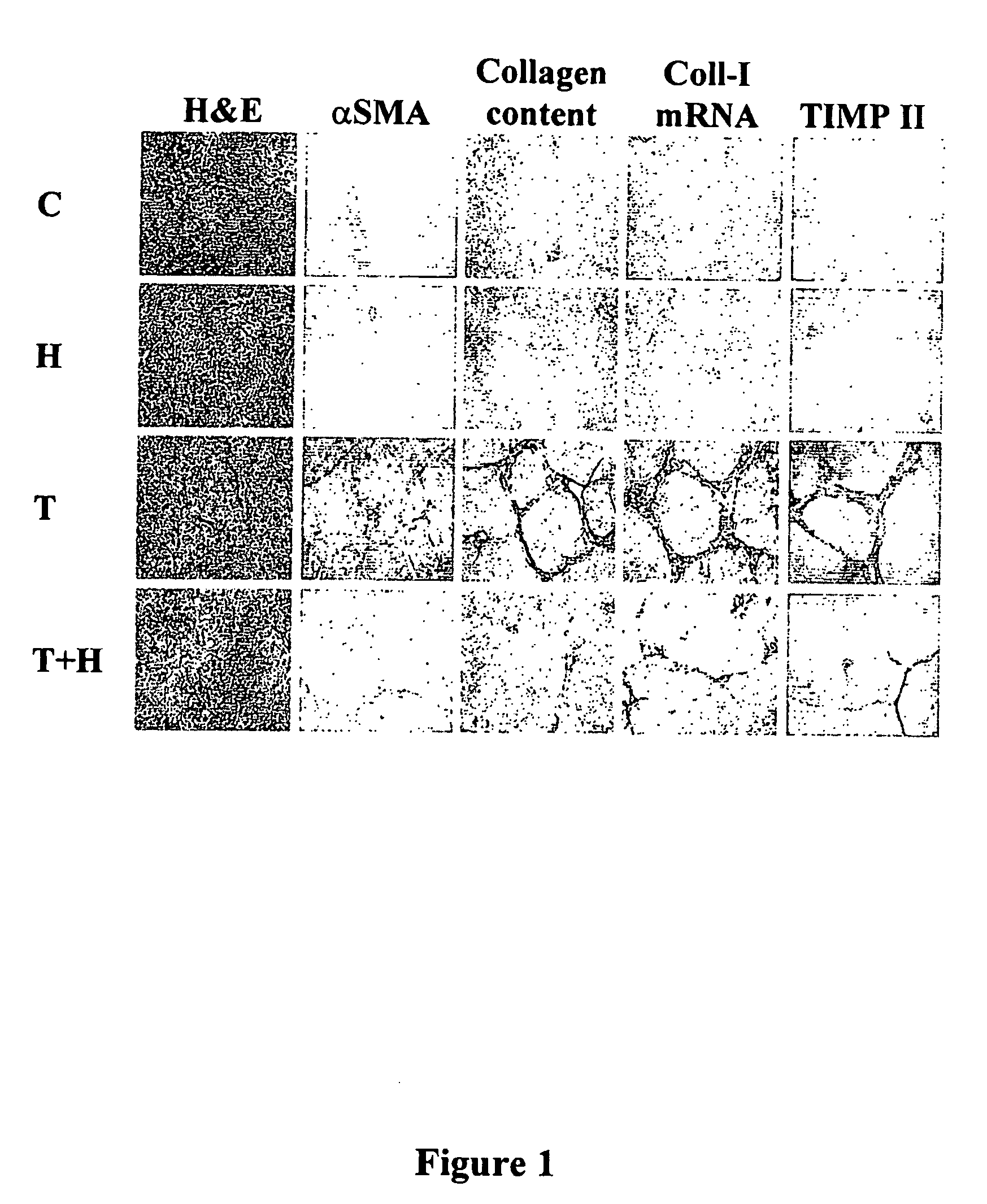
[0181] Liver sections of the control rats were devoid of ECM in general (H&E staining) and of collagen in particular (Sirius red staining). When ASMA antibodies were used, no stellate cells were detected, which suggests that the latter were in their quiescent state. No cells expressing the collagen α1(I) gene or synthesizing TIMP-2 were detected by in situ hybridization or immunohistochemistry, respectively (FIG. 1). No changes in the above parameters were observed in rats treated with halofuginone alone. When treated for 4 weeks with TAA, the livers exhibited a marked increase in ECM content, and displayed bundles of collagen that surrounded the lobules and resulted in large fibrous septa and distorted tissue architecture. These septa were populated by αSMA-positive cells expressing high levels of the collagen α1(I) gene and containing high levels of TIMP-2, all of which are characteristic of advanced fibrosis. These sections wer...
example 2
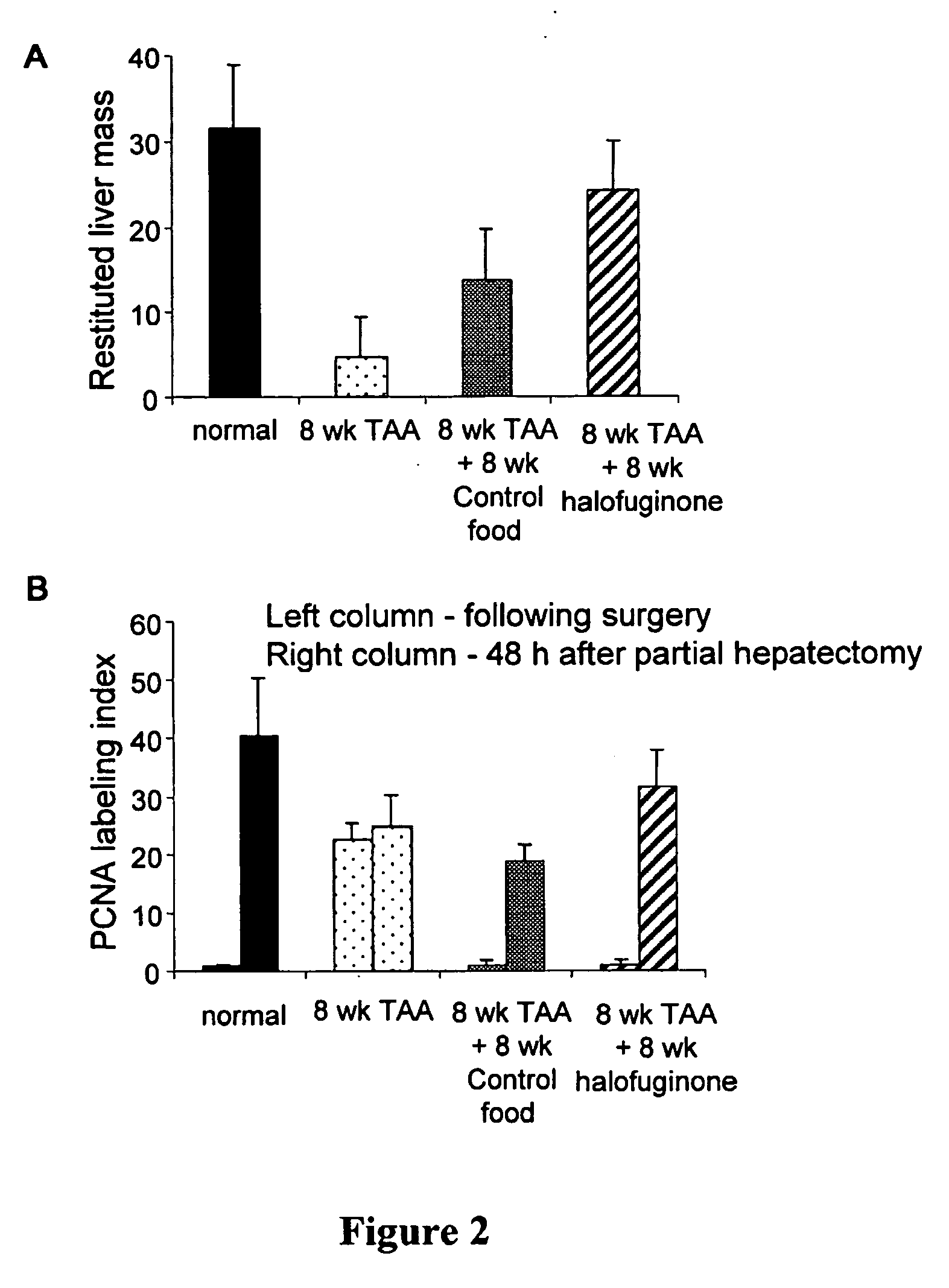
[0182] The halofuginone-dependent decrease in liver Ishak staging was accompanied by an improved regenerative capacity. Eight weeks of halofuginone treatment resulted in close to normal values in liver mass, significantly higher than the values recorded in the control food treated group (24.2±5.7 vs.13.7±4.5, p<0.05) (FIG. 2A). This increase was associated with PCNA labeling index of 31.4±6.4 as compared to 18.8±2.9 in untreated animals (FIG. 2B).
[0183] It is worth noting that the levels of PCNA prior to PHx varied between the above groups. PCNA staining before PHx was negligible in the healthy control group. TAA feeding was characterized as expected by a large number of proliferating cells. TAA removal either in the presence or absence of halofuginone resulted in a low labeling index despite the histopathology noted in the non-treated group. The ability of the two groups however to respond to 70% PHx was different, demonstrating a significant improved capacity t...
example 3
Effect of Halofuginone on IGFBP-1 Synthesis
[0186] Rat primary hepatocytes, HepG2, Hep3B, Huh-7 and HSC were used to identify the source of the halofuginone-dependent synthesis of IGFBP-1. In addition, cell-lines derived from other tissues (fibroblasts and osteoblasts) were used as well. Only cells of the hepatocyte origin demonstrated increased IGFBP-1 gene expression and synthesis in response to halofuginone (FIG. 5A). In rat primary hepatocytes, insulin caused reduction in IGFBP-1 synthesis in agreement with other studies (Ishak K. et al., J Hepatol; 22:696-699) while halofuginone, at concentration as low as 1 nM, increased the synthesis of IGFBP-1 (FIG. 5B). In HepG2, no expression of the IGFBP-1 gene was detected without halofuginone (FIG. 6A). Halofuginone, at concentrations of 10 nM, increased IGFBP-1 gene expression and a further increase was observed at higher concentrations. Without halofuginone, very low (in some cases undetectable) levels of IGFBP-1 were detected in the ...
PUM
| Property | Measurement | Unit |
|---|---|---|
| Composition | aaaaa | aaaaa |
| Gene expression profile | aaaaa | aaaaa |
Abstract
Description
Claims
Application Information
 - R&D
- Intellectual Property
- Life Sciences
- Materials
- Tech Scout
- Unparalleled Data Quality
- Higher Quality Content
- 60% Fewer Hallucinations
Browse by: Latest US Patents, China's latest patents, Technical Efficacy Thesaurus, Application Domain, Technology Topic, Popular Technical Reports.
© 2025 PatSnap. All rights reserved.Legal|Privacy policy|Modern Slavery Act Transparency Statement|Sitemap|About US| Contact US: help@patsnap.com


