Method for a fully automated monoclonal antibody-based extended differential

a monoclonal antibody and extended differential technology, applied in the field of fully automated monoclonal antibody-based extended differential, can solve the problems of inability to readily differentiate multiple subsets of cells in a single sample, difficulty in the performance of accurate 5-part white blood cell performance, and common shortcomings of current analytical systems

Active Publication Date: 2006-11-30
BECKMAN COULTER INC
View PDF25 Cites 23 Cited by
  • Summary
  • Abstract
  • Description
  • Claims
  • Application Information

AI Technical Summary

Benefits of technology

[0016] In yet another aspect, a method for the enumeration of multiple cell populations in a biological sample is performed by mixing or reacting the sample, with one of the compositions described herein in a single reaction mixture. These compositions are further elucidated in the following detailed description. In one embodiment of the method, the single reaction mixture is contacted with an optional lytic reagent that differentially lyses non-nucleated red blood cells present in the sample. The resulting single reaction mixture is then passed through a sensing region in a cell analyzer. The sensing region in one embodiment may be a single flow aperture; and the cell analyzer may be a flow hematology analyzer. This sensing region measures the mixture for at least two of the same or different parameters selected from one or more channels of fluorescence, one or more optical parameters, one or more electrical parameters, and combinations thereof, preferably in a single step. Populations of cells in the sample are enumerated by analyzing at least two parameters for each of the cell populations. Use of a composition as above herein permits the enumeration of multiple hematologic cell populations in a biological sample. When utilizing the compositions described herein consisting of fluorescent dyes and fluorochrome labeled antibodies with overlapping spectra that cannot be separated or distinguished based upon optical or electronic compensation means, a new fluorescent footprint is established. This new fluorescent footprint is a result of the overlapping spectra and the combined cellular staining patterns of the dyes and fluorochrome labeled antibodies chosen for the composition. The new fluorescent footprint results in histogram patterns that are useful for the identification of additional cell populations or subtypes in hematological disease.

Problems solved by technology

Conventional hematology instruments, while capable of differentiating and enumerating the vast majority of cell types and subsets normally present in a peripheral blood sample, cannot readily differentiate multiple subsets of cells in a single sample, particularly those cells that are atypical or immature.
The requirement to correlate the respective outputs of multiple transducers in order to report certain characteristics of a cell type or subset can, under certain circumstances, be problematic, in that it introduces uncertainty in the analytical results (U.S. Pat. Nos. 5,631,165 and 5,565,499; see, e.g., Thomas et al., J. Histochem. Cytochem., 25(7): 827-835 (1977)).
Despite the application of these technologies, the currently available analytic systems suffer from common shortcomings, including difficulty in the performance of an accurate 5-part white blood cell differential in the presence of various atypical leukocyte populations or other abnormal conditions (cellular / non cellular) that interfere with performance of the 5-part differential.
In addition, the conditions that permit the detection of, or flagging for, the presence of atypical cell types suffer from high false positive or high false negative rates.
These shortcomings are unacceptable because they either result in an unnecessarily high manual review rate or the failure to detect clinically significant abnormalities.

Method used


Image

  • Method for a fully automated monoclonal antibody-based extended differential
  • Method for a fully automated monoclonal antibody-based extended differential
  • Method for a fully automated monoclonal antibody-based extended differential

Examples


example 1

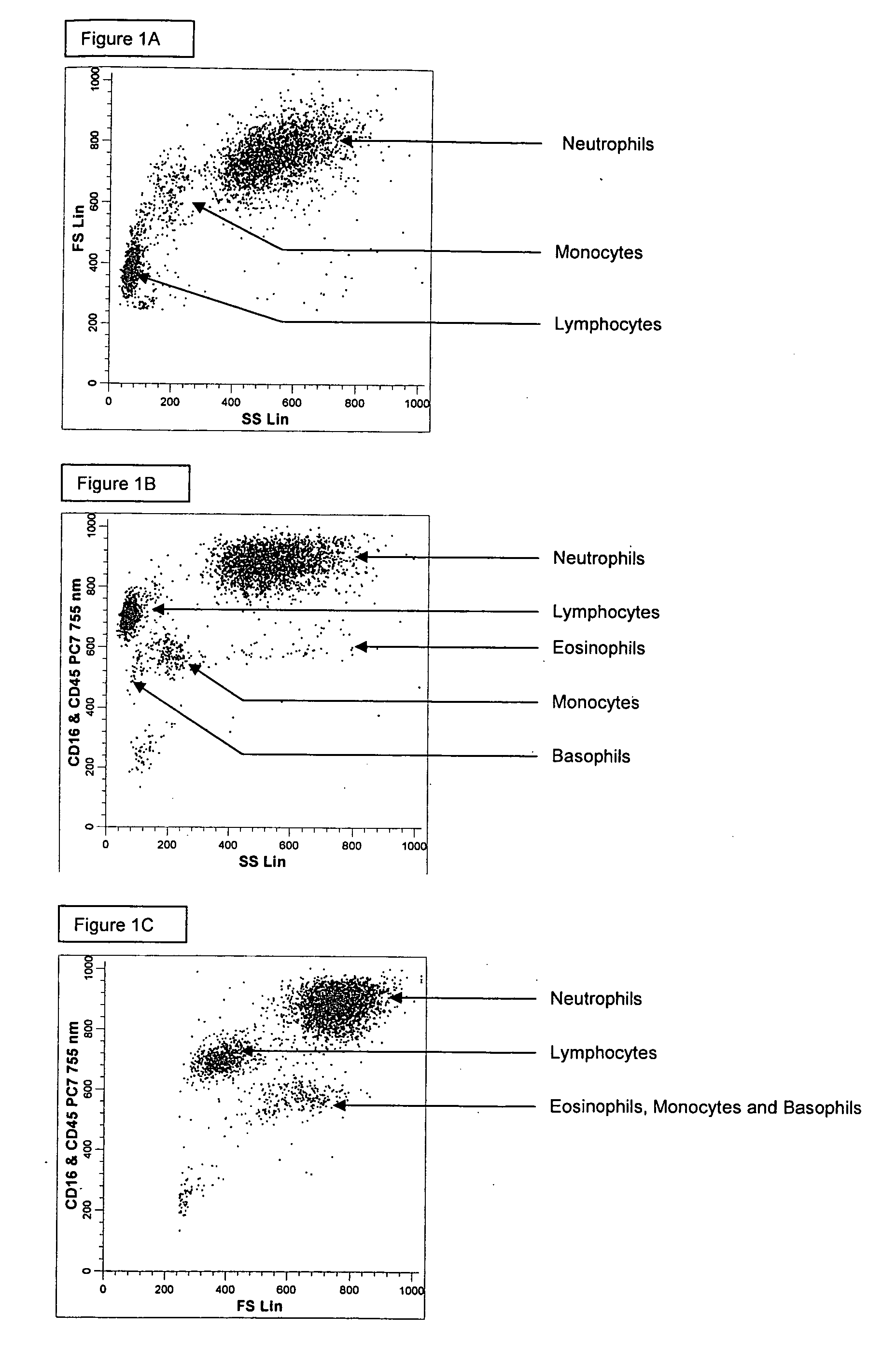
[0184] A single reaction mixture was prepared by reacting 100 μL of normal human peripheral blood with about 1 μg of anti-CD45-PC7, i.e., a first antibody labeled with a first fluorochrome having a first emission spectrum, the first antibody binding to an antigenic determinant that is differentially expressed on populations of leukocytes and non-leukocytes and about 1 μg of anti-CD16-PC7, i.e., an additional antibody labeled with same fluorochrome having the same emission spectrum. The anti-CD16 antibody binds to an antigenic determinant that is differentially expressed on populations of mature and immature granulocytes or myeloid cells. This reaction mixture is mixed briefly and incubated at room temperature for approximately 10 minutes. The reaction was performed in the absence of a fluorescent dye.

[0185] This reaction mixture is then reacted for about 8 seconds with the lytic system (about 600 μL of Immunoprep™ reagent A; see U.S. Pat. No. 5,030,554) that differentially lyses th...

example 2

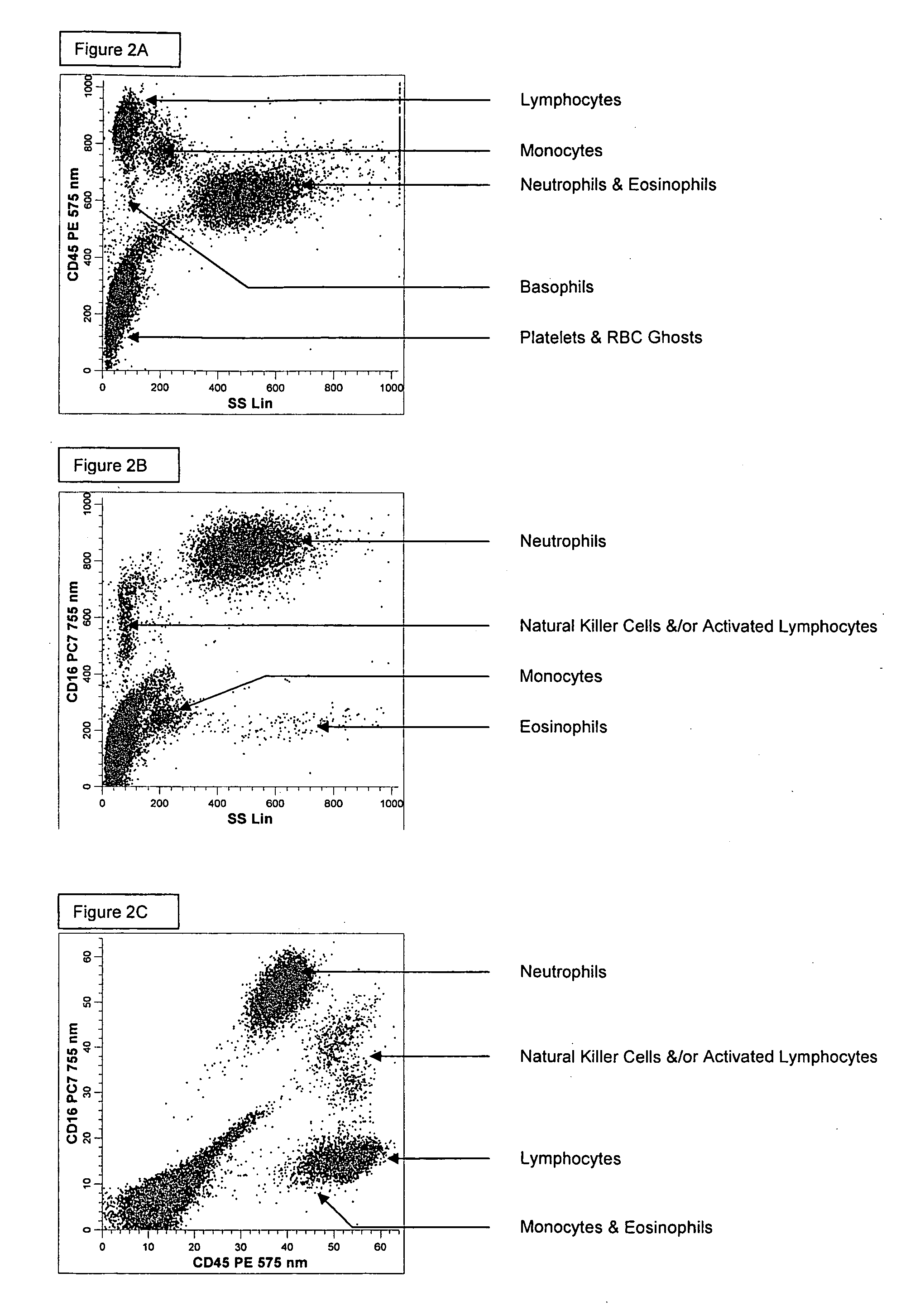
[0190] A single reaction mixture was prepared by reacting 100 μL of normal human peripheral blood with about 1 μg of anti-CD45-PE, i.e., a first antibody labeled with a first fluorochrome having a first emission spectrum, the first antibody binding to an antigenic determinant that is differentially expressed on populations of leukocytes and non-leukocytes and about 1 μg of anti-CD16-PC7, i.e., an additional antibody labeled with a second fluorochrome that has an emission spectrum distinguishable from the emission spectrum of the fluorochrome PE. The anti-CD16 antibody binds to an antigenic determinant that is differentially expressed on populations of mature and immature granulocytes or myeloid cells. This reaction mixture is mixed briefly and incubated at room temperature for approximately 10 minutes. The reaction was performed in the absence of a fluorescent dye.

[0191] This reaction mixture is then reacted for about 8 seconds with the lytic system (about 600 μL of Immunoprep™ rea...

example3

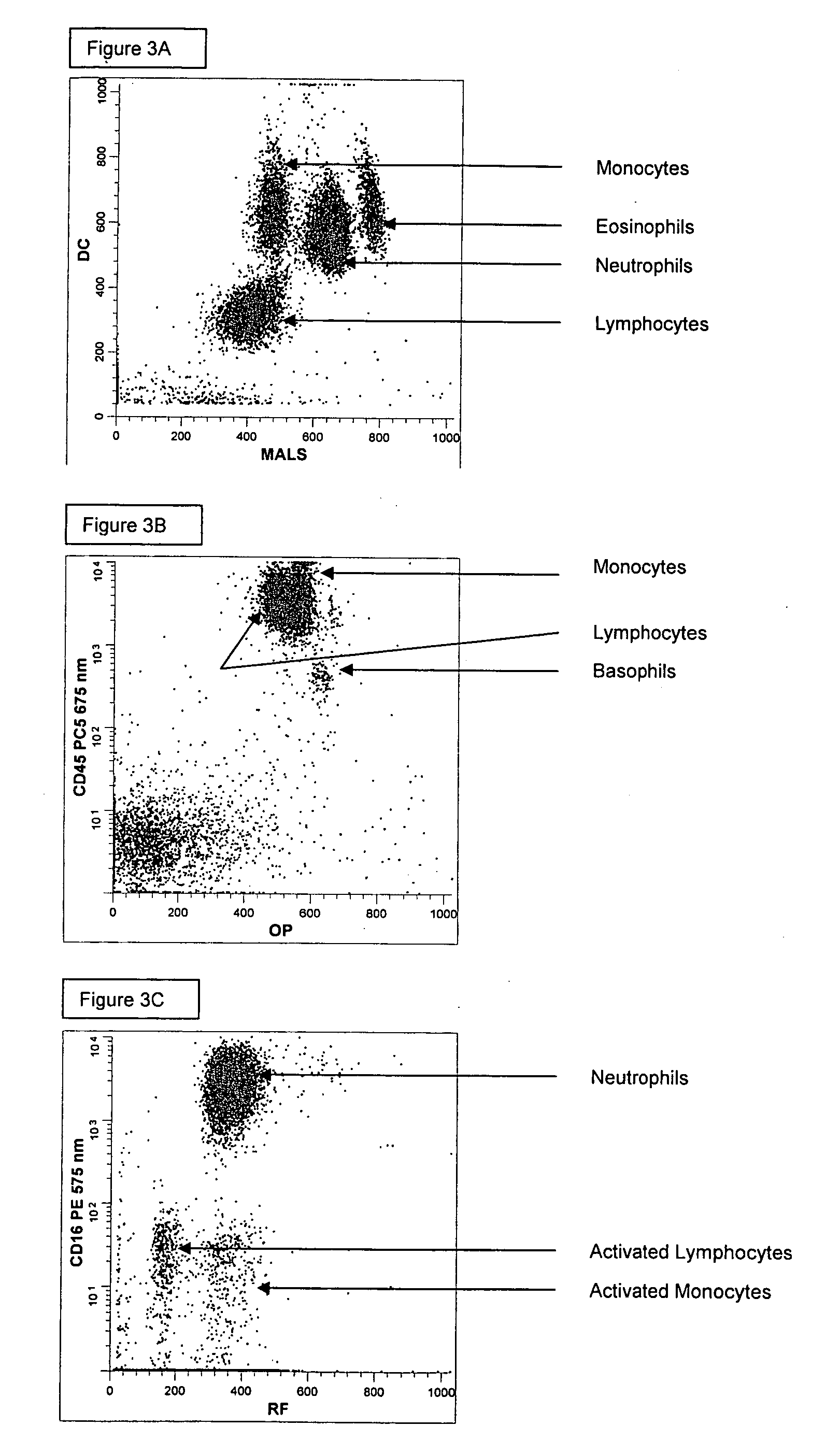
[0194] A single reaction mixture was prepared by reacting 200 μL of normal human peripheral blood with about 1 μg of anti-CD45-PC5, i.e., a first antibody labeled with a first fluorochrome having a first emission spectrum, the first antibody binding to an antigenic determinant that is differentially expressed on populations of leukocytes and non-leukocytes and about 1 μg of anti-CD16-PE, i.e., an additional antibody labeled with a second fluorochrome that has an emission spectrum distinguishable from the emission spectrum of the fluorochrome PC5. The anti-CD16 antibodybinds to an antigenic determinant that is differentially expressed on populations of mature and immature granulocytes or myeloid cells. This reaction mixture is mixed briefly and incubated at room temperature for approximately 10 minutes. The reaction was performed in the absence of a fluorescent dye.

[0195] A portion (about 34 μL) of this reaction mixture is then reacted for about 6 seconds with the lytic system (abou...


PUM

PropertyMeasurementUnit
forward angleaaaaaaaaaa
peak emission wavelengthaaaaaaaaaa
wavelengthaaaaaaaaaa

Abstract

Methods for differentially identifying cells in an instrument employ compositions containing a combination of selected antibodies and fluorescent dyes having different cellular distribution patterns and specificities, as well as antibodies and fluorescent dyes characterized by overlapping emission spectra which form non-compensatable spectral patterns. When utilizing the compositions described herein consisting of fluorescent dyes and fluorochrome labeled antibodies with overlapping spectra that cannot be separated or distinguished based upon optical or electronic compensation means, a new fluorescent footprint is established. This new fluorescent footprint is a result of the overlapping spectra and the combined cellular staining patterns of the dyes and fluorochrome labeled antibodies chosen for the composition. The new fluorescent footprint results in histogram patterns that are useful for the identification of additional cell populations or subtypes in hematological disease.

Description

CROSS REFERENCE TO RELATED APPLICATIONS [0001] This application is a continuation-in-part of U.S. patent application Ser. No. 11 / 130,492, filed May 17, 2005, which claims the benefit of the priority of U.S. Provisional Patent Application No. 60 / 573,167, filed May 21, 2004, now abandoned.BACKGROUND OF THE INVENTION [0002] Whole blood and peripheral blood samples from human subjects suffering from a variety of diseases can contain both blood cells or non-blood cells (e.g., tumor cells, bacteria, etc.) suspended in a liquid medium or plasma. The blood cells include red blood cells (erythrocytes or RBCs), white blood cells (leukocytes or WBCs), and platelets. Depending on the level of maturity of the cells, red cells are further classified into nucleated RBCs (NRBCs), reticulated RBCs (reticulocytes), and mature RBCs. Mature white cells fall into one of five different “normal” categories, namely, monocytes, lymphocytes, eosinophils, neutrophils and basophils. Each of the white cell subs...

Claims


Application Information

Patent Timeline
Patent Type & Authority Applications(United States)
IPC IPC(8): G01N33/567C07K16/46G01N33/53G01N33/569
CPCG01N33/56966G01N33/56972G01N2015/1006G01N2015/1402Y10T436/107497Y10T436/25125Y10T436/13Y10T436/101666G01N15/1459
Inventor PAUL, RONALD D.RUBIO, OILDACAREAGA, DIANA B.LOPEZ, LIDICE L.MYLVAGANAM, RAVINDRA
Owner BECKMAN COULTER INC
Why Eureka
Try Eureka