

Immunological assay and antibodies for Anti-Mullerian Hormone
a technology of immunological assay and anti-mullerian hormone, which is applied in the field of immunological assay and methods to measure biological compounds, can solve the problems of inability to use amh assays to measure human amh and in rodent samples
- Summary
- Abstract
- Description
- Claims
- Application Information
AI Technical Summary
Benefits of technology
Problems solved by technology
Method used
Image

Examples
example 1
Antibody Production
[0038] Female AMH null mice on a C57B1 / 6J background were immunized with recombinant human AMH prepared as described previously (Hudson et al., 1990). An initial injection of 50 μg of recombinant human AMH was given subcutaneously in Ribi adjuvant (Sigma M6536) and two similar booster injections were given to each animal at 4-week intervals. Four days after the third boost, 5 μl of blood was taken from each animal for screening by ELISA. Each sample was diluted in 2 ml of PBS containing 1% BSA (w / v) and 0.1% (w / v) NaN3 as a preservative and screened as follows: Nunc Maxisorb plates (SLS, UK) were coated with recombinant human AMH supplied by Dr Richard Cate (Biogen, Cambridge, Mass. USA) at 0.1 μg / ml in 0.2M sodium carbonate-bicarbonate buffer, pH 9.4 (Perbio) overnight (50 μl / well). Following adsorption, the plates aspirated and blocked with 100 μl per well of 1% (w / v) BSA in PBS for an hour. The plates were then washed with wash buffer (0.05M Tris-HCl buffer c...
example 2
[0039] Antibody screening. One aliquot of splenocytes was fused with SP2 / 0 myeloma cells in the presence of polyethylene glycol (Boehringer Mannheim), according to Köhler and Milstein (Lee et al., 1996). Fusions were screened first by ELISA on AMH which is an E. coli expressed version of only the human mature region (R & D Systems). A second round of screening was performed on ELISA plates coated with a preparation of intact rat AMH, as described by Weenen et al. (Weenen et al, Mol Hum Reprod 2004). High responding clones were recloned on methylcellulose medium (Clonacell HY) and cell lines were grown up in growth medium containing low concentrations of IgG. Antibodies were purified by protein G chromatography and biotinylated with water soluble LC biotin ester (Pierce Chemicals). Procedures used were from standard protocols (Harlow and Lane). These clones selected for immunoassay studies were thus screened to provide epitopes in AMH that are at least partially in the mature region ...
example 3
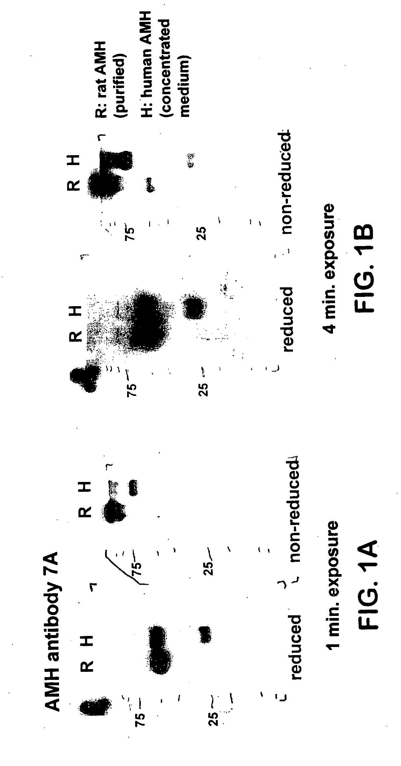
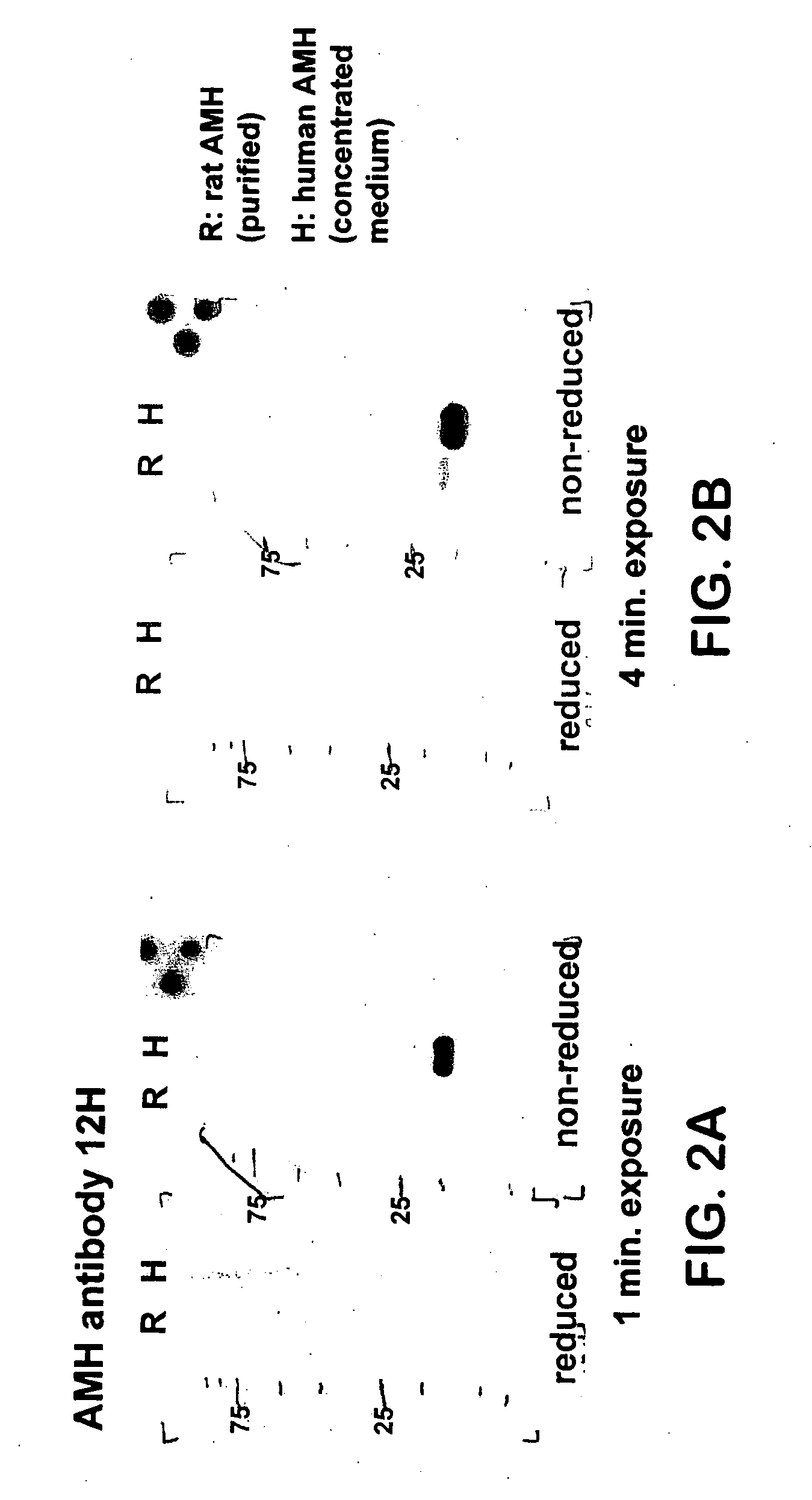
[0040] Western Blotting. Western blot analysis was performed using the mouse monoclonal antibodies 5 / 6A, 9 / 6, 2 / 6, F2B12H (antibody 12H) and F2B7A (antibody 7A). The 5 / 6A, 9 / 6, and 2 / 6 antibodies are antibodies to human AMH described previously (Al-Qahtani et al., 2005); results obtained using these antibodies are shown here for the purpose of comparison to the characteristics of the 12H and 7A antibodies. The 5 / 6A antibody binds to the mature region of human AMH; the 2 / 6 and 9 / 6 antibodies bind to the pro region of human AMH. Recombinant rat and human AMH were separated using 10% polyacrylamide gel electrophoresis under reducing or non-reducing conditions. Proteins were transferred to nitrocellulose membranes and incubated with the antibody at a 1:1000 dilution, followed by a secondary peroxidase-conjugated goat anti-mouse antibody at a 1:10000 dilution. Proteins were visualized by the ECL plus Western blotting detection system (Amersham Biosciences).
[0041] FIGS. 1(A-B) show Weste...
PUM
| Property | Measurement | Unit |
|---|---|---|
| Fraction | aaaaa | aaaaa |
| Fraction | aaaaa | aaaaa |
| Fraction | aaaaa | aaaaa |
Abstract
Description
Claims
Application Information