

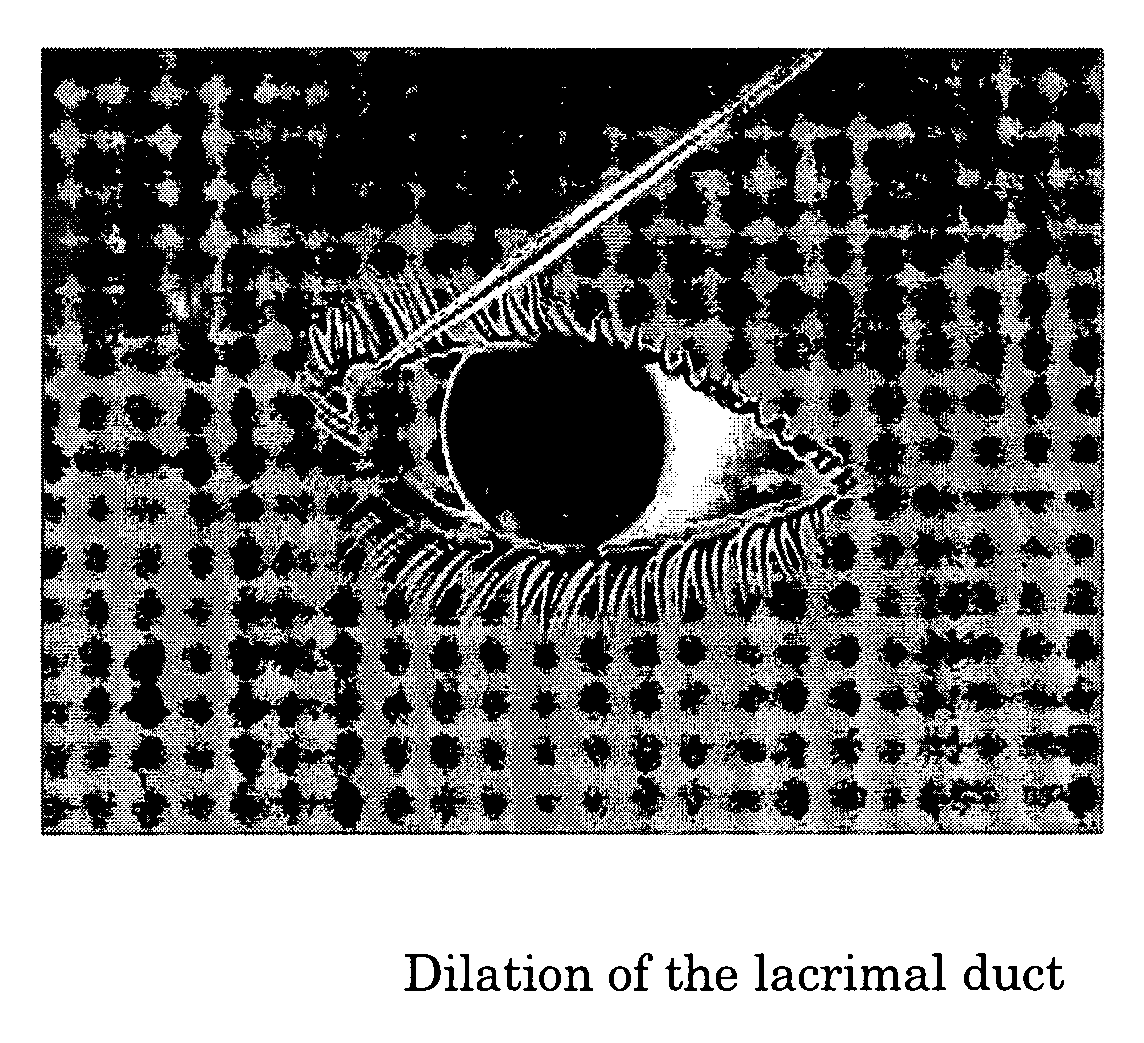
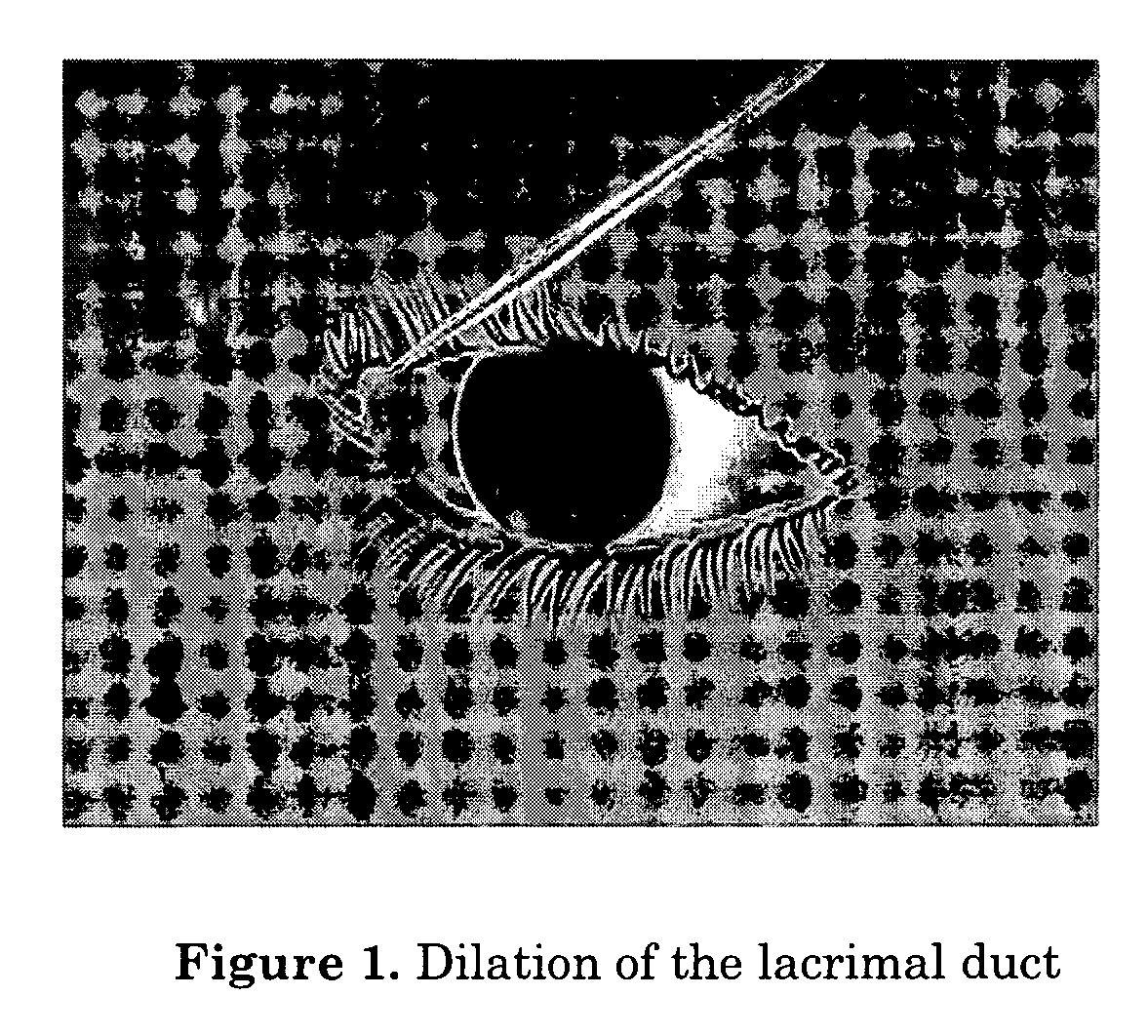
Method of intracanalicular laser dacryocystorhinostomy
a laser and cannula technology, applied in the field of surgical techniques, can solve the problems of long surgical time, red and painful, and swollen lacrimal sac, and achieve the effect of reducing the time of surgery
- Summary
- Abstract
- Description
- Claims
- Application Information
AI Technical Summary
Benefits of technology
Problems solved by technology
Method used
Image

Examples
example 1
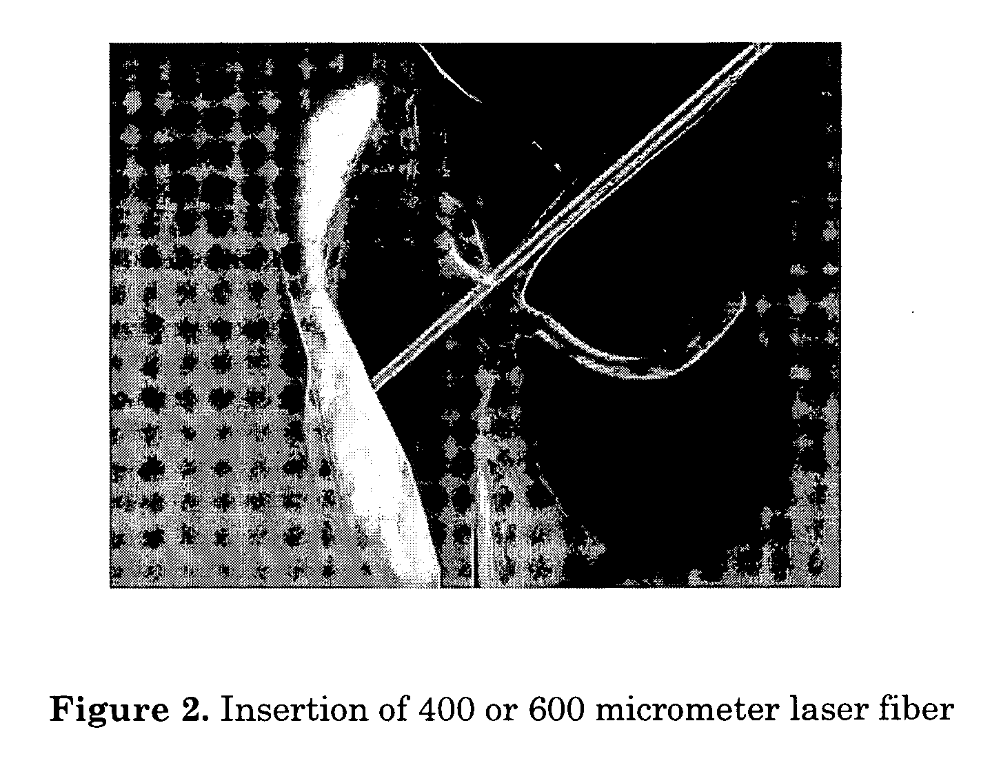
[0046] Summary: Forty-three transcanalicular DCR were performed using a 980 nm diode laser (Varius, Intermedic, Barcelona, Spain) and bicanalicular intubation with silicone tube and prolene filament, both PVP-covered (PVP Ritleng lacrimal intubation set, FCI, Issy-les-Moulineaux Cedex, France). The results were analysed using a prospective, interventional, non randomized and non comparative study. Local and topical anaesthesia were used in patients with a clinical history of epiphora or dacryocystitis for nasolacrimal obstruction. A diode laser was used to effect a vaporization of the lacrimal sac, osteotomy and vaporization with coagulation of nasal mucosa. The mean duration of surgery was 14 minutes (range 7 to 29 minutes). In all cases, and during a two-month period, bicanalicular intubation was carried out using a silicone tube and prolene filament. Follow-up was between 4 to 38 months. The degree of epiphora was evaluated using the Munk scale, and lacrimal permeability was eval...
PUM
 Abstract
Description
Claims
Application Information