
Tactile breast imager and method for use

a breast imager and tactile technology, applied in the field of biological objects imaging, can solve the problems of limiting such frequency, high cost of frequent examination, and the possibility of small tumors in mammography, and achieve the effect of higher sensitivity and accuracy

Inactive Publication Date: 2007-02-15
SARVAZYAN ARMEN P +2
View PDF18 Cites 11 Cited by
  • Summary
  • Abstract
  • Description
  • Claims
  • Application Information

AI Technical Summary

Benefits of technology

The device enhances the sensitivity and accuracy of breast tissue examination, facilitating early detection of breast cancer through objective analysis of pressure patterns and automatic data recording, making it suitable for regular home use and improving survival rates by enabling more frequent and accurate self-examinations.

Problems solved by technology

However the cost of frequent examinations, plus the accumulated radiation exposure from frequent mammograms tend to limit such frequency.
In addition, mammography may miss small tumors, especially in the dense breasts of younger women.
The primary criticism of manual examination is that women do not examine their breasts properly.
But nevertheless most women have difficulty in detecting small lesions and differentiating between harmful and harmless lesions and they have little or no knowledge of the various types of lumps, which may occur in the breast.
Again, the challenge is to differentiate significant palpable findings from the nonsignificant ones that do not feel much different.
In many patients, the findings are not conclusive and the breast examiner / physician has difficulty in interpreting what his fingers feel in the breast.
Unfortunately, the human fingertip may not be sensitive enough to measure the difference.
In fact, it is believed that palpation is not able to detect tumors of less than about one centimeter in size.
Specifically, these devices cannot be used for a regular home use in a repeatable pattern that allows for accurate and reproducible serial examinations.

Method used


Image

  • Tactile breast imager and method for use
  • Tactile breast imager and method for use
  • Tactile breast imager and method for use

Examples


first embodiment

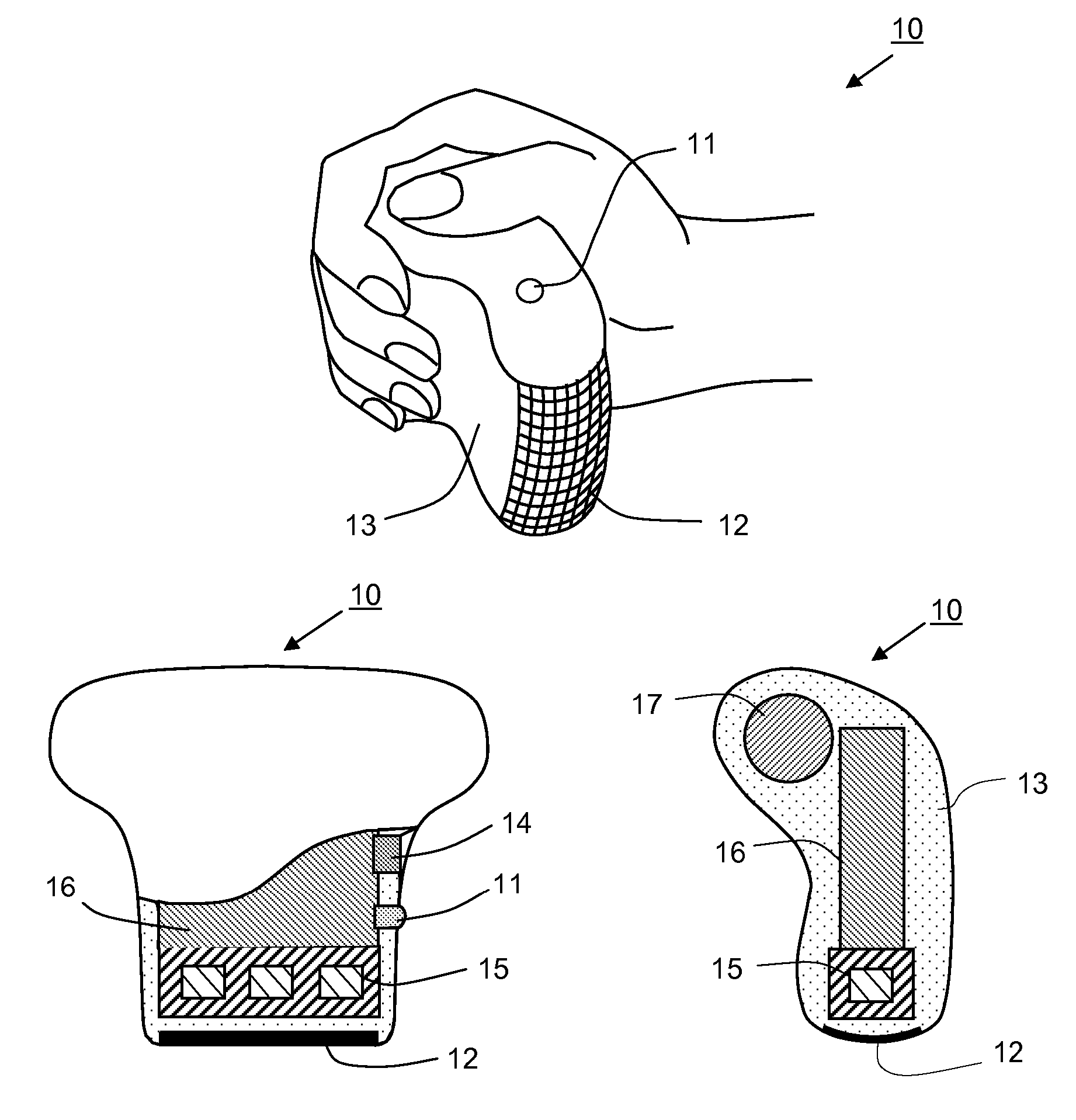
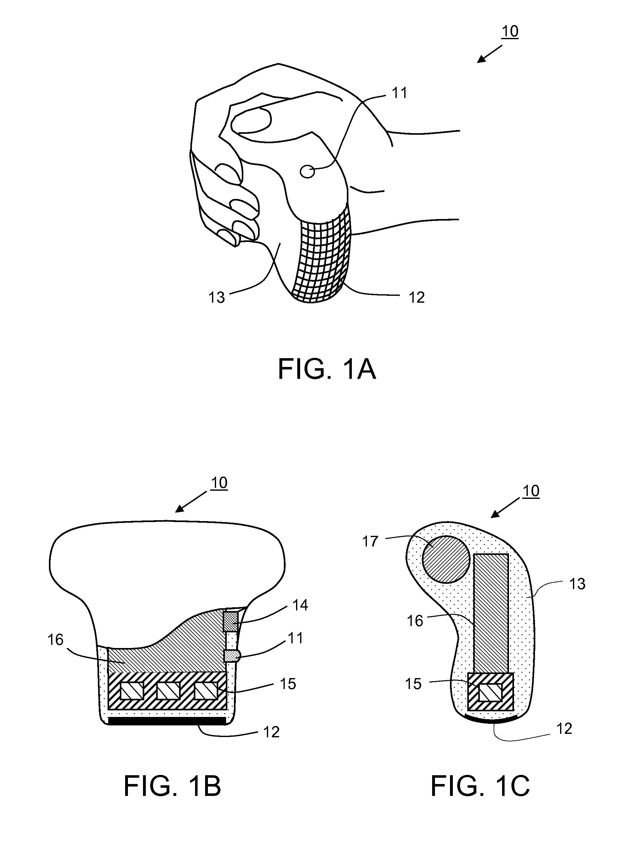
[0036]FIGS. 1A, 1B and 1C show a hand-held self-palpation tactile imaging device 10 for detecting and locating a lesion in breast tissue in accordance with the present invention. The device 10 comprises pressure sensing means such as a 2-D tactile sensor array 12, a positioning data means such as an inertial motion tracking sensor 15, an electronic unit 16, a power supply 17, a computer connector 14, and an output signal source 11, all of which are mounted in a housing 13 adapted for easy grip by a human hand. Tactile sensor array 12 generates signals in response to pressure imposed on a pressure-sensing surface as it is pressed against and moved over the breast or another type of soft tissue. Tactile sensor array 12 comprises a matrix of tactile sensing elements measuring discretely contact pressure between the surfaces of the sensing elements and breast tissue. Motion tracking system 15 for generating signals in response to motion of the device 10, can include accelerometers, magn...

second embodiment

[0042]FIGS. 4A and 4B show a hand-held self-palpation device 20 for detecting and locating a lesion in breast tissue in accordance with the present invention. The device 20 comprises 2-D tactile sensor array 41, a patchboard 43 with buttons 44-46, an output signal source 47, all of which are mounted in a housing 42. During the breast self-examination, a patient is putting this device on her fingertips. Tactile sensor array 41 generates signals in response to pressure imposed on a pressure-sensing surface as it is pressed against and moved over the breast. The buttons 44, 45, 46 are intended for hand entering information about which breast and which quadrant of the breast is under examination. The patchboard 43 and housing 42 include all necessary electronics for storing and preliminary processing acquired tactile data. Scanning area location data are stored in a device recording system. After the breast examination is complete, the examination data can be transmitted to a home compu...

third embodiment

[0043]FIG. 5 represents a hand-held self-palpation device 30 for detecting and locating a lesion in breast tissue in accordance with the present invention. The device 30 comprises 2-D tactile sensor array 51 mounted on a touch pad 52 and an electronic unit 56 with a display means 55 optionally secured on a patient's wrist or hand by a strap 54. During the breast self-examination, the patient can in real-time environment observe the results of breast examination and communicate with device 30 by means of control buttons 53. Along with that the patient can enter the information about position of the local scanning zone using data entry means such as buttons 57. The device 30 allows transmission of received breast examination data into a home computer.


PUM


Abstract

A method and device for breast examination adapted for easy home use including a tactile imager probe equipped with a pressure sensor array and a motion tracking system for simultaneously recording pressure and positioning data during the examination of a breast or any other soft tissue. The method includes a step of starting the examination from a known point or an anatomical mark such as a nipple or a sternum; moving the probe towards the area of interest and oscillating it thereabout while manually or automatically recording pressure and positioning data. Subsequent data analysis identifies the presence of a lesion, calculates its location relative to the probe, estimates the location of the probe relative to the anatomical landmark and finally calculates the location of the lesion relative to that anatomical landmark. The method allows repeating examinations over time with great accuracy as they all start from the same anatomical landmark. The probe includes provisions for easy hand grip or attaching to fingers of a patient, a cable connector or a wireless transmitter for transmitting the data out to a computer or another data analysis device, as well as manual data entry means allowing the patient herself to enter the positioning data.

Description

CROSS-REFERENCE DATA [0001] A priority date benefit is claimed herein from a U.S. Provisional Patent Application No. 60 / 477,740 filed by the same inventors on Jun. 12, 2003 and entitled “Tactile breast imager”, which is incorporated herein in its entirety by reference. This application is a divisional application of a co-pending U.S. patent application Ser. No. 10 / 866,395 filed Jun. 12, 2004 with the same title and incorporated herewith in its entirety by reference.[0002] This invention was made with government support under SBIR Grants No. R43 CA91392 and No. R43 / 44 CA69175 awarded by the National Institutes of Health, National Cancer Institute. The government has certain rights in this invention.BACKGROUND OF THE INVENTION [0003] This invention relates generally to imaging of biological objects and, more particularly, to a method and apparatus for mass breast screening and detecting early changes in mechanical properties of breast tissue that are indicative of breast cancer and ot...

Claims


Application Information

Patent Timeline
Patent Type & Authority Applications(United States)
IPC IPC(8): A61B5/103A61B5/00
CPCA61B5/0002A61B5/0053A61B5/4312A61B2562/046A61B5/6843A61B2560/045A61B2562/0247A61B5/681
Inventor SARVAZYAN, ARMEN P.EGOROV, VLADIMIRSON, JAES
Owner SARVAZYAN ARMEN P
Learn More