Method for acquiring and evaluating vascular examination data

a technology of vascular examination and data acquisition, applied in the field of acquiring and evaluating vascular examination data, can solve the problems of difficult and inaccurate precise determination and acquisition of contours, limited local resolution of ivus images, and problematic automatic evaluation of ivus images

Inactive Publication Date: 2007-02-22
SIEMENS AG
View PDF12 Cites 111 Cited by
  • Summary
  • Abstract
  • Description
  • Claims
  • Application Information

AI Technical Summary

Benefits of technology

[0006] The invention therefore has to deal with the problem of creating a method for acquiring and evaluating vascular examination data, which allows a reliable and automatic analysis of image data.

Problems solved by technology

In practice however the automatic evaluation of IVUS images has proven problematic.
Such images do indeed show the individual tissue or vessel structures, such as intima, media and plaque layers, but the precise determination and acquisition of contours is difficult and inaccurate, as the structures to be identified are small and the local resolution of the IVUS images is inadequate for this purpose.
In practice therefore contours are input manually to some degree but this is very time-consuming, as the user has to draw in the contours by hand on every image.
One disadvantage of this is that the local resolution of the IVUS images is limited by the systems used, such that incorrect or inaccurate results are obtained in many instances.

Method used


Image

  • Method for acquiring and evaluating vascular examination data
  • Method for acquiring and evaluating vascular examination data

Examples


Embodiment Construction

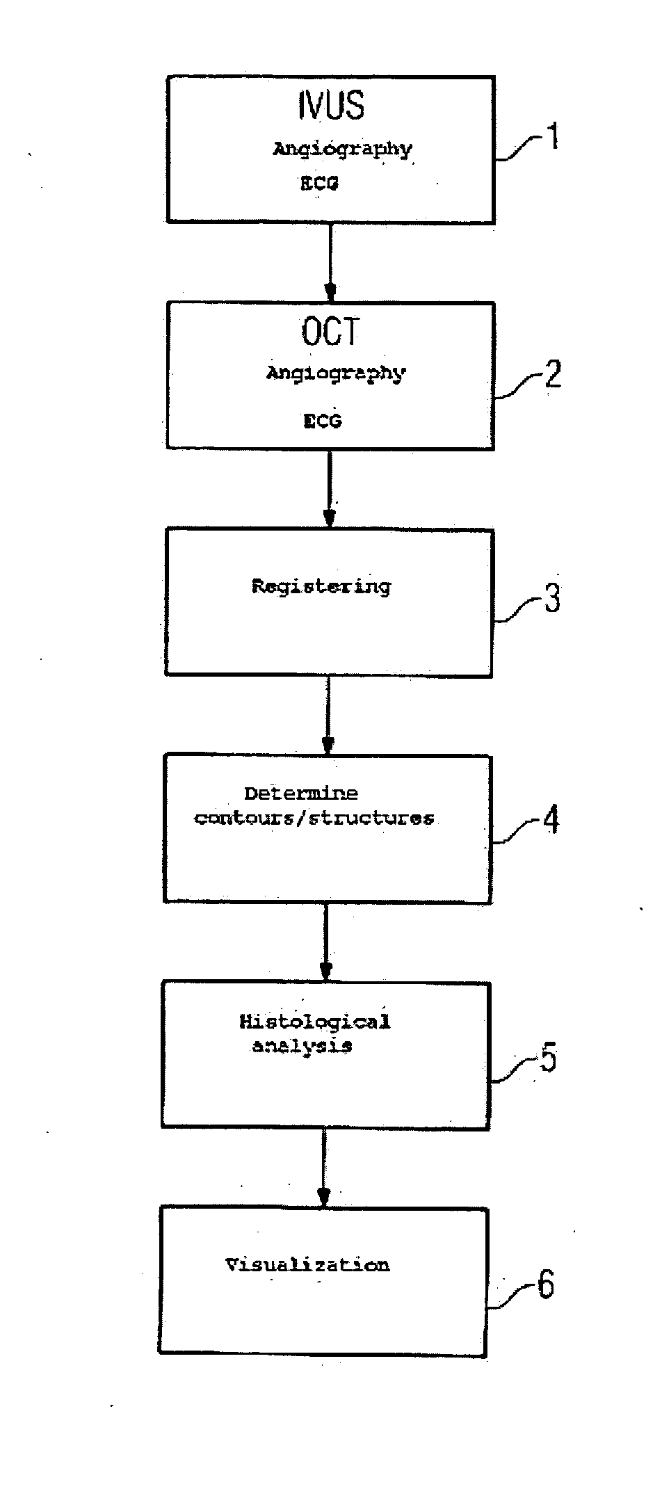
[0019] In step 1 the IVUS catheter is inserted into the vessel to be examined. IVUS images and x-ray projections (angiography data) are then recorded at the same time. These two recordings are ECG-triggered, such that both the angiography data and the IVUS images are recorded at the same point of the cardiac cycle. It is ensured with these recordings that the IVUS catheter is clearly visible on the x-ray projections. It is possible to record whole sections of vessels using the pull-back method.

[0020] The method can also be configured such that catheters with one or more position sensors are used, such that it is possible to assign the recorded images based on the catheter positions that are recorded at the same time.

[0021] After the IVUS catheter has been removed, in step 2 an OCT catheter is inserted into the same vessel and moved into the same position the IVUS catheter was in before. ECG triggering is used in the same manner to record angiography data at the same time as the OC...


PUM


Abstract

The invention relates to a method for acquiring and evaluating vascular examination data, comprising: acquisition of IVUS images of a vessel to be examined using an IVUS catheter; simultaneous acquisition of angiography data of the IVUS catheter having at least one angiography marker; acquisition of OCT images of the same point of the vessel to be examined using an OCT catheter; simultaneous acquisition of angiography data of the OCT catheter having at least one angiography marker; registering the IVUS and OCT images; determination of contours of the structures of the vessel under examination based on the OCT images; arithmetic histological analysis of the co-registered IVUS and OCT images using the information about the contours; and display the results.

Description

CROSS REFERENCE TO RELATED APPLICATIONS [0001] This application claims priority of German application No. 10 2005 037 897.8 filed Aug. 10, 2005, which is incorporated by reference herein in its entirety. FIELD OF THE INVENTION [0002] The invention relates to a method for acquiring and evaluating vascular examination data. BACKGROUND OF THE INVENTION [0003] Methods are already known, with which catheters provided with sensors can be used for vessel imaging. These include ultrasound catheters, for example IVUS (intravascular ultrasound) catheters, which are used to detect atherosclerotic plaque for example. In practice however the automatic evaluation of IVUS images has proven problematic. Such images do indeed show the individual tissue or vessel structures, such as intima, media and plaque layers, but the precise determination and acquisition of contours is difficult and inaccurate, as the structures to be identified are small and the local resolution of the IVUS images is inadequat...

Claims


Application Information

Patent Timeline
Patent Type & Authority Applications(United States)
IPC IPC(8): A61B8/00
CPCA61B5/0066A61B5/02007A61B5/7285A61B6/541A61B8/12A61B6/5247A61B2090/364
Inventor CAMUS, ESTELLEREDEL, THOMAS
Owner SIEMENS AG
Why Eureka
Try Eureka