Non-cylindrical prosthetic valve system for transluminal delivery

a prosthetic valve and non-cylindrical technology, applied in the field of prosthetic cardiac valve and related deployment system, can solve the problems of complex complex structure, prolonged patient hospitalization, and high cost, and achieve the effects of avoiding substantial migration of the prosthetic valve assembly, eliminating problems, and optimizing function and durability

Inactive Publication Date: 2007-02-22
MEDTRONIC COREVALVE
View PDF101 Cites 1046 Cited by
  • Summary
  • Abstract
  • Description
  • Claims
  • Application Information

AI Technical Summary

Benefits of technology

[0009] The object of the present invention is to transluminally provide a prosthetic valve assembly that includes features for preventing substantial migration of the prosthetic valve assembly once delivered to a desired location within a body. The present invention aims to remedy these significant problems. Another objective of the invention is to provide a support at the time of positioning of the replacement valve that makes it possible to eliminate the problem caused by the native valve sheets, which are naturally calcified, thickened and indurated, or by the residues of the valve sheets after valve resection. Yet another objective of the invention is to provide a support making possible complete sealing of the replacement valve, even in case of an existing cardiac ring which has a surface which is to varying degrees irregular and / or to varying degrees calcified. Another objective of the invention is to have a device that can adapt itself to the local anatomy (i.e. varying diameters of the ring, the subannular zone, the sino-tubular junction) and maintain a known diameter of the valve prosthesis to optimize function and durability. The invention also has the objective of providing a support whose position can be adapted and / or corrected if necessary at the time of implantation.

Problems solved by technology

This procedure generally requires prolonged patient hospitalization, as well as extensive and often painful recovery.
It also presents advanced complexities and significant costs.
The stent-supported systems designed for the positioning of a heart valve introduce uncertainties of varying degree with regard to minimizing migration from the target valve site.
A cardiac valve that is not adequately anchored in place to resist the forces of the constantly changing vessel wall diameter, and turbulent blood flow therethrough, may dislodge itself, or otherwise become ineffective.
In particular, the known stents do not appear to be suited to sites in which the cardiac wall widens on either proximally and / or distally of the valve annulus situs.
Furthermore, the native cardiac ring remaining after ablation of the native valve can hinder the positioning of these stents.
These known systems also in certain cases create problems related to the sealing quality of the replacement valve.
In effect, the existing cardiac ring can have a surface that is to varying degrees irregular and calcified, which not only lessens the quality of the support of the stent against this ring but also acts as the source of leaks between the valve and this ring.
Also, these systems can no longer be moved at all after deployment of the support, even if their position is not optimal.
Furthermore, inflating a balloon on a stented valve as described by Andersen may traumatize the valve, especially if the valve is made from a fragile material as a living or former living tissue.
Also, the existing techniques are however considered not completely satisfactory and capable of being improved.
In particular, some of these techniques have the problem of involving in any case putting the patient under extracorporeal circulation or peripheral aorto-venous heart assistance and temporary stopping of the heart; they are difficult to put into practice; they do not allow precise control of the diameter according to which the natural valve is cut, in view of the later calibration of the prosthetic valve; they lead to risks of diffusion of natural valve fragments, often calcified, into the organism, which can lead to an embolism, as well as to risks of perforation of the aortic or cardiac wall; they moreover induce risks of acute reflux of blood during ablation of the natural valve and risk of obstruction of blood flow during implantation of the device with a balloon expandable stent for example.

Method used


Image

  • Non-cylindrical prosthetic valve system for transluminal delivery
  • Non-cylindrical prosthetic valve system for transluminal delivery
  • Non-cylindrical prosthetic valve system for transluminal delivery

Examples


Embodiment Construction

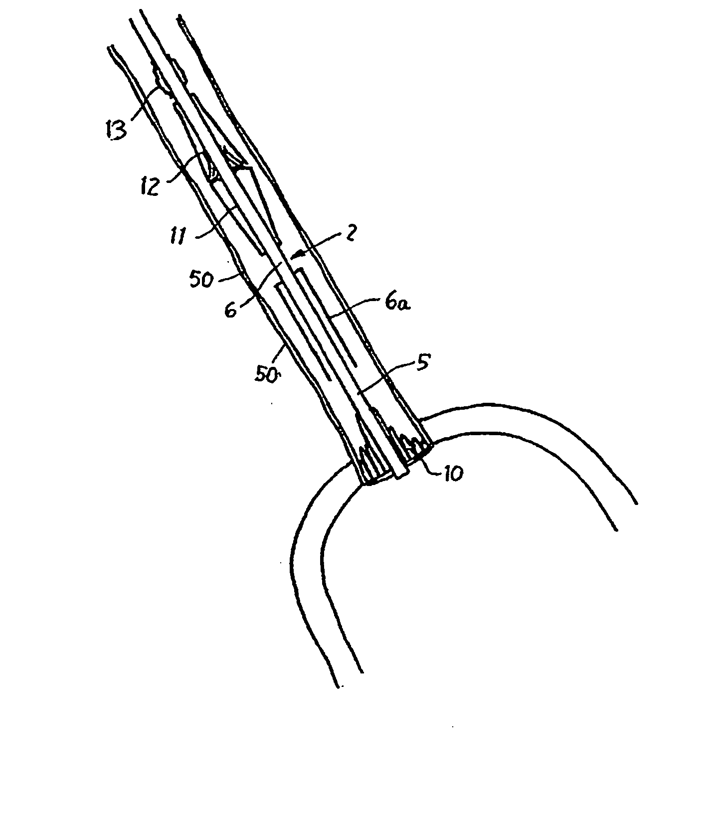
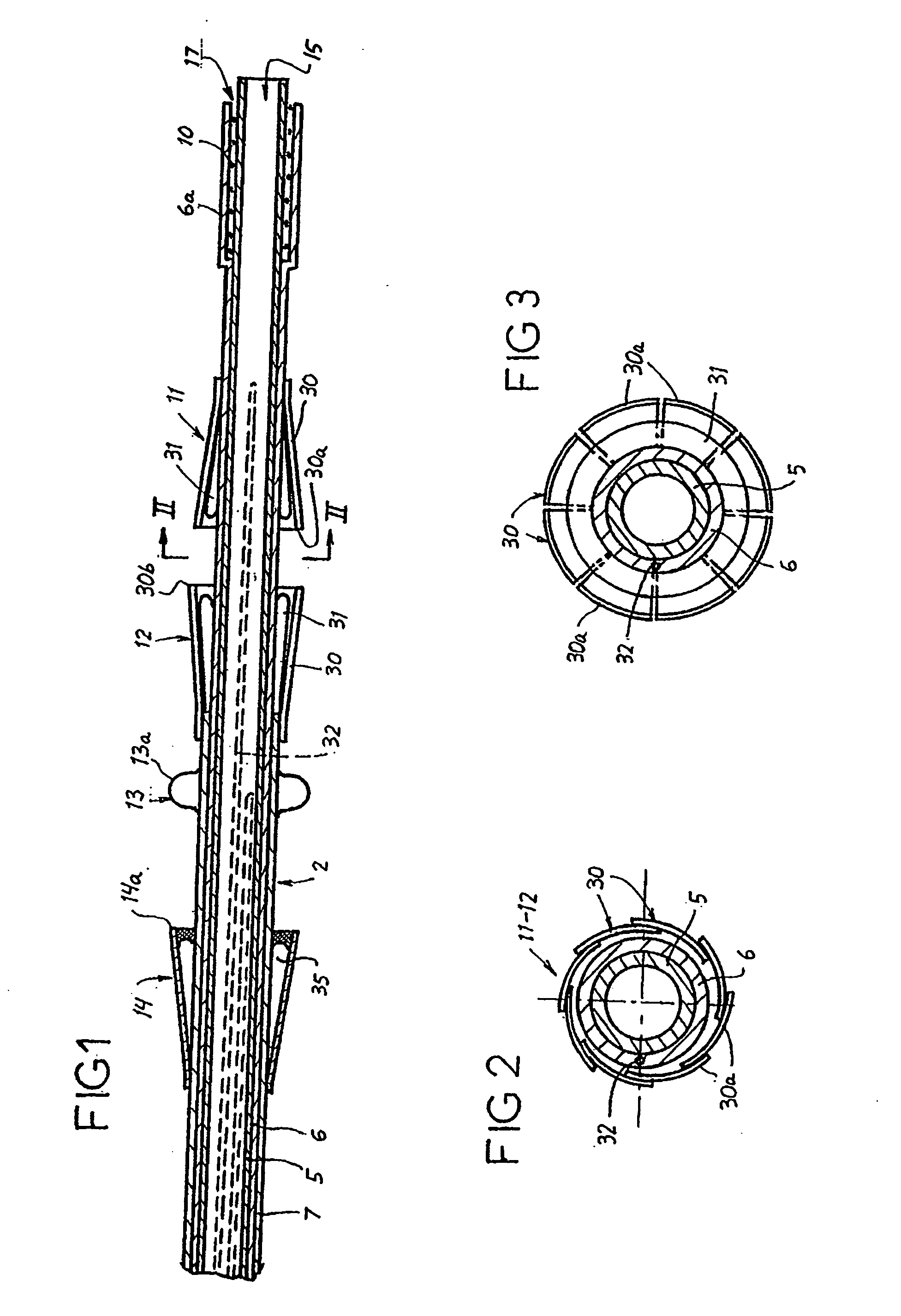
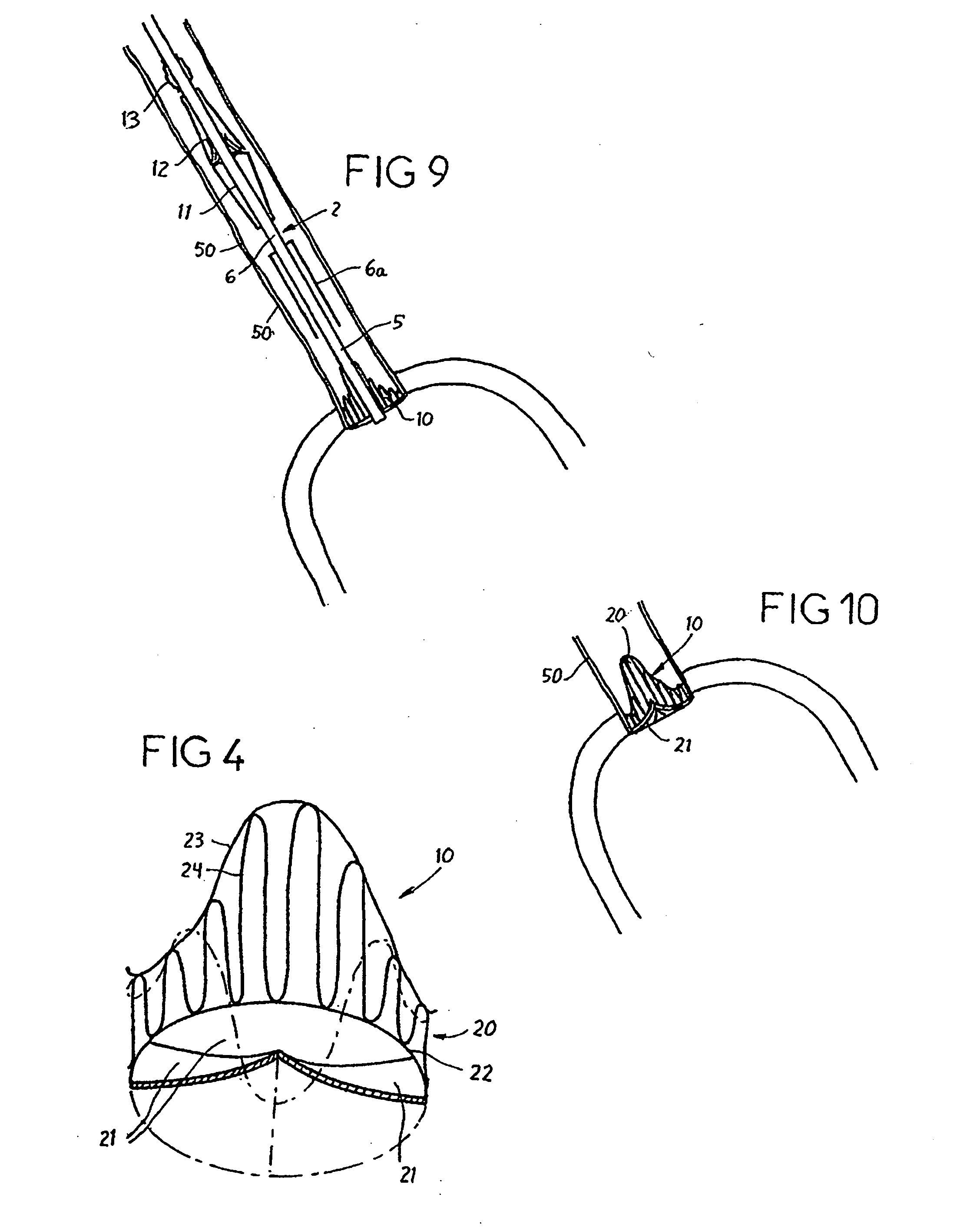
[0136] Reference is now made to the figures wherein like parts are designated with like numerals throughout. FIGS. 1 to 3 represent a device 1 for replacing a heart valve by a percutaneous route. This device comprises a tubular catheter 2 formed from three tubes 5, 6, 7 engaged one inside the other and on which there are placed, from the proximal end to the distal end (considered with respect to the flow of blood, that is to say from right to left in FIG. 1), a prosthetic valve 10, two series of blades 11, 12, a balloon 13 and a filter 14. The three tubes 5, 6, 7 are mounted so that they can slide one inside the other. The interior tube 5 delimits a passage 15, the cross section of which is large enough to allow blood to flow through it. At the proximal end, the intermediate tube 6 forms a bell housing 6a delimiting, with the interior tube 5, an annular cavity 17 in which the prosthetic valve 10 is contained in the furled condition.

[0137]FIG. 4 shows that this valve 10 comprises an...


PUM


Abstract

A prosthetic valve assembly for use in replacing a deficient native valve comprises a replacement valve supported on an expandable prosthesis frame. If desired, one or more expandable anchors may be used. The prosthesis frame, which entirely supports the valve annulus, valve leaflets, and valve commissure points, is configured to be collapsible for transluminal delivery and expandable to contact the anatomical annulus of the native valve when the assembly is properly positioned. Portions of the prosthesis frame may expand to a preset diameter to maintain coaptivity of the replacement valve and to prevent occlusion of the coronary ostia. The prosthesis frame is compressible about a catheter, and restrained from expanding by an outer sheath. The catheter may be inserted inside a lumen within the body, such as the femoral artery, and delivered to a desired location, such as the heart. When the outer sheath is retracted, the prosthesis frame expands to an expanded position such that the valve and prosthesis frame expand at the implantation site and the anchor engages the lumen wall. The prosthesis frame has a non-cylindrical configuration with a preset maximum expansion diameter region about the valve opening to maintain the preferred valve geometry. The prosthesis frame may also have other regions having a preset maximum expansion diameter to avoid blockage of adjacent structures such as the coronary ostia.

Description

CROSS-REFERENCE TO RELATED APPLICATIONS [0001] The present application A) claims priority under 35 U.S.C. §119(e) to U.S. Provisional Application No. 60 / 684,192 filed on May 24, 2005, and B) is a continuation-in-part of U.S. Ser. No. 10 / 772,101 filed on Feb. 4, 2004, which is a continuation-in-part of U.S. Ser. No. 10 / 412,634 filed on Apr. 10, 2003, now U.S. Pat. No. 7,018,406, which is a continuation-in-part of U.S. Ser. No. 10 / 130,355, now U.S. Pat. No. 6,830,584, which is the U.S. national phase under § 371 of International Application No. PCT / FR00 / 03176, filed on Nov. 15, 2000, which was published in a language other than English and which claimed priority from French Application No. 99 / 14462 filed on Nov. 17, 1999, now French Patent No. 2,800,984, herein incorporated by reference in their entirety.FIELD OF THE INVENTION [0002] The present invention relates to a prosthetic cardiac valve and related deployment system that can be delivered percutaneously through the vasculature, a...

Claims


Application Information

Patent Timeline
Patent Type & Authority Applications(United States)
IPC IPC(8): A61F2/06
CPCA61F2/2418A61F2230/008A61F2/2436A61F2/2439A61F2250/0036A61F2250/0037A61F2250/0069A61F2220/0008A61F2220/0016A61F2220/005A61F2220/0075A61F2230/0013A61F2230/005A61F2230/0054A61F2230/0067A61F2230/0078A61F2/2433
Inventor SEGUIN, JACQUESBORTLEIN, GEORGNGUYEN, THANPANNEK, EDWARD
Owner MEDTRONIC COREVALVE
Why Eureka
Try Eureka