Method for correcting a deformity in the spinal column and its corresponding implant

a spinal column and implant technology, applied in the field of spinal column deformation correction, can solve the problems of increasing mechanical stabilization difficulties of the region, high risk of fragment displacement, and stabilizing the spinal curvature, and achieve the effect of increasing the stability of the implant and reducing the siz

Inactive Publication Date: 2007-03-01
CARRASCO MAURICIO RODOLFO
View PDF10 Cites 20 Cited by
  • Summary
  • Abstract
  • Description
  • Claims
  • Application Information

AI Technical Summary

Benefits of technology

[0074] In accordance with the present disclosure, the invention provides a new and improved spinal device and the surgical method thereof correcting spinal deformities. It is a method for causing an increase in the spine curvature by using a fish-shaped implant and said method consists in preparing the area by means of the osteotomy on the articular apophysis and spine apophysis of both vertebrae, following a skimming line over the area determined by the vertebra edge corresponding to the affected intervertebral space; a wedge osteotomy on the spine apophysis of the lower vertebra, wherein the angle determined by the cutting line of the spine apophysis of the lower vertebra and the cutting line of the spine apophysis of the top vertebra is an angle similar to the correction angle that will be obtained between the two vertebral bodies and corresponding to the angle formed by the upper and lower areas of the implant. After that, the disc is removed and the intervertebral space is prepared to receive the implant. The implant is placed with its vertex towards the posterior area of the vertebral spine and the rounded area towards the anterior area of the vertebral spine, without protruding from the perimeter of the vertebral bodies, said vertex completely positioned in the posterior edge of the intervertebral space allowing the existence of a free space in the anterior area of the intervertebral space; a compression force of the vertebral bodies is then caused on the upper and lower areas of the implant, which is used as a fulcrum for this step so that an open angle is formed towards the anterior area of the vertebral column, being said the angle formed bigger than that previously existing; osteosynthesis material is placed and finally the vertebrae are fixed with pedicular screws and osteosynthesis material is placed within the intervertebral space.
[0075] The implant is basically a trapezoid or acute-angled isosceles triangle where the area opposite the vertex or shortest base is a rounded surface, according to a vertical and perpendicular plan section view of the implant and it could be defined as an isosceles trapezoid which longest base corresponds to a semi circumference; a horizontal and perpendicular section to the implant sides will show a curved surface representing about a quarter of the circumference as a basically rectangular-shaped end (shortest side); the top and bottom surfaces include protuberances capable of penetrating into the vertebral mass through the vertebral plates. Said protuberances having a triangular shape are curved and bent (as a sea wave) in order to increase the implant stability after having been inserted. The protuberances extend from the beginning of the vertex to the maximum height of the implant gradually reducing their size.

Problems solved by technology

Likewise, the need of stabilizing the spinal curvature may result from an operation on said region, especially when metal assemblies have been used without correcting the defect.
The sharp lordotic angle and elongation of the anterior column occurring in this procedure were assumed to be associated with serious vascular and neurological complications.
In this way, two osteotomies are performed and the mechanical stabilization difficulties of the region are increased, there being a high risk of displacement of the fragments for having caused instability in two levels.
Furthermore, there is also the risk that only one of the osteotomies is fixed while the other one remains unfixed.
Moreover, said surgical technique is very aggressive there being a considerable blood loss as a consequence of the fracture that has been made, the vertebral bone is removed and the muscles fixed in that part of the vertebra are detached.
If the osteotomy were performed in two vertebrae of the same region, the shortening of the spine would be equal to the length of the base of both wedges, thus considerably increasing the morbidity of the proceeding and surgical risks as well.
The different techniques of vertebral osteotomies have shown technical inconvenient and complications derived from the surgical technique itself due to the bone resection and the necessary mobilization of the trunk and lower extremities during operations.
None of them could establish a predetermined correction angle.
Full angular correction of the lumbar spine in PWO was not always achieved, thus resulting in a decreased correction or monosegmental correction.
The reduction of the vertebral mass increases the risk of instability and infection.
Likewise, there is a considerable continuous blood loss after the operation.
Their conclusion was “This structured review of the literature concerning three methods of lumbar osteotomy for correction of thoracolumbar kyphosis deformity showed that reports are limited and provide scant information on clinical data.
Statistical analysis of the technical resulting data from these surgical methods was therefore not possible.
Although the available data from the current literature suggest that CWO causes less serious complications and has better results, these data are not suitable for decision making with regard to which surgical treatment is preferable.
Since the damaged disc material has been removed, something must be positioned within the intervertebral space, otherwise the space may collapse resulting in damage to the nerves extending along the spinal column.
By means of this technique, once fusion of vertebrae occurred, the material used to maintain the stability of the segment became superfluous.
Moreover, the surgical procedures necessary to implant a rod or plate to stabilize the level during fusion were frequently lengthy and complicate.
Another problem that is not addressed by the above prior devices concerns maintaining or restoring the normal anatomy of the fused spinal segment.
With the prior devices, the need to restore this curvature is neglected.
However, this over-reaming of the posterior portion is generally not well accepted because of the removal of the load bearing bone of the vertebrae, and because it is typically difficult to ream through the posterior portion of the lower lumbar segment where the lordosis is greater.
In most cases, with the use of implants of this type, no effort is made to restore the lordotic curvature, so that the cylindrical implant is likely to cause a kyphotic deformity as the vertebra settles around the implant.
This phenomenon can often lead to revision surgeries because the spine becomes imbalanced.
By way of example, said implants show that they may be used to separate vertebrae in a limited degree and consequently the correction achieved by the current devices that are inserted between the vertebrae is minimal and said implants cannot be used for important deformities in the curvature of the lumbar spine caused by the generalized disease of the intervertebral discs.
It is obvious that these procedures are used to separate vertebrae but the curvature is not corrected in the usually necessary grades.
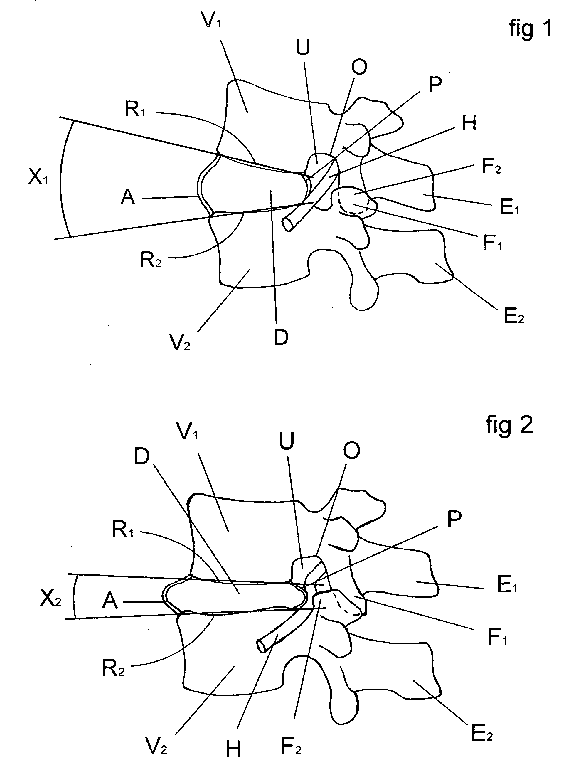
However, said separation of the vertebral bodies also separates the posterior edges of said vertebral bodies so that said implants cannot cause greater corrections of lordosis since they do not allow a convergence of the posterior area of the vertebral bodies.
This aspect of the surgical technique related to the preservation of the articular facets is in detriment to obtain a surgical field broad enough.
This limitation has been noted in the literature about complications of the method.
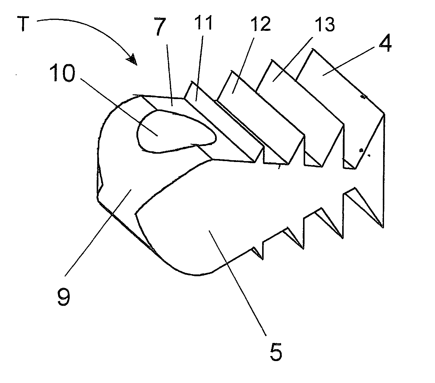
But the slots disclosed by Bryan's does not preserve the bone end-plate to maintain the structural integrity and discourage subsidence; moreover such cage is being supported by soft bone within its critical area.
This method may not ensure a pre-determined lordosis correction that may vary upon the surgeon artisan's skills.
There is another important inconvenience about the use of slots, and comprises the way of handling rachidian nerves.
Since no special technique is disclosed in this respect, we understand that the method involves great risk of damage to the rachidian nerve, considering the distance necessary to produce a slot and to handle a cutting instrument so close to the nerve.
It is not physically possible to obtain a lordotic angle correction above 8 degrees with Marion's insert and method, without dissecting the facet joint capsules.
To conclude, Marino's insert cannot be used, it is not design and do not teach the way to obtain a lordotic angle correction above 8 degrees and do not have the same constructive aspects and features that Carrasco's cage has.
And mainly the content of the prior art discussion is about how difficult and rare is no obtain permanent lordotic angle corrections above 8 degrees, and how unpredictable a lordotic angle correction is before and after surgery.
“By way of example, said implants show that they may be used to separate vertebrae in a limited degree and consequently the correction achieved by the current devices that are inserted between the vertebrae is minimal and said implants cannot be used for important deformities in the curvature of the lumbar spine caused by the generalized disease of the intervertebral discs.
In the same way, you do not achieve 300 miles per hour with a car just with a bigger motor engine.
Moreover, we have demonstrated with cited documents that such kind of surgeries [for corrections above 8 degrees] are major surgeries with high death risk and undetermined results [not possible to pre-schedule the final curvature correction achieved].
Above 8 degrees, existing cages and methods will fail to put the posterior annular fibers under tension and could limit the degree of facet realignment and neuroforaminal widening.

Method used


Image

  • Method for correcting a deformity in the spinal column and its corresponding implant
  • Method for correcting a deformity in the spinal column and its corresponding implant
  • Method for correcting a deformity in the spinal column and its corresponding implant

Examples


embodiment

Preferred Embodiment

[0108] Surgical intervention. In the surgical technique to be used with an intervention posterior to the spine, it is necessary to expose at least two adjacent vertebrae, which in their posterior aspect must remain exposed in a length no lesser than the distance between their transverse apophysis I and laterally up to the ends of the same transverse apophysis I.

[0109] In FIG. 3, the osteotomy of the upper vertebra V1 is performed on the articular apophysis F1 and the spine apophysis E1 if it is necessary according to the deformity of the latter, following the line L-L skimming the lower edge of the upper vertebra V1. The osteotomy of the lower vertebra is carried out on the lower articular apophysis F2 and the spine apophysis E2 following a line skimming the upper vertebral edge N-N of the lower vertebra V2.

[0110] In FIG. 4, the scheme view of the posterior part of the vertebrae shows the cutting line L-L of the upper vertebra V1 and the cutting line N-N of the...


PUM


Abstract

An implant to be inserted in a disc space between two adjacent vertebrae for the correction of vertebral spine curvature and method related thereto. A lateral configuration of the invention is a wedge or acute-angled isosceles triangle, wherein the base of the isosceles triangle is a rounded pyramid-like surface, and upper and lower surfaces of the isosceles triangle include fixation protuberances for fixing the implant to vertebral plates of the adjacent vertebrae.

Description

CROSS-REFERENCE TO RELATED APPLICATIONS [0001] This application claims the benefit of U.S. application Ser. No. 10 / 674,019, filed Sep. 30, 2003, which is incorporated herein by reference in its entirety.BACKGROUND OF THE INVENTION [0002] 1. Field of the Invention [0003] This invention refers to a method for correcting a deformity in the spinal column comprising the steps of resecting part of the intervertebral disc with its ring, resecting the joints joining said adjacent vertebrae, resecting at least part of the spinal apophysis of said adjacent vertebrae, providing an implant to be inserted between said adjacent vertebrae, inducing the separation of only the vertebral edges of the anterior part of said vertebrae; wherein by means of other devices adaptable to the external parts of the spinal column, the vertebral plates are compressed against the supporting faces of the implant, wherein the vertebral plates, when standing on the supporting faces of said implant, form an open angle...

Claims


Application Information

Patent Timeline
Patent Type & Authority Applications(United States)
IPC IPC(8): A61F2/44A61B17/70A61F2/00A61F2/28A61F2/30A61F2/46
CPCA61B17/7059A61F2310/00011A61F2/4455A61F2/447A61F2/4611A61F2002/2835A61F2002/30281A61F2002/30616A61F2002/30774A61F2002/30785A61F2002/30841A61F2002/30879A61F2002/30904A61F2002/4475A61F2002/448A61F2002/4629A61F2230/0086A61F2/442A61F2002/30593
Inventor CARRASCO, MAURICIO RODOLFO
Owner CARRASCO MAURICIO RODOLFO
Why Eureka
Try Eureka