Light-activated cation channel and uses thereof

a technology of cation channels and light-activated ions, which is applied in the field of light-activated ion channel proteins, can solve the problems of changing the voltage across the membrane, the difficulty of driving reliable and precisely sized subthreshold depolarizations, and the need for expensive, custom-synthesized exogenous compounds for existing genetically-targeted approaches

Active Publication Date: 2007-03-08
THE BOARD OF TRUSTEES OF THE LELAND STANFORD JUNIOR UNIV
View PDF98 Cites 225 Cited by
  • Summary
  • Abstract
  • Description
  • Claims
  • Application Information

AI Technical Summary

Benefits of technology

[0010] Accordingly, one aspect of the invention provides for compositions and methods related to light-activated cation channel (LACC) proteins with millisecond-scale opening kinetics that provide genetically targeted photostimulation at fine temporal resolution, enabling elucidation of the temporal activity patterns in specific neurons that suffice to drive circuit dynamics, information processing, and plasticity. The composition and methods herein offer optical control of the electrical and ionic milieu of neurons and a variety of other cell types, both in vitro and in vivo, at the rapid timescales important for many biological processes—including ion channel modulation, signal transduction, neural coding via temporal spiking and synaptic activation patterns, sensory and motor processing, interneuron modulation of circuit dynamics, and neuropsychiatric dysfunction. Additional aspects of the invention include compositions and methods relating to light-activated cation channel proteins for drug discovery and biotechnological and neuropsychiatric applications, advancing the ability to characterize, detect, and treat a variety of medical disorders.

Problems solved by technology

The flow of ions across the membrane can also result in a change in the voltage across the membrane.
But compared with driving spiking, it is in principle a more difficult task to drive reliable and precisely sized subthreshold depolarizations.
Thus, existing genetically-targeted approaches require expensive, custom-synthesized exogenous compounds, and operate on the scale of seconds to minutes (Lima et al., Cell 121:141 (2005), Banghart et al., Nat Neurosci 7:1381 (2004), Zemelman et al., Proc Natl Acad Sci USA 100:1352 (2003), Foster et al., Nature 433:698 (2005)).

Method used


Image

  • Light-activated cation channel and uses thereof
  • Light-activated cation channel and uses thereof
  • Light-activated cation channel and uses thereof

Examples


example 1

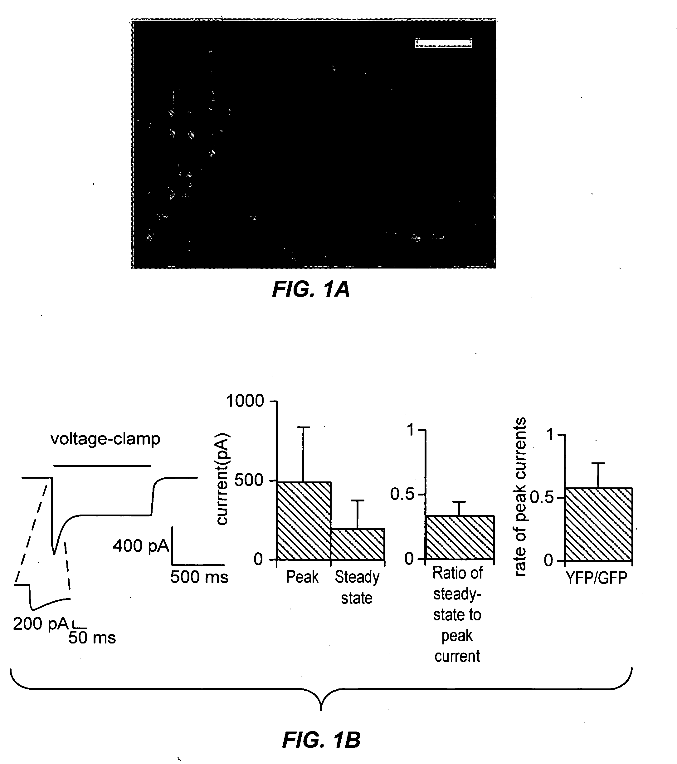
[0243] To obtain stable and reliable ChR2 expression for coupling light to neuronal depolarization, we constructed lentiviruses containing a ChR2-YFP fusion protein for genomic modification of neurons. Infection of cultured rat CA3 / CA1 neurons led to appropriately membrane-localized and well-tolerated expression of ChR2 for days to weeks after infection (FIG. 1a). There was no evidence of toxicity due to the expressed protein, even at high fusion protein expression levels. Whole-cell voltage-clamp recording of neurons showed that conventional GFP illumination in the bandwidth 450-490 nm (300 W xenon lamp (Sutter DG-4), via Chroma excitation filter HQ470 / 40x) induced depolarizing currents with fast rise rates—reaching a maximal rise rate of 160+111 pA / ms within 2.3+1.1 ms after the onset of the light pulse (mean+standard deviation reported throughout, n=18; FIG. 1b, left). Mean whole-cell inward currents were large, 496 pA±336 pA at peak and 193 pA±177 pA at steady-state (FIG. 1b, mi...

example 2

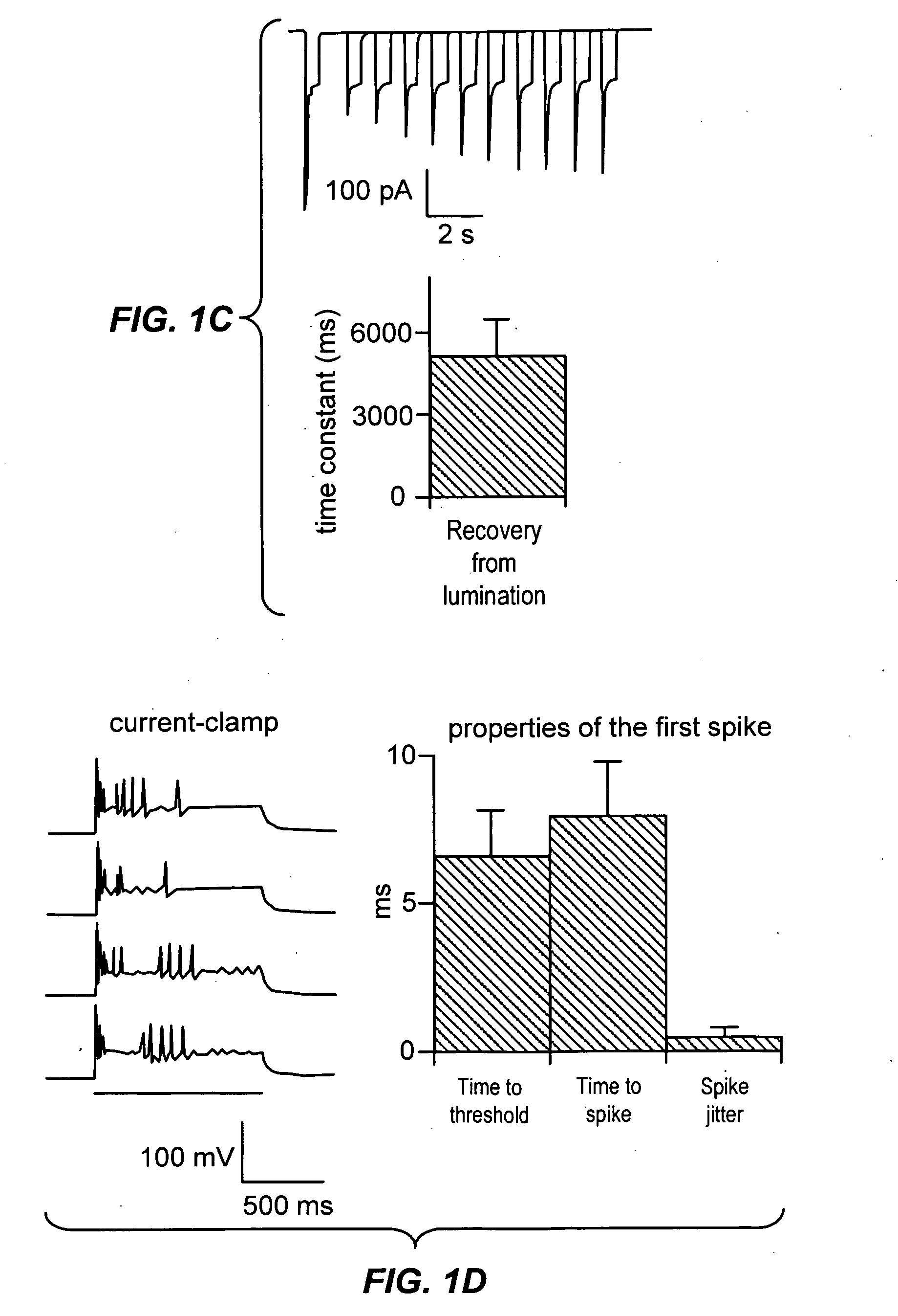
[0244] We examined whether ChR2 could drive actual depolarization of neurons held in current-clamp mode, with the same steady illumination protocol we used for eliciting ChR2-induced currents (FIG. 1d, left). Early in an epoch of steady illumination, single neuronal spikes were rapidly and reliably elicited (8.0+1.9 ms latency to spike peak, n=10; FIG. 1d, right), consistent with the fast rise times of ChR2 currents described above. However, for these cells, at these specific conditions, any subsequent spikes elicited during steady illumination were poorly timed (FIG. 1d, left). Thus, for this particular sample, steady illumination was not adequate for controlling the timing of ongoing spikes with ChR2, despite the reliability of the first spike. Earlier patch-clamp studies using somatic current injection showed that spike times were more reliable during periods of rapidly rising membrane potential than during periods of steady high-magnitude current injection. This is consistent wi...

example 3

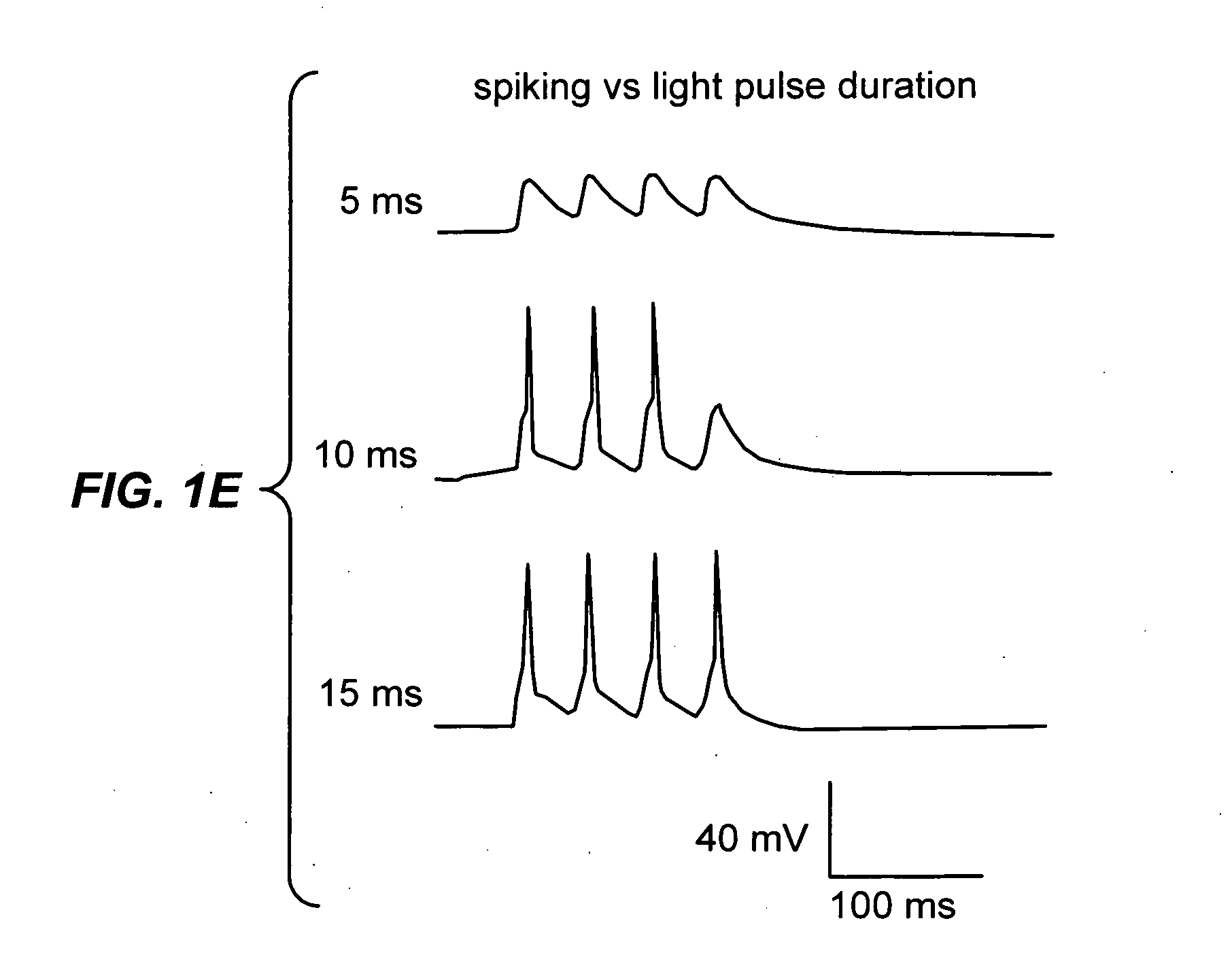
[0245] We found that the single spike reliably elicited by steady illumination had extremely low temporal jitter from trial to trial (FIG. 1d, right; 0.5+0.3 ms; n=10 neurons). This observation led us to devise a pulsed-light paradigm, which takes advantage of the low jitter of the single reliable spike evoked at light pulse onset. But in order for such a pulsed-light paradigm to work, the conductance and kinetics of ChR2 would have to permit peak currents of sufficient amplitude, during light pulses shorter than the desired interspike interval. Indeed, using fast optical switching, we found that multiple pulses of light with interspersed periods of darkness could elicit reliable and well-timed trains of spikes (FIG. 1e; shown for 25 Hz trains of four pulses). FIG. 1e highlights the fact that longer light pulses evoke single spikes with greater probability than short light pulses. The ability to easily alter light pulse duration with fast optical switching suggests a straightforward...


PUM

PropertyMeasurementUnit
wavelengthaaaaaaaaaa

Abstract

The present invention provides compositions and methods for light-activated cation channel proteins and their uses within cell membranes and subcellular regions. The invention provides for proteins, nucleic acids, vectors and methods for genetically targeted expression of light-activated cation channels to specific cells or defined cell populations. In particular the invention provides millisecond-timescale temporal control of cation channels using moderate light intensities in cells, cell lines, transgenic animals, and humans. The invention provides for optically generating electrical spikes in nerve cells and other excitable cells useful for driving neuronal networks, drug screening, and therapy.

Description

CROSS-REFERENCE [0001] This application is related to and claims priority to U.S. Provisional Application No. 60 / 701,799, filed Jul. 22, 2005, which is incorporated herein by reference in its entirety.STATEMENT AS TO FEDERALLY SPONSORED RESEARCH [0002] This invention was made with the support of the United States government under Contract numbers K08 MH071315 and F31DC007006 from the National Institutes of Health, as well as support from the Schools of Medicine and Engineering at Stanford University.FIELD OF THE INVENTION [0003] The invention relates to light-activated ion channel proteins that can generate millisecond-timescale electrical spikes when incorporated into neurons and illuminated with rapid pulses of light. In particular the invention provides millisecond-timescale temporal control of cation channels using moderate light intensities in cells, cell lines, transgenic animals, and humans. The invention is useful for driving neuronal networks, for drug screening, and for th...

Claims


Application Information

Patent Timeline
Patent Type & Authority Applications(United States)
IPC IPC(8): G01N33/53G06F19/00A61K35/30
CPCA01K2217/05Y10S530/82A01K2227/40A01K2227/703A01K2227/706A61K31/137A61K31/353A61K31/405A61K31/4172A61K38/00A61K48/00A61N5/0622C07K14/705C07K2319/60C12N2740/15043C12N2740/15071G01N33/6872G01N2500/04A61K35/30A01K2227/105A61L27/3604A61F2/18A61K2300/00A61P17/00A61P21/00A61P25/00A61P25/02A61P25/16A61P25/24A61P25/28A61P25/30A61P27/02A61P29/00A61P3/04A61P31/12A61P31/18A61P31/20A61P35/00A61P43/00A61P5/00A61P5/02A61P5/06A61P5/48A61P5/50A61P7/00A61P7/06A61P9/00Y02A90/10A61N5/062C07K14/405A61N5/0601G01N2500/10H05K999/99
Inventor BOYDEN, EDWARD S.DEISSEROTH, KARL
Owner THE BOARD OF TRUSTEES OF THE LELAND STANFORD JUNIOR UNIV
Why Eureka
Try Eureka