Apparatus and method to acquire data for reconstruction of images pertaining to functional and anatomical structure of the breast

a breast and functional technology, applied in the field of breast functional and anatomical structure reconstruction, can solve the problems of difficult to determine the biopsy site, the risk of potentiating the metastatic spread of cancer, and the loss of the 3d location of the tumor or the depth effect, so as to minimize patient motion and complete the scan relatively quickly

Inactive Publication Date: 2007-03-22
IMAGING DIAGNOSTIC SYST
View PDF10 Cites 43 Cited by
  • Summary
  • Abstract
  • Description
  • Claims
  • Application Information

AI Technical Summary

Benefits of technology

[0008] It is an object of the present invention to provide an apparatus for providing functional and anatomical images of t...

Problems solved by technology

A major drawback of this imaging modality is that each pixel of a mammography image represents a ray sum of attenuation coefficients of the tissue along the ray.
Therefore, the 3D location of the tumor or the depth effect is lost.
The compression employed in mammography causes pain and disturbs the breast, making it difficult to determine biopsy sites and has the risk of potentiating the metastatic spread of cancer.
Unfortunately, a much higher x-...

Method used


Image

  • Apparatus and method to acquire data for reconstruction of images pertaining to functional and anatomical structure of the breast
  • Apparatus and method to acquire data for reconstruction of images pertaining to functional and anatomical structure of the breast
  • Apparatus and method to acquire data for reconstruction of images pertaining to functional and anatomical structure of the breast

Examples


Embodiment Construction

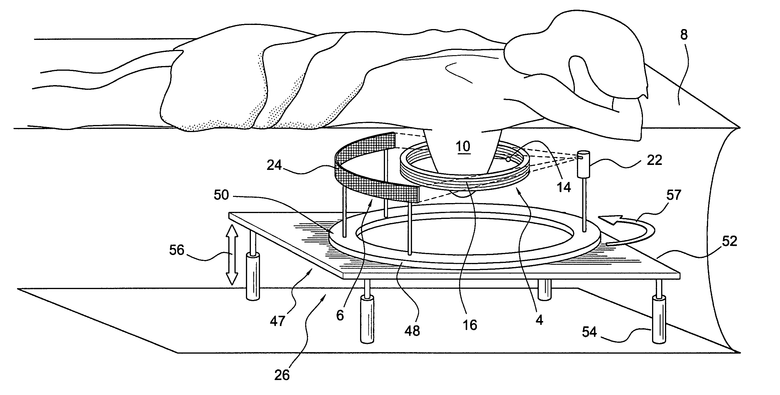
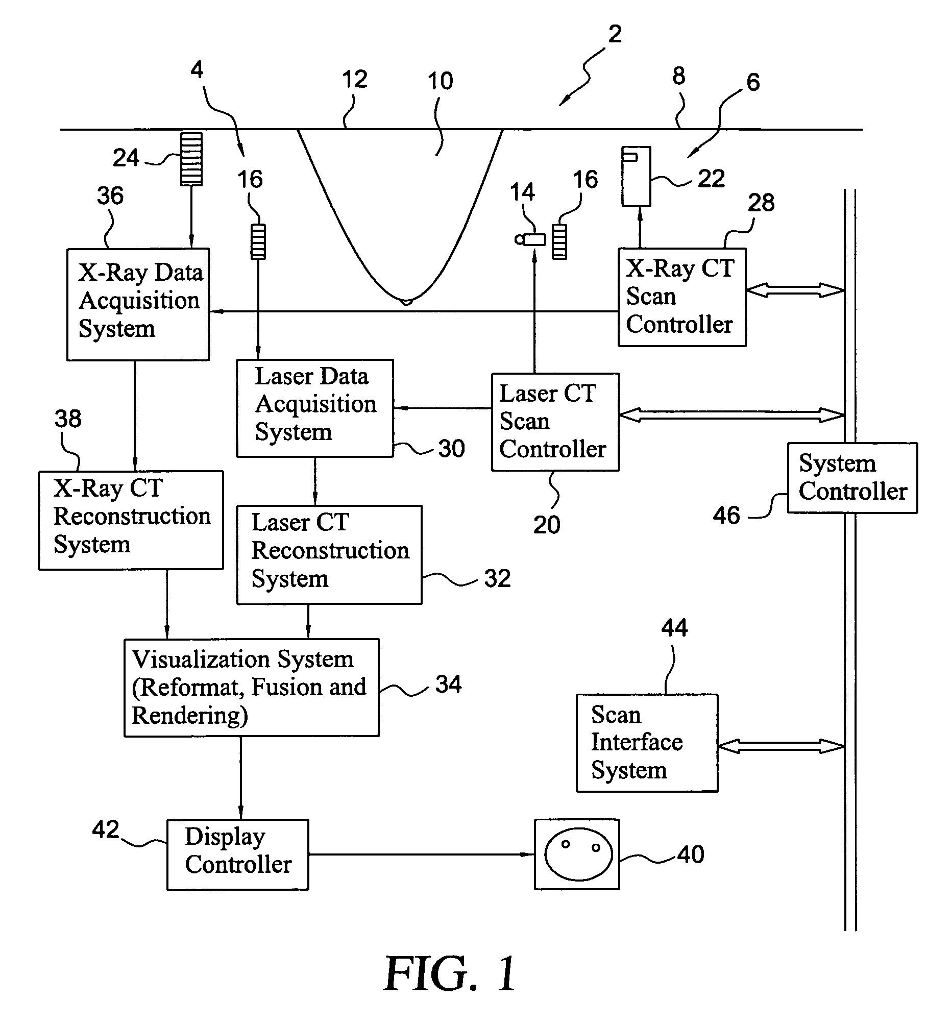
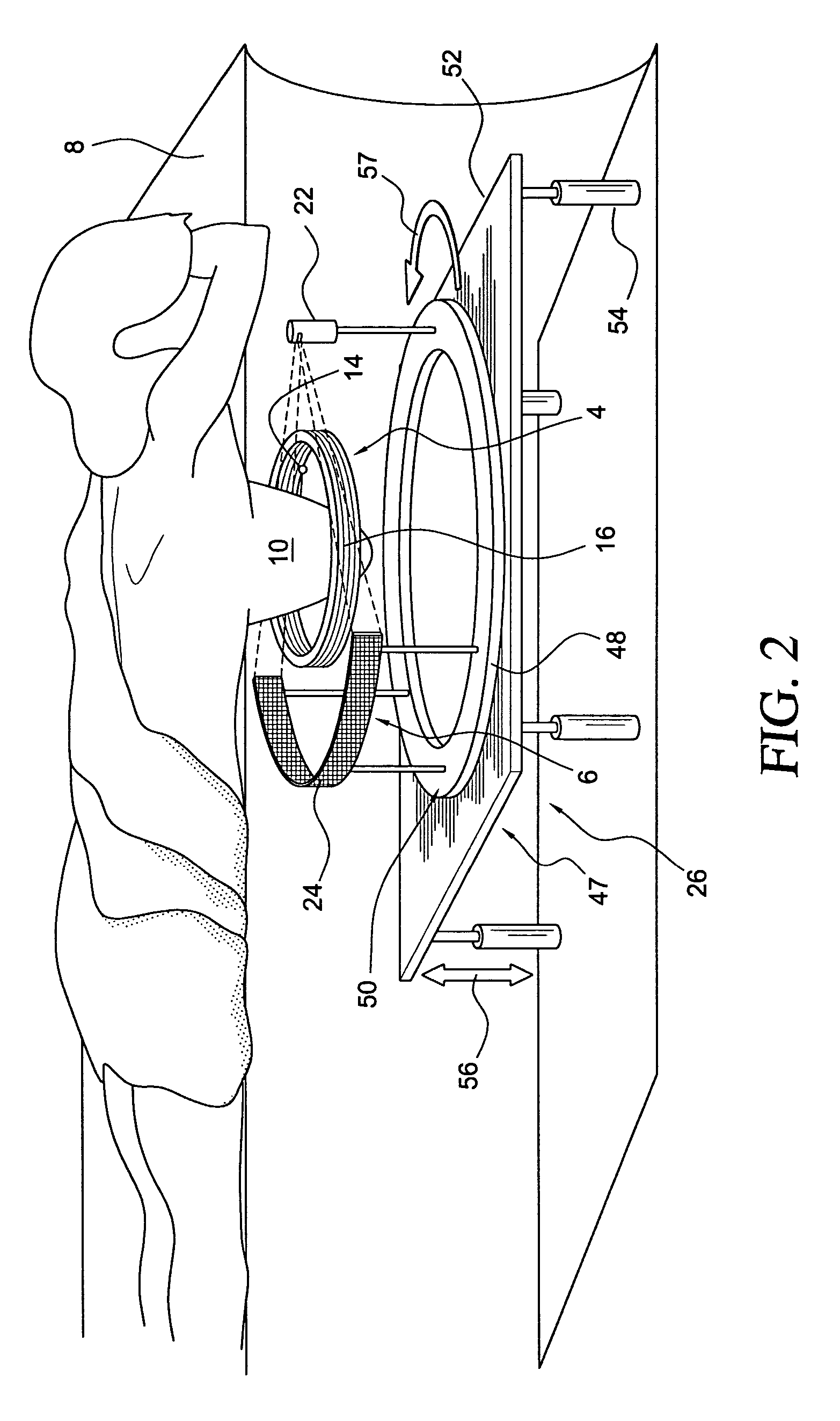
[0023] A scanning apparatus made in accordance with the present invention comprises two independent CT scanners sharing a patient couch. The patient lies on the couch in a prone position, with one of the breasts vertically pendent through an opening in the couch for scanning. The two scanners share a common patient couch to facilitate a direct correlation of the reconstructed images representing the functional and anatomical structures of the breast that are derived from the data collected by the two scanners respectively. The laser scan of the breast is followed immediately by an x-ray scan or vice-versa in order to keep the patient position invariant. To advantageously shorten the total scan time, the two scans may be performed concurrently. Two independent image reconstruction systems are provided. The first is for the reconstruction of functional images from data collected by the laser CT scanner, and the other for the reconstruction of anatomical images from data collected by t...


PUM


Abstract

An apparatus for breast scanning to obtain functional and anatomical images of the breast comprises a patient support for a patient to rest in a prone position, the support having an opening with one of her breasts vertically pendent through the opening for scanning; a laser CT scanner disposed below the support for generating a first set of data for reconstruction of functional images of the breast; an X-ray CT scanner disposed below the support for generating a second set of data for reconstruction of anatomical images of the breast; and a display to visualize at least one of the functional and anatomical images. A method for acquiring data for reconstruction of images pertaining to functional and anatomical structures of a breast comprises positioning a patient in a prone position on a support having an opening through which a breast of the patient is pendant; scanning the breast with a laser CT scanner to obtain data of the breast for functional image reconstruction of the breast; and while the patient is still prone on the support, scanning the breast with an X-ray CT scanner to obtain data of the breast for anatomical image reconstruction of the breast.

Description

RELATED APPLICATIONS [0001] This is a nonprovisional application, which claims the priority benefit of provisional application Ser. No. 60 / 718,307, filed Sep. 20, 2005, herein incorporated by reference.BACKGROUND OF THE INVENTION [0002] It is vital to be able to detect cancers in the breast at their early stage. Moreover, it is very important to discern malignant from benign lesions. For biopsy and surgery purposes, it is essential to know the location and the extent of the lesion. For therapeutic purposes, it is useful to track tumor response to neoadjuvant and radiation therapy. [0003] Current commercial scanners or medical diagnostic equipments available in the market provide only partial solutions to the above goals. For instance, it is current practice to use mammography to detect tumors. A major drawback of this imaging modality is that each pixel of a mammography image represents a ray sum of attenuation coefficients of the tissue along the ray. Therefore, the 3D location of ...

Claims


Application Information

Patent Timeline
IPC IPC(8): A61B6/04
CPCA61B5/0091A61B5/4312A61B6/0414A61B6/027A61B6/466A61B6/502A61B6/5235A61B6/0435
Inventor HANSEN, TIMOTHY B.TUY, HEANG K.WAKE, ROBERT H.PONDER, STEVEN L.OLIVIER, PATRICK
Owner IMAGING DIAGNOSTIC SYST
Why Eureka
Try Eureka