System and method for providing slant-angle collimation for nuclear medical imaging

a technology of nuclear medical imaging and collimation, which is applied in the field of radiation imaging systems and methods, can solve the problems of relatively expensive diagnostic tool subsystems of the gantry system, and achieve the effects of reducing the length of the translational pass, cost saving, and simplifying setup and operation

Active Publication Date: 2007-03-29
SIEMENS MEDICAL SOLUTIONS USA INC
View PDF6 Cites 9 Cited by
  • Summary
  • Abstract
  • Description
  • Claims
  • Application Information

AI Technical Summary

Benefits of technology

[0011] The present invention relies on a collimator in which the angle of view varies across the collimator. With this type of collimator, SPECT systems based on substantially complete angular sampling can be devised for single and multiple headed camera systems that require as few as a single pass of the camera along the patient, without relative rotation between the patient and camera, while also minimizing the length of the translational pass required of the camera. Important advantages will be seen in cost savings for the gantry, simplification of setup and operation, and for some configurations, significantly smaller space requirements for the incorporating systems. To this end, one embodiment of the invention takes the form of a system utilizing slant-angle collimation for SPECT radiation sampling. The system comprises (includes, but is not necessarily limited to) a collimator positioned between a radiating mass and a radiation detector. The collimator is spaced apart from a translational path of the radiating mass at a predetermined distance that defines a patient accommodation space. Apertures extend through the collimator and form passageways for gamma (radiation) rays emanating from the radiating mass to strike the radiation detector.
[0012] In summary, the provision and utilization of collimators configured according to the teachings above facilitate enhanced radiological imaging quality, while at the same time simplifying and reducing the cost of the support structures required to carry the necessary instruments and which affect relative, longitudinal motion between those instruments and the patient. Among other benefits, the method and arrangement of the present invention permits the utilization of exclusively longitudinal relative motion between the patient and imaging instruments compared to the orbital motion about the patient which has been previously required when conducting such procedures as full body scanning.

Problems solved by technology

One drawback associated with this requirement however, is that such gantry systems are relatively expensive subsystems of the diagnostic tool because they must be capable of providing rapid rotation is of the large and heavy camera heads through very precise orbits about the patient.

Method used


Image

  • System and method for providing slant-angle collimation for nuclear medical imaging
  • System and method for providing slant-angle collimation for nuclear medical imaging
  • System and method for providing slant-angle collimation for nuclear medical imaging

Examples


Embodiment Construction

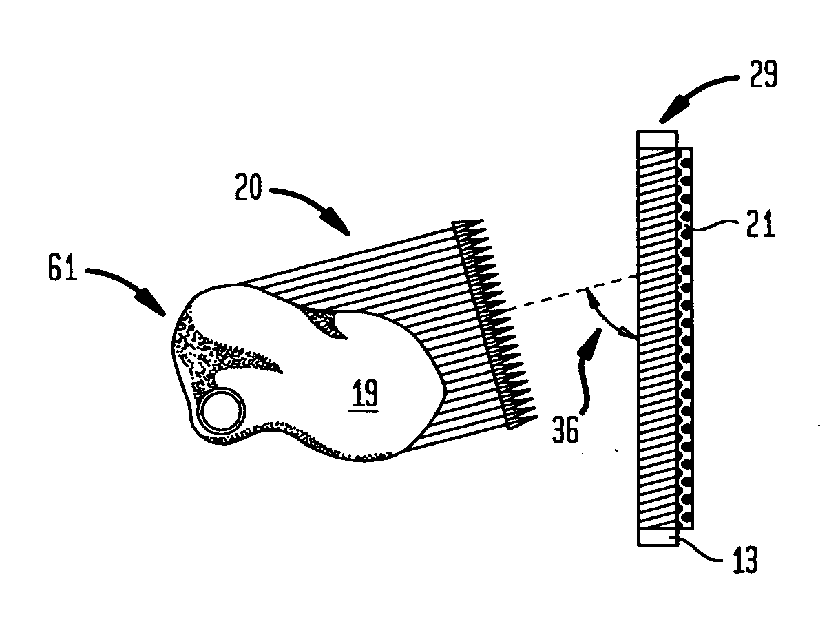
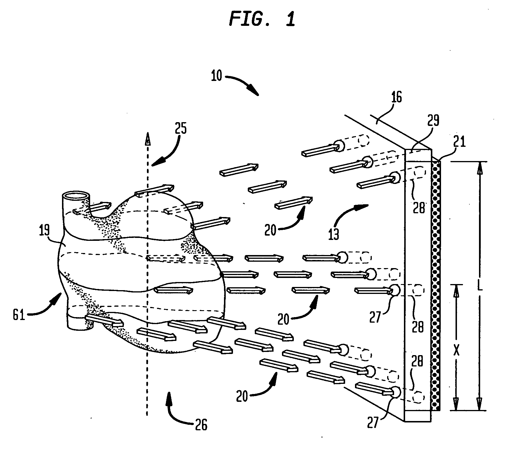
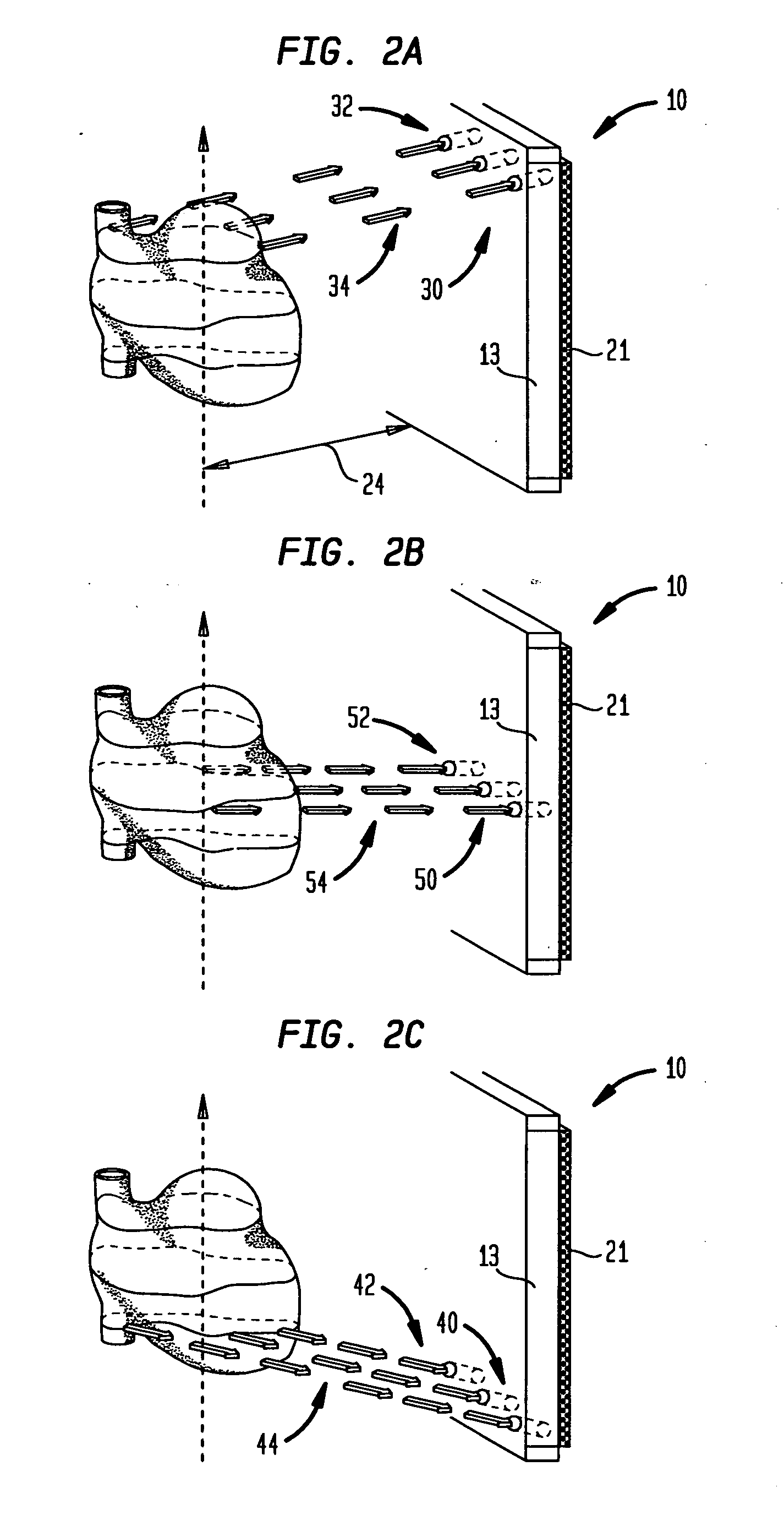
[0021] When considering full angular gamma camera sampling utilizing SPECT parallel projection views, or their equivalents, the image samples should be obtained over one hundred and eighty or three hundred and sixty degrees. According to the present invention, a system (10) is provided that includes a collimator (13) adapted with apertures (27) that serve as openings for slant-angle passageways (28) which accommodate views of a radiating area or object-of-interest (61), such as the heart of a patient (19), from a specific slant-angle.

[0022] Referring to FIG. 1, the specific angle at which the slotted passageway (28) is canted within the collimator (13) is dependent on the relative position (x) of the passageway (28) in the collimator (13). In the instance of FIG. 1, the radiation source is the radiating mass (19) shown as the radioactively infused heart (19) of a patient. For each row of passageways (28) through the collimator (13), the associated collimated (gamma) rays travel alo...


PUM


Abstract

Method and arrangement for implementing a system (10) for providing multi-angular SPECT radiation sampling utilizing slant-angle collimation. The system (10) includes a collimator (13) positioned between a radiating mass (19) within a patient (60) and a radiation detector (21). The collimator (13) is spaced apart from a translational path (25) of the radiating mass (19) at a predefined distance (24). A plurality of apertures (27) extend through the collimator (13) and each forms a passageway (28) for radiation rays (20) emanating from the radiating mass (19) in a direction substantially aligned with a longitudinal axis (29) of the respective passageway (29) and in this manner enables the aligned radiation rays (20) to strike the radiation detector (21). The plurality of passageways (28) include a first group (30) of passageways adjacently aligned in a first row (32) and arranged so that the longitudinal axes (29) of the first group (30) of passageways (32) are substantially contained in a first plane (34) oriented substantially perpendicularly to a central plane (16) of the collimator (13). Each of the parallel longitudinal axes (29) of the first row (32) of passageways are obliquely oriented with respect to the central plane (16) of the collimator (13) with an included angle (36) therebetween. Each of the included angles, when measured clockwise from the central plane (16) or face of the collimator (13) to a respective longitudinal axis, is an acute angle.

Description

BACKGROUND OF THE INVENTION [0001] 1. Field of the Invention [0002] The present invention relates to systems and methods used in radiation imaging; and more particularly, to collimators utilized in cooperation with radiation detectors that permit only desired radiation to strike the detector thereby producing a more accurate image-of-interest, typically of an internal portion of a patient when medical and diagnostic applications are considered. [0003] 2. Description of the Background Art [0004] In conventional radiation imaging arrangements, collimators are used in a wide variety of equipment in which it is desired to permit only beams of radiation emanating along a particular path to pass a selected point or plane. Collimators are frequently used in radiation imagers to ensure that only radiation beams passing along a direct path from the known radiation source strike the detector thereby minimizing detection of beams of scattered or secondary radiation. [0005] Particularly in radi...

Claims


Application Information

Patent Timeline
Patent Type & Authority Applications(United States)
IPC IPC(8): G21K1/02
CPCG21K1/025
Inventor HAWMAN, ERIC G.
Owner SIEMENS MEDICAL SOLUTIONS USA INC
Why Eureka
Try Eureka