Apparatus for obtaining an image of a blood cell and method for obtaining an image of a blood cell

a technology for blood cells and apparatus, applied in the field of apparatus a method for obtaining an image of blood cells, can solve the problems of unsuitable setting of conditions, unsuitable change of conditions, and inability to suitably perform imaging of blood cells. to achieve the effect of efficient operation of blood cell imaging

Active Publication Date: 2007-04-05
SYSMEX CORP
View PDF6 Cites 44 Cited by
  • Summary
  • Abstract
  • Description
  • Claims
  • Application Information

AI Technical Summary

Benefits of technology

[0020] An object of the present invention is to set optimum imaging conditions to efficiently perform imaging of blood cells based on blood analysis result.

Problems solved by technology

In this blood cell analyzing device, an abnormal cell is determined based on the “cell number” calculation information, the number of cells of the blood cell classification is designated two or three times higher, or changes the blood cell examination method for the preparation, however, a problem arises inasmuch as the abnormal sample determination via the number of blood cells may not have suitable condition settings for the designated blood cell numbers and the changed blood cell examination method.
Specifically, when a sample is determined to be abnormal based on the blood cell count, a sample which has abnormal blood cells but the blood cell count is normal can not be determined as an abnormal sample, such that the conditions can not be suitably changed.
Further, when a sample is determined to be abnormal based on a “blood cell count,” a sample that is normal is normal but has a blood cell count that somewhat exceeds the normal range with the appearance of abnormal cells may be determined to be an abnormal sample, such that the condition are unsuitably set.
That is, the blood cell count is one standard for determining an abnormal sample (for example, there may be suspicion of leukemia when the white blood cell count exceeds a normal number), but the blood cell count is not direct information indicating the possibility of the occurrence of abnormal cells in a sample, and is not a reliably standard for determining whether or not a sample is abnormal.
A white cell count that exceeds the normal range may occur even in healthy people without leukemia depending on their condition, and it is not desirable that the blood classification condition settings should unsuitably determine these as abnormal samples when the condition settings for blood classification might determine them as normal samples.
However, the following problems arise when the blood cell number is less than the normal range.
However, when a predetermined number of white blood cells must be imaged and there are few white blood cells in a sample, a long time is required to retrieve the required number of white blood cells in the sample, thereby lengthening the imaging time.
Moreover, when the required number of white blood cells are not present in the sample, the imaging process is not completed.
Additionally, when imaging blood imaging is performed with auto focusing of the optical microscope using the red blood cell, which are the most prevalent component in the sample, as a standard; however, when there are very few red blood cells, such auto focusing can not be performed.

Method used


Image

  • Apparatus for obtaining an image of a blood cell and method for obtaining an image of a blood cell
  • Apparatus for obtaining an image of a blood cell and method for obtaining an image of a blood cell
  • Apparatus for obtaining an image of a blood cell and method for obtaining an image of a blood cell

Examples


Embodiment Construction

[0043] An embodiment of the present invention is described in detail below with reference to the accompanying drawings.

General System Structure

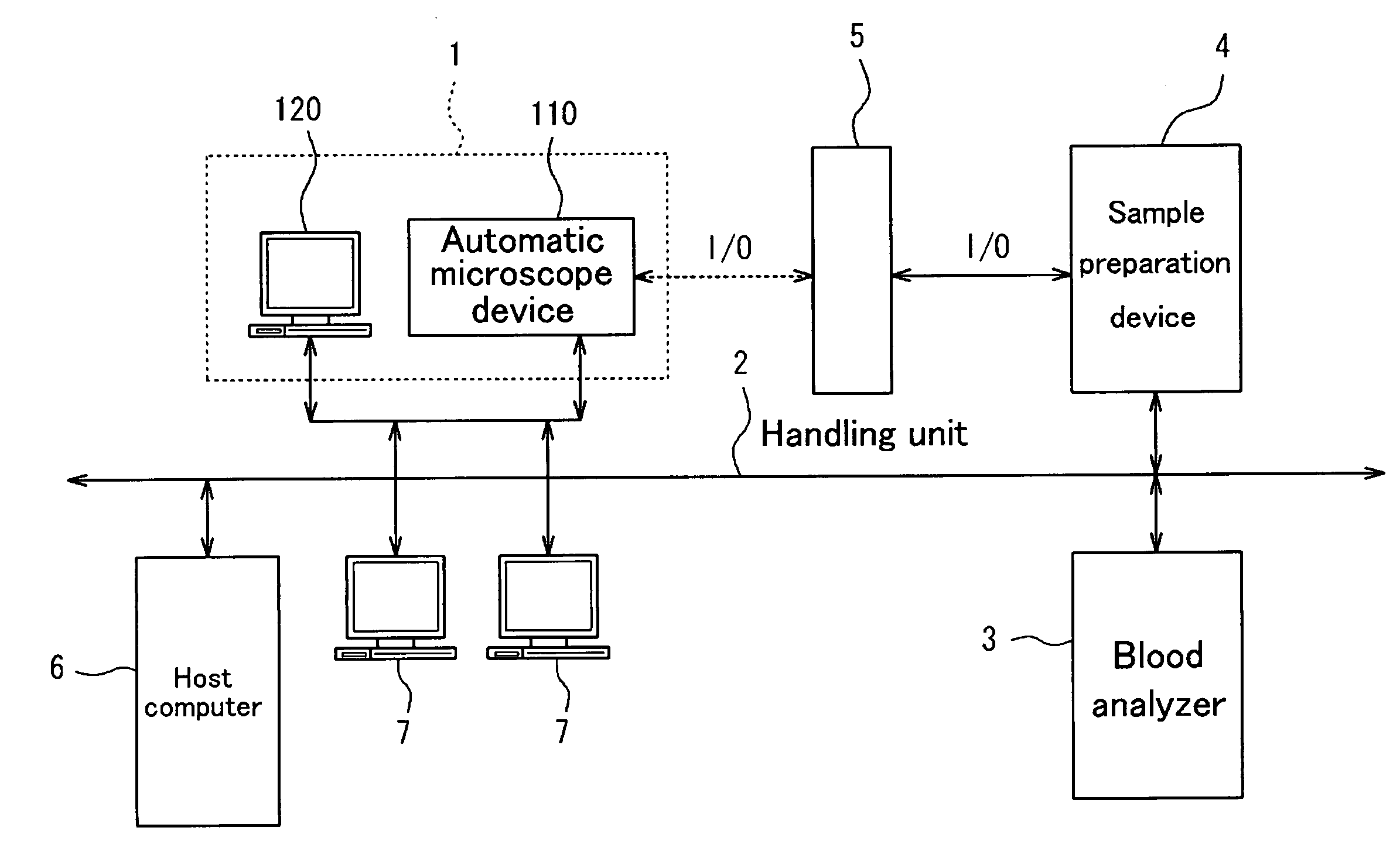
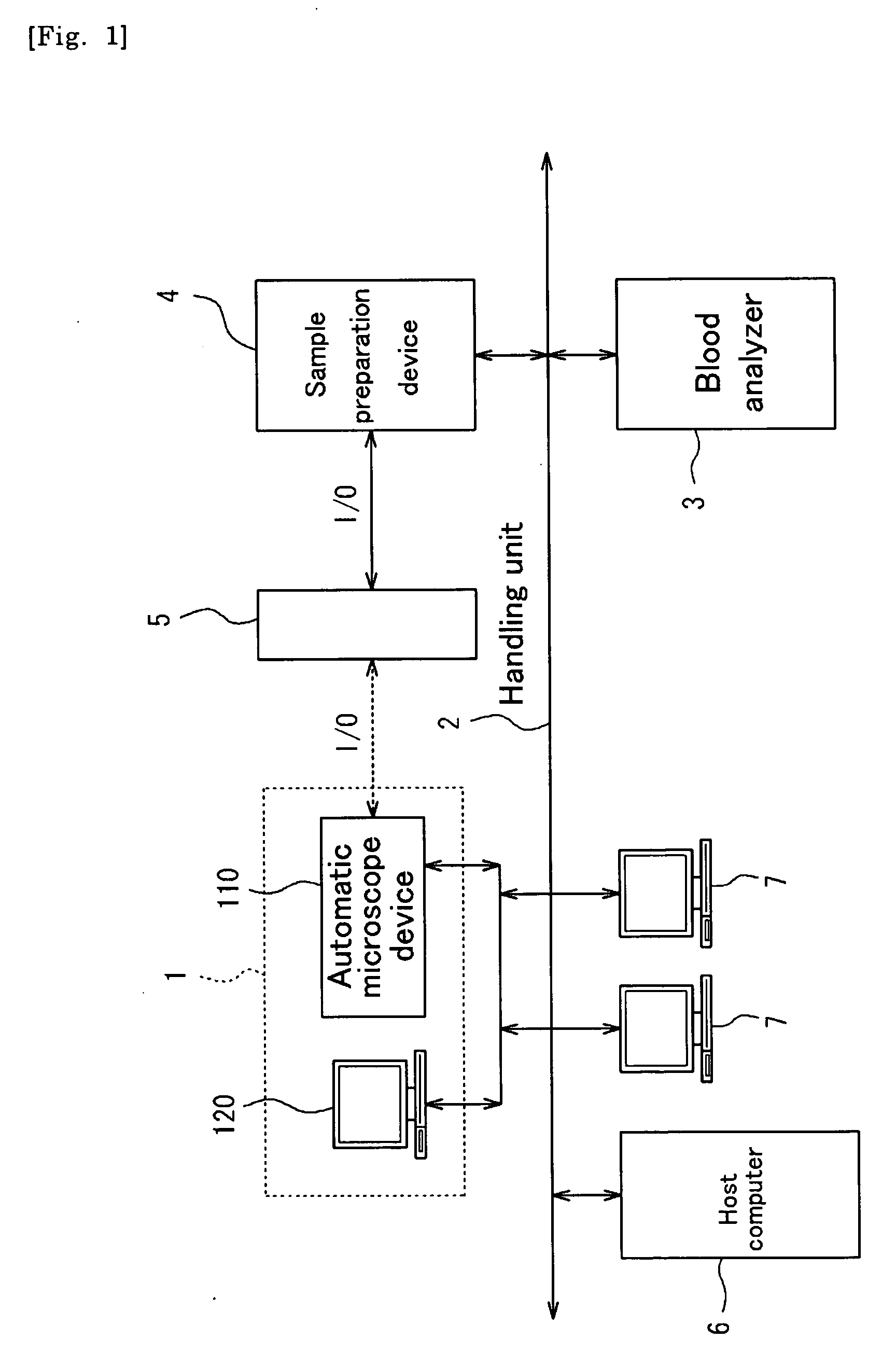
[0044] In the present embodiment, a blood image analyzer 1, which images and analyzes blood, is offered as an example of an apparatus for imaging blood. FIG. 1 shows the general system structure that includes the blood imaging analyzer 1. This system is installed in a facility that performs blood examinations, such as a hospital or the like, and has various types of devices connected thereto via a network (LAN) 2.

[0045] Devices in this system in addition to the blood image analyzer 1 include a blood analyzer 3, sample preparation device 4, handling unit 5, host computer 6, comprehensive review terminal 7 and the like.

Structure of the Blood Analyzer

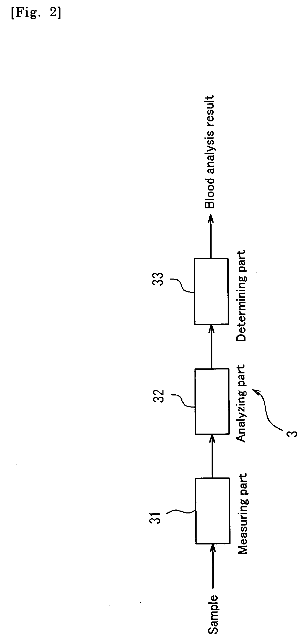
[0046] The blood analyzer 3 is configured as an automatic blood cell analyzer that measures predetermined items (multiple items) in blood and analyzes the results.

[0047] As shown in FIG. 2, ...


PUM

PropertyMeasurementUnit
optical microscopeaaaaaaaaaa
concentrationaaaaaaaaaa
whiteaaaaaaaaaa

Abstract

A apparatus for obtaining an image of a blood cell is described, a representative one of which includes image obtainer for obtaining an image of a predetermined blood cell in a blood sample smeared on a sample holder; an analysis result obtainer for obtaining an analysis result of the blood sample; and a controller for controlling the image obtainer such that the image obtainer obtains the image under a first imaging condition when the analysis result does not indicate a presence of a predetermined anomalous cell, and obtains the image under a second imaging condition to be different from the first imaging condition when the analysis result indicates the presence of the predetermined anomalous cell.

Description

[0001] This application claims priority under 35 U.S.C. § 119 to Japanese Patent Application Nos. JP2005-282291 filed Sep. 28, 2005 and JP2005-285067 filed Sep. 29, 2005, the entire content of which is hereby incorporated by reference. FIELD OF THE INVENTION [0002] The present invention relates to an apparatus for obtaining an image of a blood cell and a method for obtaining an image of a blood cell. BACKGROUND OF THE INVENTION [0003] Japanese Laid-Open Patent Publication No. 60-162955 discloses an automatic analysis device for blood cells capable of automatically and simultaneously performing blood cell calculations and morphological cell classifications from the same sample. [0004] This automatic cell analysis device is provided with an automatic cell classifying system for preparing a blood preparation by smearing a slide glass with blood from a blood sample and staining the smear then identifying and classifying blood cell images of a blood preparation, and an automatic cell cal...

Claims


Application Information

Patent Timeline
Patent Type & Authority Applications(United States)
IPC IPC(8): G01N33/48G06K9/00
CPCG01N15/147G01N15/1475G01N2015/1486
Inventor NAKAYA, MASANORITOHMA, RYUICHI
Owner SYSMEX CORP
Why Eureka
Try Eureka