
Endoscopic bite block

a bit block and endoscope technology, applied in the field of endoscope bit block, can solve the problems of difficult access to oral suction, increased risk of aspiration and potential procedure related complications, and difficult suction secretions, and achieves the effects of preventing complete occlusion of the suction device, reducing the risk of aspiration and potential procedure related complications, and being highly effective and efficient in gastrointestinal endoscopy procedures

Inactive Publication Date: 2007-05-24
GARREN MARY L +2
View PDF22 Cites 30 Cited by
  • Summary
  • Abstract
  • Description
  • Claims
  • Application Information

AI Technical Summary

Benefits of technology

[0006] Still another object of the invention is an endoscopic bite block which is simple in construction, but highly effective and efficient in gastrointestinal endoscopy procedures.
[0007] Basically, the bite block of the present invention is extremely useful for performing endoscopy. The block provides protection for the endoscope while simultaneously allowing the evacuation of pooled intra-oral secretions without the labor of an additional person.
[0008] Fundamentally, the bite block of the present invention comprises a unitary body fabricated from plastic that is rigid enough to withstand pressure from apposition of the mandible to maxilla and having an exterior peri-labial component which is flexible enough to slightly mold to the exterior of the lips. The labial component has strap retainers at the sides thereof to allow attachment of an elastic strap for securing for the bite block (via an elastic strap around the head connecting one side to the other). In addition, the bite block has a suction device extending from the exterior labial surface to the intra-oral surface. The suction device at the labial surface has the ability to be connected to a commercial suction source by flexible tubing or with an intermediary hollow suction wand. The terminal intra-oral portion of the suction device has an angle which allows suctioning where secretions pool during endoscopy which is usually conducted with the patient in the left lateral position. In addition, the terminal portion of the intra-oral suction device has slits circumferentially located and surrounding a circular terminal opening. This design prevents complete occlusion of the suction device by oral tissues.

Problems solved by technology

This method makes it difficult to suction secretions and increases the risk of aspiration and potential procedure related complications.
In addition, access for oral suction is difficult to obtain with currently available bite blocks.

Method used


Image

  • Endoscopic bite block
  • Endoscopic bite block
  • Endoscopic bite block

Examples


Embodiment Construction

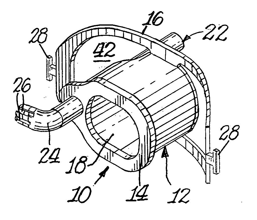
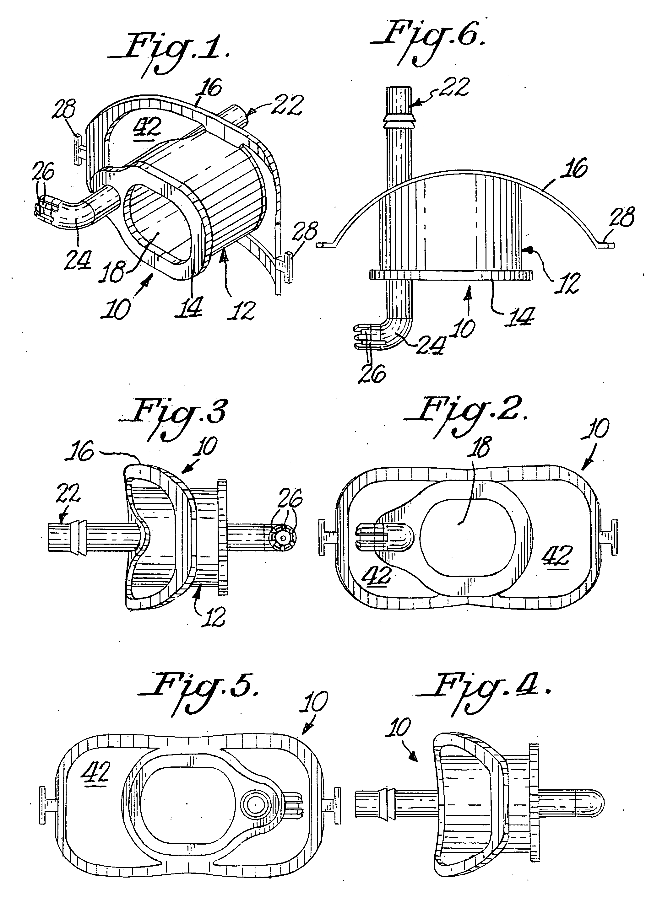
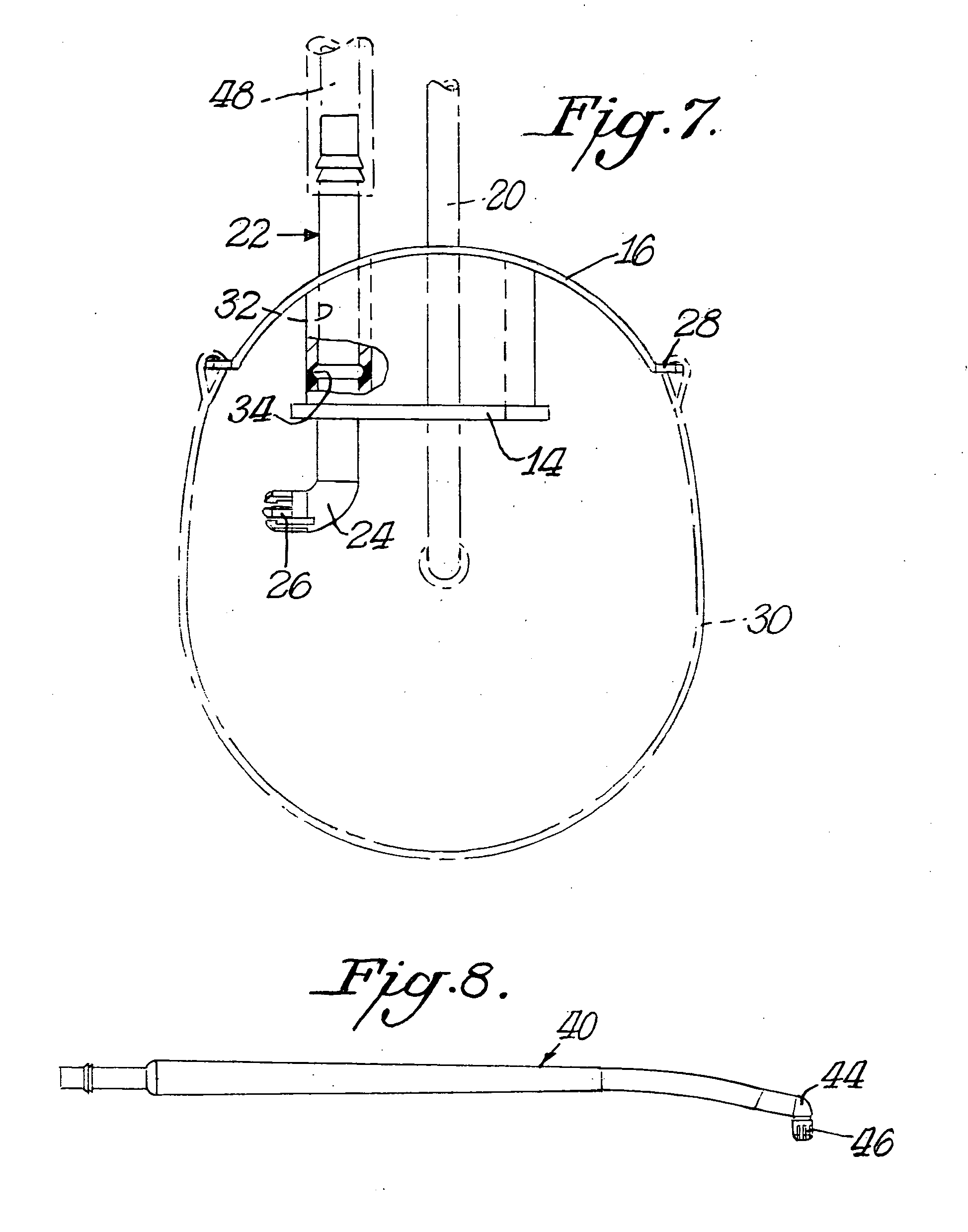
[0022] Referring in more particularity to the drawings, FIG. 1-6 illustrate an endoscopic bite block 10 for a person undergoing upper gastrointestinal endoscopy. The bite block comprises a unitary body 12 having an intra-oral portion 14 and an exterior portion 16. The unitary body may be formed from thermoplastic material by molding techniques known in the art. A central passageway 18 extends through the unitary body 12, and the passageway is arranged to receive an endoscope 20, such as shown in FIG. 7.

[0023] A suction device 22 extends through the unitary body 12 from the exterior portion 16 to the intra-oral portion 14 of the unitary body 12. The suction device 22 includes an angled intra-oral end 24 generally pointing to the left check cavity of a person undergoing gastrointestinal endoscopy for the suction removal of pooled oral fluids.

[0024] The angled intra-oral end 24 of the suction device 22 includes multiple circumferential slit like openings 26 that allow suction drainag...


PUM


Abstract

A bite block intended primarily for use with upper gastrointestinal endoscopy. The bite block comprises a unitary fabrication that fits into the mouth between the teeth or dental ridges and has a central passageway large enough to accommodate a gastroscope. The bite block also has a surface which lies exterior to the oral cavity and extends around the outer surface of the lips. The bite block also has a suction device which can be attached to a standard suction machine via flexible tubing or with an intermediary hollow wand. The suction device extends through the bite block into the intra-oral portion thereof where it forms an angle and allows suction drainage of pooled oral fluids from the cheek cavity. The interior tip of the suction device has multiple slit like openings circumferentially arranged to allow suction drainage of oral fluids without becoming occluded by the oral tissues.

Description

BACKGROUND OF THE INVENTION [0001] The present invention relates to an endoscopic bit block, and more particularly to a bite block that comprises a unitary body which allows for easy passage of an endoscope and which includes an integrated mouth suction angled in a manner that catches and removes pooled oral fluids that accumulate during endoscopic procedures. [0002] Instruments, such as endoscopes, are inserted through a person's mouth into the human body, in medical procedures. To prevent the patient from biting into the instrument, a bite block is used which is generally a plastic tube positioned in the patient's open mouth. The instrument is then inserted through the bite block opening into the stomach or other areas. [0003] Presently, endoscopy is usually performed with the patient in the left lateral position using a bite block which maintains the teeth apart to protect the endoscope from damage by the teeth. This method makes it difficult to suction secretions and increases t...

Claims


Application Information

Patent Timeline
Patent Type & Authority Applications(United States)
IPC IPC(8): A61M16/00
CPCA61B1/2736A61M16/0488A61M16/0497A61M16/0493
Inventor GARREN, MARY L.GARREN, LLOYD R.LUM, SILAS
Owner GARREN MARY L
Learn More