
Robot for minimally invasive interventions

Inactive Publication Date: 2007-05-31
ENHANCED MEDICAL SYST
View PDF8 Cites 231 Cited by
  • Summary
  • Abstract
  • Description
  • Claims
  • Application Information

AI Technical Summary

Benefits of technology

[0010] The ability of the device to move to any desired location in the region of interest from any starting point enables minimally invasive surgery to become independent of the location of the incision. Use of the device also allows a subxiphoid transpericardial approach to any intrapericardial procedure, regardless of the location of the treatment site. As a result, deflation of the left lung is no longer needed, and it becomes feasible to use local or regional rather than general anesthetic techniques. These advantages provide a system for ambulatory outpatient cardiac surgery.
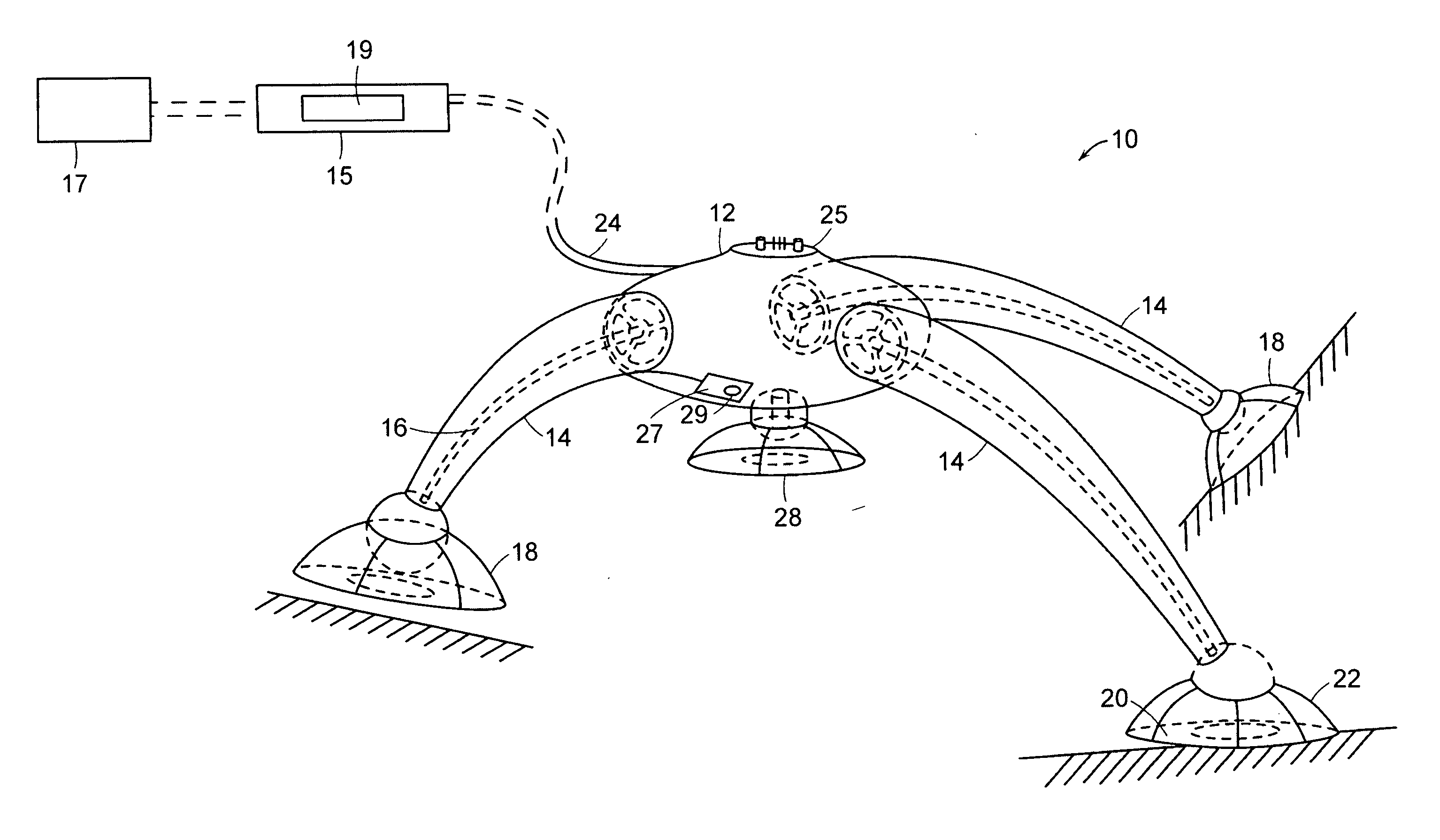
[0011] For arrhythmia treatment procedures, the device approaches the heart from the outer surface, placing a walking unit upon the epicardium upon which it moves with the beating heart while navigating across it. The device gains access to the epicardium by crossing through the pericardial sac. The devices uses a minimally invasive approach such as a sub-xiphoid incision combined with endoscopic insertion that provides both visualization during access and a means to safely transect the sac without harming the epicardium. Sub-xiphoid access will place the device initially upon the heart apex to begin its navigation over the cardiac surface. The small size of device, typically 6 mm or smaller in cross section and 20 mm or shorter in length, allows it to use a small diameter access channel to the pericardium, further lessening side effects from tissue damage along the access path to the heart. A preferred embodiment employs a device having dimensions of 10 mm or less in every dimension with a cross sectional diameter of 3 mm or less.
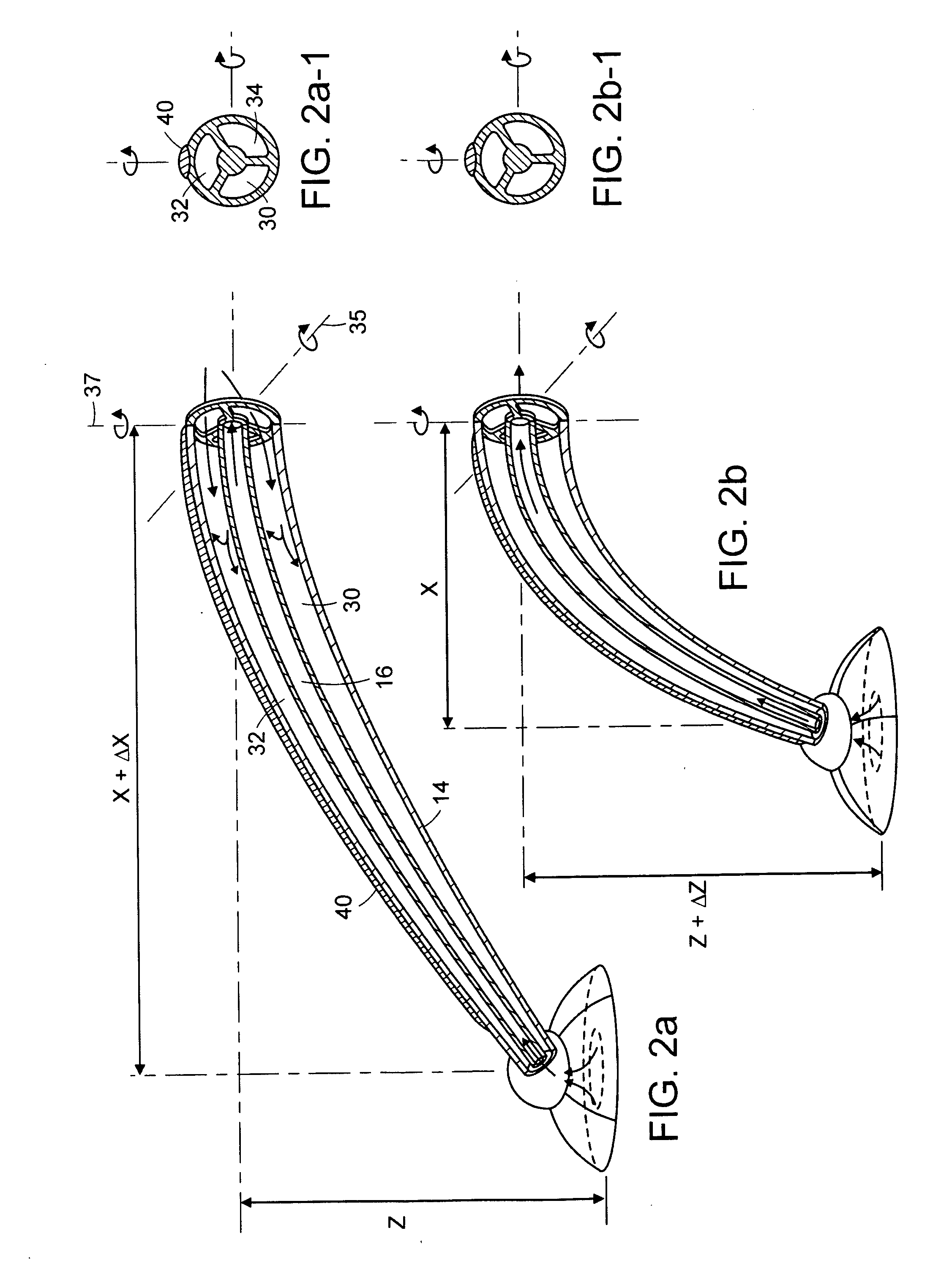
[0013] The device moves across the surface of the beating heart by having at least two feet that independently make contact with and hold onto the surface. When configured with two feet, the device can move in a manner similar to an inchworm where the front and the back of the device alternately attach to the heart surface and the relative distance between the ends is changed as one of the feet is attached. Thus, with the back foot in place the front can extend away from it while providing the ability to change the direction of movement by pointing the front in desired travel path. When the front finds its attachment, the back foot can detach and contract to bring itself closer to the now attached front foot. When the device is configured with more than 2 feet it can move lateral to the direction it is pointed allowing additional mobility options.
[0015] A unique, but common situation, is for the device to encounter fat attached to the epicardium or other internal body surface. In this case, the device's foot configuration allows it to maintain suction upon the fat without tearing it loose from its attachment. The device can detect the presence of fat underfoot by, for example, sensing an impedance change and shift its attachment strategy to achieve this connection without loosening itself or the fat. Another strategy that the device can employ when traversing the heart should the fat prove to be unstable is to maintain an attachment to the pericardial surface while crossing fatty areas. The device can carry this out by having an alternate set of suction connections on the side away from the epicardium which can be used instead of the usual epicardial feet. The device also contains mitigation elements in its suction system to prevent fat from being pulled into its system and plugging it. This includes the specific configuration of the feet and a flushing system that removes the fat should it get into the vacuum system.

Problems solved by technology

The challenges of minimally invasive access are further complicated by the goal of avoiding cardiopulmonary bypass.

Method used


Image

  • Robot for minimally invasive interventions
  • Robot for minimally invasive interventions
  • Robot for minimally invasive interventions

Examples


Embodiment Construction

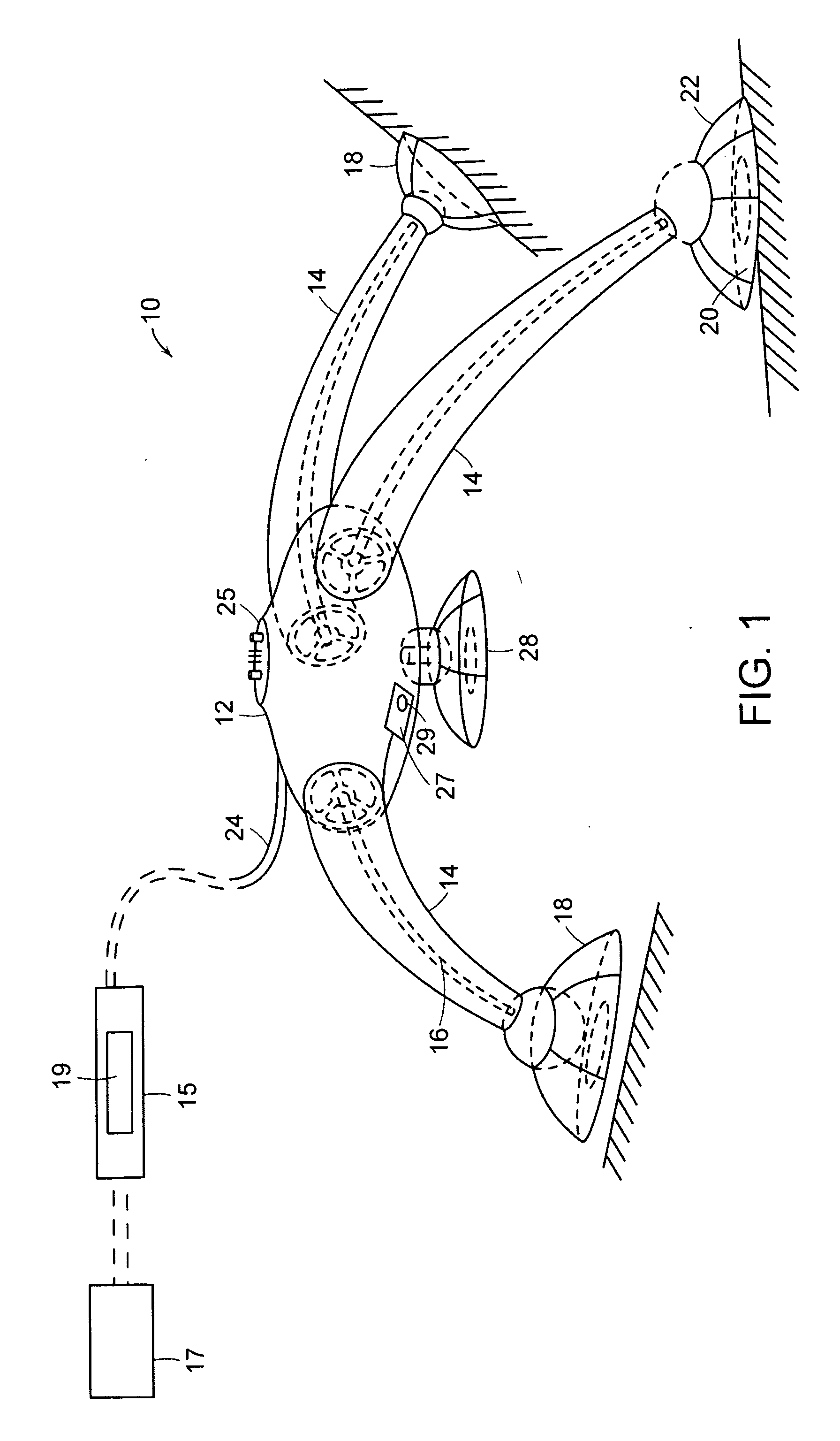
[0033] A preferred embodiment of a robot constructed according to the present invention is illustrated in FIG. 1. The device 10 includes forming a central body 12 and a plurality of members or legs 14. The device can have a 6-20 mm cross sectional footprint and a length of 5-20 mm, for example. That size allows the device 10 to fit within a standard 20 mm diameter cannula or endoscope channel. Each of the body sections 14 is equipped with an independent suction line 16 and a foot 18 with one or more suction pad or pads 20, 22, respectively, for gripping to biological tissue. The suction lines 16 and suction pads 20, 22 illustrate a preferred system for prehension.

[0034] The translation and rotation of the body section 12 is controlled from an external control system, in this embodiment a handle 15. This can be controlled remotely by RF transmission to the robot and / or by a single or multi-lumen sheath 24. A single or three independently actuated lumens in the sheath 24 provide at l...


PUM


Abstract

The present invention relates to a miniature robotic device to be introduced, in the case of the heart, into the pericardium through a port, attach itself to the epicardial surface, and then, under the direct control of the user or physician, travel to the desired location for diagnosis or treatment.

Description

CROSS REFERENCE TO RELATED APPLICATIONS [0001] This application claims the priority of U.S. Provisional Application No. 60 / 699,087 filed Jul. 14, 2005 entitled, ROBOT FOR MINIMALLY INVASIVE INTERVENTIONS. The entire content of the above application is being incorporated herein by reference.BACKGROUND OF THE INVENTION [0002] Heart surgery is typically done by opening the chest cavity or by a minimally invasive procedure using the intercostal spacing to access the heart, or endoscopically in which surgical tools can be introduced via an endoscope channel. [0003] Closed-chest endoscopic visualization of the epicardium utilizes techniques for evaluation of blunt chest trauma, pericardial effusion, lung cancer, staging, and epicardial implantation of ventricular pacing leads. Endoscope access can require thoracotomy with breach of the left pleural space. Direct access to the pericardial space via subxiphoid puncture is an increasingly practiced technique for epicardial procedures. In suc...

Claims


Application Information

Patent Timeline
IPC IPC(8): A61B1/00A61B5/00A61B17/00
CPCA61B1/00149A61B1/04A61B1/041A61B18/22A61B19/22A61B2017/00247A61B2017/00703A61B2018/00392A61B2018/00982A61B2019/2215A61B2019/2249A61B2019/465A61B2034/302A61B34/70A61B34/72A61B2090/065A61B34/30A61B2018/00351
Inventor MEGLAN, DWIGHT
Owner ENHANCED MEDICAL SYST
Learn More