Apparatus and formulations for suprachoroidal drug delivery

Inactive Publication Date: 2007-08-30
CLEARSIDE BIOMEDICAL
View PDF24 Cites 188 Cited by
  • Summary
  • Abstract
  • Description
  • Claims
  • Application Information

AI Technical Summary

Benefits of technology

[0012] In another embodiment, devices are provided for minimally invasive delivery of a drug formulation into the suprachoroidal space of the eye comprising a needle having a leading

Problems solved by technology

Accessing the eye for medical treatment is hindered by the small size and delicate nature of the tissues.
The posterior region of the eye, including the retina, macula and optic nerve, is especially

Method used


Image

  • Apparatus and formulations for suprachoroidal drug delivery
  • Apparatus and formulations for suprachoroidal drug delivery
  • Apparatus and formulations for suprachoroidal drug delivery

Examples


example 1

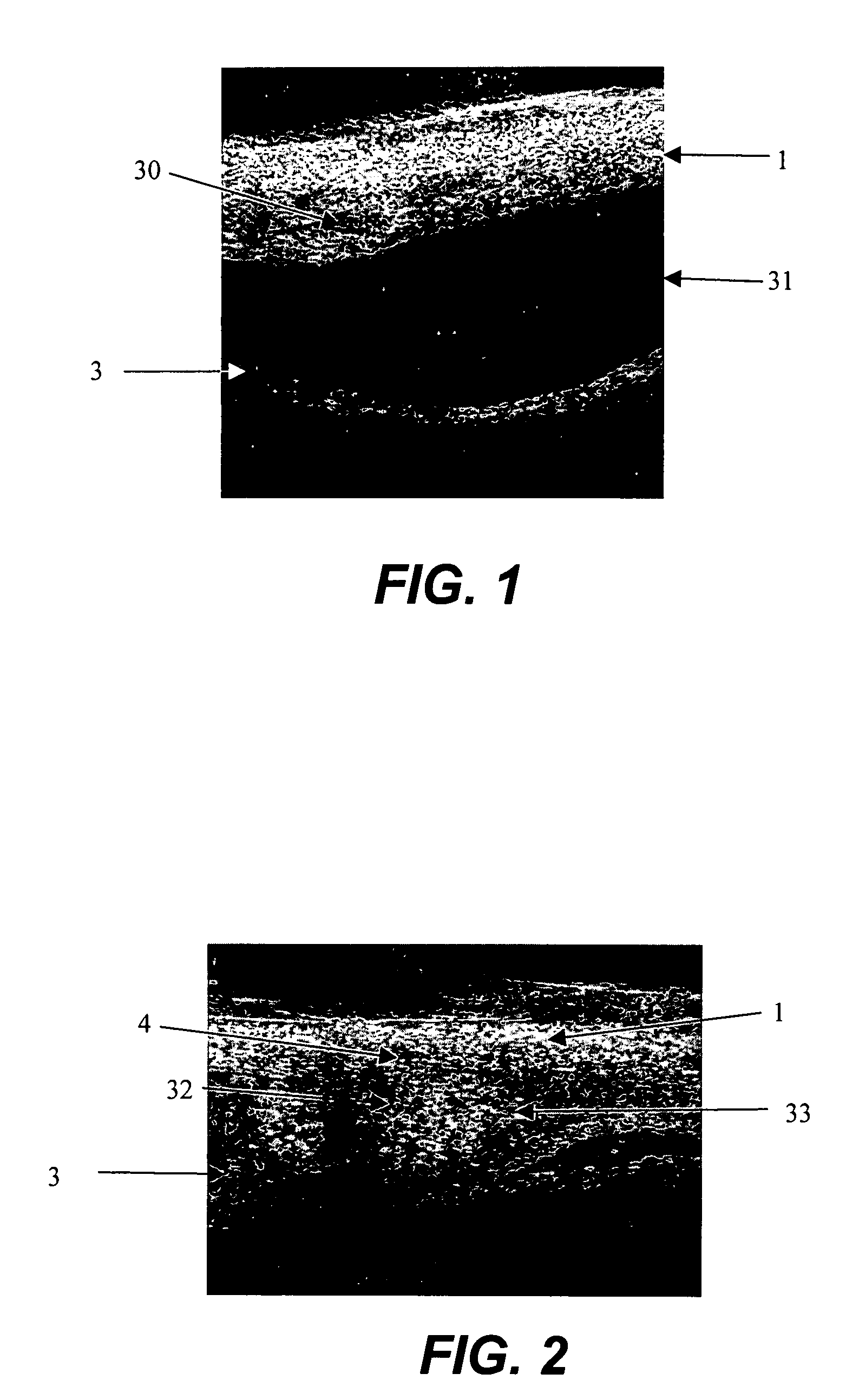
[0054] Fluorescent dyed polystyrene microspheres (Firefli™, Duke Scientific, Inc., Palo Alto, Calif.) suspended in phosphate-buffered saline were used as model drug to evaluate the size range in which particulates will migrate in the suprachoroidal space from the anterior region to the posterior region.

[0055] An enucleated human cadaver eye was radially incised to the choroid in the pars plana region, which is in the anterior portion of the eye. Using a syringe terminated with a blunt 27 gauge needle, 0.15 mL of a 1% by volume microsphere suspension (mean diameter 6 micron) was delivered into the anterior region of the suprachoroidal space. The needle was withdrawn and the incision sealed with cyanoacrylate adhesive.

[0056] The eye was then perfused for 24 hours with phosphate buffered saline at 10 mm Hg pressure by introducing into the anterior chamber a 30 gauge needle attached to a reservoir via infusion tubing. The reservoir was placed on a lab jack and elevated to provide cons...

example 2

[0059] The experiment of Example 1 was repeated, except that a mixture of 6 and 33 micron diameter fluorescent microspheres as a model drug was suspended in a polymeric excipient comprising a surgical viscoelastic (Healon 5, Advanced Medical Optics, Inc.), a 2.3% concentration of sodium hyaluronic acid of 4,000,000 Daltons molecular weight, with thixotropic properties of a zero shear viscosity of 7,000,000 mPas and 400 mPas viscosity at 1000 s−1 shear rate. The mixture was introduced into the suprachoroidal space in the manner of Example 1. After 24 hour perfusion, the microspheres resided solely in the suprachoroidal space at the anterior instillation site and did not show evidence of migration, demonstrating the localizing effect of the thixotropic polymeric excipient.

example 3

[0060] To demonstrate the effect of polymeric excipient viscosity on drug localization, the experiment of Example 1 was repeated, except that bevacizumab (Avastin™, Genentech), an anti-VEG antibody, was adsorbed onto 5 micron diameter carboxylated fluorescent microspheres and mixed at equal volumes with one of three hyaluronic acid based surgical viscoelastics (Healon, Healon GV, Healon 5, Advanced Medical Optics, Inc.), each with a different viscosity and thixotropic properties. (Healon, 300,000 mPas viscoscity at zero shear rate, 150 mPas viscosity at 1000 s−1 shear rate; Healon GV, 3,000,000 mPas viscosity at zero shear rate, 200 mPas at 1000 s−1 shear rate; Healon 5, 7,000,000 mPas viscosity at zero shear rate, 400 mPas viscosity at 1000 s−1 shear rate.) Each mixture was introduced into the anterior region of the suprachoroidal space at the pars plana in the anterior region of the eye in the manner of Example 1. After 24 hours perfusion, the microspheres in Healon and Healon GV ...


PUM

PropertyMeasurementUnit
Diameteraaaaaaaaaa
Frequencyaaaaaaaaaa
Thicknessaaaaaaaaaa

Abstract

Drug formulations, devices and methods are provided to deliver biologically active substances to the eye. The formulations are delivered into scleral tissues adjacent to or into the suprachoroidal space without damage to the underlying choroid. One class of formulations is provided wherein the formulation is localized in the suprachoroidal space near the region into which it is administered. Another class of formulations is provided wherein the formulation can migrate to another region of the suprachoroidal space, thus allowing an injection in the anterior region of the eye in order to treat the posterior region.

Description

CROSS-REFERENCE TO RELATED APPLICATIONS [0001] The priority of provisional U.S. application Ser. No. 60 / 776,903, filed Feb. 22, 2006 is claimed pursuant to 35 USC 119(e). The provisional application is incorporated herein by reference in its entirety.FIELD OF THE INVENTION [0002] The invention relates to the field of drug delivery into the eye. BACKGROUND OF INVENTION [0003] The eye is a complex organ with a variety of specialized tissues that provide the optical and neurological processes for vision. Accessing the eye for medical treatment is hindered by the small size and delicate nature of the tissues. The posterior region of the eye, including the retina, macula and optic nerve, is especially difficult to access due to the recessed position of the eye within the orbital cavity. In addition, topical eye drops penetrate poorly into the posterior region, further restricting treatment options. [0004] The suprachoroidal space is a potential space in the eye that is located between th...

Claims


Application Information

Patent Timeline
IPC IPC(8): A61K38/39A61K31/728A61K31/722A61K31/737A61K31/718A61K31/717A61K9/50
CPCA61K9/0048A61K9/1635A61K31/717A61K31/718A61K31/722A61F9/0017A61K31/737A61K47/10A61K47/32A61K47/36A61M5/31501A61K31/728A61K9/0051A61K9/14A61K9/50A61K31/716A61K38/39A61F9/00736A61M5/486A61M2005/3103A61M2210/0612A61F9/0026A61F9/007
Inventor YAMAMOTO, RONALDCONSTON, STANLEY R.SIERRA
Owner CLEARSIDE BIOMEDICAL
Why Eureka
Try Eureka