
Endoscope system and observation method using the same

Active Publication Date: 2007-09-13
OLYMPUS CORP
View PDF5 Cites 60 Cited by
  • Summary
  • Abstract
  • Description
  • Claims
  • Application Information

AI Technical Summary

Benefits of technology

[0014] The present invention has been conceived in light of the circumstances described above, and an object thereof is to provide an endoscope system and an observation method using the same in which a fluorescent dye / probe that can quickly identify the presence of disease is efficiently applied in a living organism and optically detected to acquire clear fluorescence images.
[0023] The present invention affords an advantage in that a fluorescent dye / probe that selectively dyes cells and tissue of cancer / tumors to differentiate them from normal cells and tissue and that can rapidly identify the presence of disease is efficiently applied in a living organism and optically detected to obtain clear fluorescent images.

Problems solved by technology

However, oral administration or administration by intravenous injection or the like, as typified by the method using 5-aminolevulinic acid, requires a large amount of the agent, and therefore has the drawback that it consumes a large amount of fluorescent material, which is generally expensive.
In addition, since it is administered to the whole body, it takes time to accumulate in the tumor site.
For example, because there is a time delay for accumulation at various sites in the digestive system, there is a problem in that it is not possible to observe it at the desired instant.
Furthermore, because the intensity of the fluorescence is very weak, there is a problem in that the quality of the fluorescence image tends to deteriorate due to noise and so forth.

Method used


Image

  • Endoscope system and observation method using the same
  • Endoscope system and observation method using the same
  • Endoscope system and observation method using the same

Examples


first embodiment

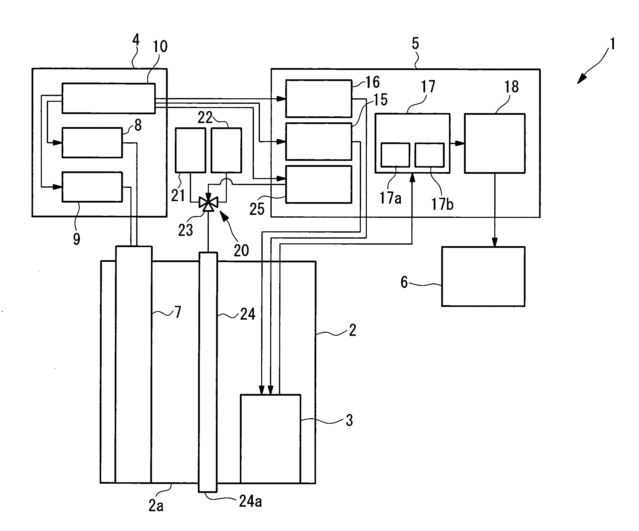
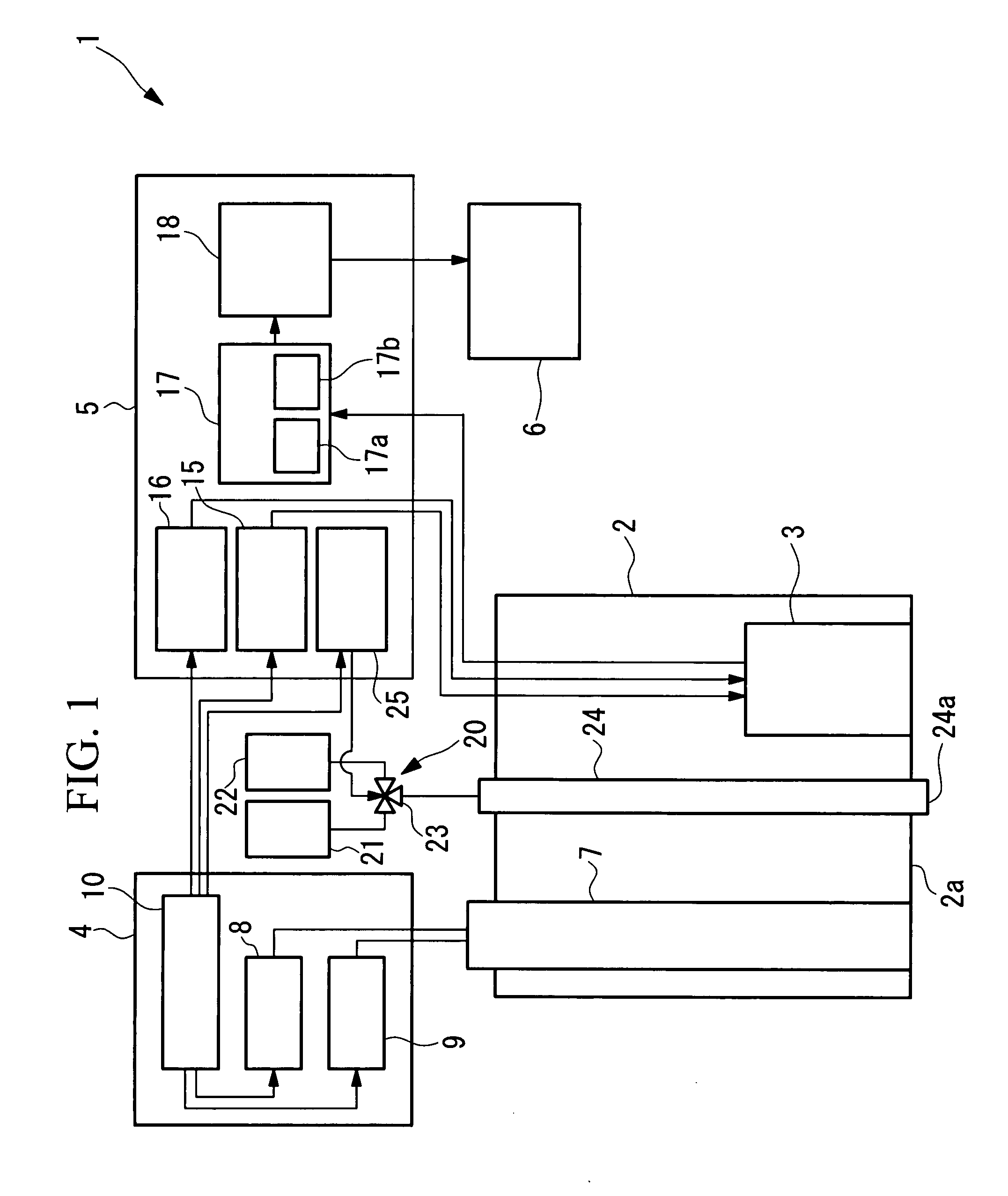
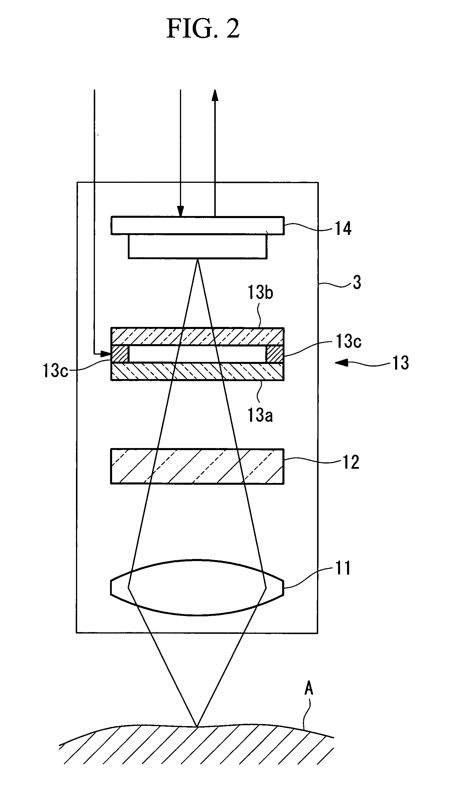
[0035] An endoscope system 1 according to the present invention will be described below with reference to FIGS. 1 to 6.

[0036] As shown in FIG. 1, the endoscope system 1 according to this embodiment includes an insertion portion 2 for insertion into a body cavity of a living organism, an image-acquisition unit (image-acquisition portion) 3 disposed inside the insertion portion 2, a light-source unit (light-source portion) 4 for emitting a plurality of types of light, a liquid-delivery unit 20 (agent dispensing portion, cleaning-water dispensing portion) for supplying liquid to be spouted out from the tip 2a of the insertion portion 2, a control unit (control portion) 5 for controlling the image-acquisition unit 3, the light-source unit 4, and the liquid-delivery unit 20, and a display unit (output portion) 6 for displaying images acquired by the image-acquisition unit 3.

[0037] The insertion portion 2 has extremely narrow outer dimensions, allowing it to be inserted inside the body c...

second embodiment

[0098] Next, an endoscope system 1′ according to the present invention will be described below with reference to FIGS. 8 to 10.

[0099] In the description of this embodiment, parts having the same configuration as those in the endoscope system 1 according to the first embodiment described above are assigned the same reference numerals, and a description thereof is omitted.

[0100] The endoscope system 1′ according to this embodiment differs from the endoscope system 1 according to the first embodiment in the configuration of a light-source unit 4′ and the transmittance characteristics of the tunable spectral device 13 and the excitation-light-cutting filter 12.

[0101] As shown in FIG. 8, the light-source unit 4′ of the endoscope system 1′ according to this embodiment includes two excitation light sources 31 and 32.

[0102] The first excitation light source 31 is a semiconductor laser emitting first excitation light with a peak wavelength of 490±5 nm. It is possible to excite the esteras...


PUM


Abstract

A fluorescent dye / probe that can rapidly identify the presence of disease is efficiently applied in a living organism and optically detected to obtain clear fluorescent images. There is provided an endoscope system and an observation method using the same, comprising agent dispensing means for dispensing towards an acquisition object a fluorescent agent that binds with a specific substance inside the acquisition object or that accumulates inside the acquisition object; control means for controlling the agent-dispensing means; a light source portion for emitting excitation light for exciting the fluorescent agent and irradiation light having different spectral characteristics from the excitation light; an optical system for transmitting the excitation light and the irradiation light towards the acquisition object; and image-acquisition means, disposed at a portion that is inserted inside a body cavity and capable of acquiring fluorescence excited from the acquisition object by the excitation light, and light in a different wavelength band, which is excited from the acquisition object by the irradiation light; wherein the control means controls the agent-dispensing means so that the acquisition object is irradiated with the irradiation light before the fluorescent agent is spouted out towards the acquisition object.

Description

BACKGROUND OF THE INVENTION [0001] 1. Field of the Invention [0002] The present invention relates to an endoscope system and to an observation method using the same. [0003] This application is based on Japanese Patent Application No. 2006-051915, the content of which is incorporated herein by reference. [0004] 2. Description of Related Art [0005] In endoscopy, observation of fluorescence images can give different information about a living organism from reflection images, and it is therefore beneficial in the diagnosis and observation of diseases. [0006] For example, if a fluorescent probe that reacts with a substance originating in a diseased area to change from a non-fluorescent substance to a fluorescent substance is administered, it is possible to observe the concentration distribution of the substance originating from the disease by examining the fluorescence image. [0007] By observing autofluorescence, that is, fluorescence originating in the living organism itself, it is poss...

Claims


Application Information

Patent Timeline
IPC IPC(8): A61B1/12A61B1/06A61B6/00
CPCA61B1/043A61B5/0084A61B5/0071A61B1/0638A61B1/0655
Inventor MORISHITA, KOKIHASEGAWA, AKIRA
Owner OLYMPUS CORP
Learn More