Method and apparatus for the guided ablative therapy of fast ventricular arrhythmia

a ventricular arrhythmia and guided ablative therapy technology, applied in the field of guided ablative therapy, can solve the problems of morbidity and mortality in the developed world, abnormal cardiac and brain electrical conduction processes, and difficulty in ventricular tachycardia ablation

Inactive Publication Date: 2007-09-20
MASSACHUSETTS INST OF TECH
View PDF8 Cites 21 Cited by
  • Summary
  • Abstract
  • Description
  • Claims
  • Application Information

AI Technical Summary

Benefits of technology

[0020] According to a first aspect, the method of the invention for locating the site of origin of an arrhythmia includes applying a multiplicity of recording electrodes to the body. Electrical activity from the recording electrodes is analyzed to identify the location of the site of origin of the arrhythmia. One or more stimulating electrodes is introduced into the body and the heart is stimulated with supra-threshold stimuli through the one or more stimulating electrodes. The electrical activity from the recording electrodes is processed to identify the locations of the one or more stimulating electrodes in relation to the location of the site of origin of the arrhythmia. In a preferred embodiment, radio frequency energy is delivered to the location of the site of origin of the arrhythmia to ablate the site. The arrhythmia may be initiated by electrical stimulation of the heart. The analyzing step may be preformed at a multiplicity of time epochs during the cardiac cycle and may involve computing the location of at least one equivalent moving dipole.

Problems solved by technology

Abnormalities in cardiac and brain electrical conduction processes are principal causes of morbidity and mortality in the developed world.
However, in contrast to supraventricular tachycardia ablation, which is highly successful because the atrio-ventricular node anatomy is known, ventricular tachycardia ablation remains difficult because the site of origin of the arrhythmia could be anywhere in the ventricles.
Sustained ventricular tachycardia is often a difficult arrhythmia to manage.
The ability of the physician to deliver radio-frequency energy through a catheter at the reentry site is restricted by the limitations of the current technology that is employed to guide the catheter to the appropriate ablation site.
The principal limitation of the radio frequency ablation technique is the determination of the correct site for delivery of the radio frequency energy.
This is a major problem, especially for those arrhythmias which compromise hemodynamic function of the patient.
Many arrhythmias for this reason are not presently amenable to radio frequency ablation treatment.
Conversely, an arrhythmia site of origin that was not initially successfully ablated may later become permanently nonfunctional if it lies within the border zone of a lesion and if microvascular injury and inflammation within this zone result in progressive necrosis [Nath, 1994].
Catheter ablation of sustained monomorphic ventricular tachycardia late after myocardial infarction has been challenging.
Mapping and ablation are further complicated by the frequent presence of multiple reentry circuits, giving rise to several morphologically different ventricular tachycardias [Wilbur, 1987; Waspel, 1985].
The mapping procedure may take many hours during which the arrhythmia must be maintained.
Furthermore, there are several difficulties in selecting a dominant, “clinical ventricular tachycardia” for ablation.
Often it is not possible to determine which ventricular tachycardia is in fact the one that has occurred spontaneously.
However, the use of fluoroscopy for these purposes may be problematic for the following reasons: (1) It may not be possible to accurately associate intracardiac electrograms with their precise location within the heart; (2) The endocardial surface is not visible using fluoroscopy, and the target sites can only be approximated by their relationship with nearby structures such as ribs and blood vessels as well as the position of other catheters; (3) Due to the limitations of two-dimensional fluoroscopy, navigation is frequently inexact, time consuming, and requires multiple views to estimate the three-dimensional location of the catheter; (4) It may not be possible to accurately return the catheter precisely to a previously mapped site; (5) It is desirable to minimize exposure of the patient and medical personnel to radiation; and (6) Most importantly, fluoroscopy cannot identify the site of origin of an arrhythmia and thus cannot be used to specifically direct a catheter to that site.
However, this technique fundamentally has the limitation that it involves sequentially sampling endocardial sites.
Also, the localization is limited to the endocardial surface and thus sites of origin within the myocardium cannot be accurately localized.
One of the drawbacks of this methodology is that the ventricular geometry is not fixed but varies during the cardiac cycle.
This leads to relative movement between the recording electrode and the tissue, significantly limiting the accuracy of the mapping method.
Also, the localization is limited to the endocardial surface, and thus sites of origin within the myocardium cannot be accurately localized.
It is not possible to construct the three-dimensional distribution of cardiac electrical sources from a two-dimensional distribution of ECG signals obtained on the body surface.

Method used


Image

  • Method and apparatus for the guided ablative therapy of fast ventricular arrhythmia
  • Method and apparatus for the guided ablative therapy of fast ventricular arrhythmia
  • Method and apparatus for the guided ablative therapy of fast ventricular arrhythmia

Examples


Embodiment Construction

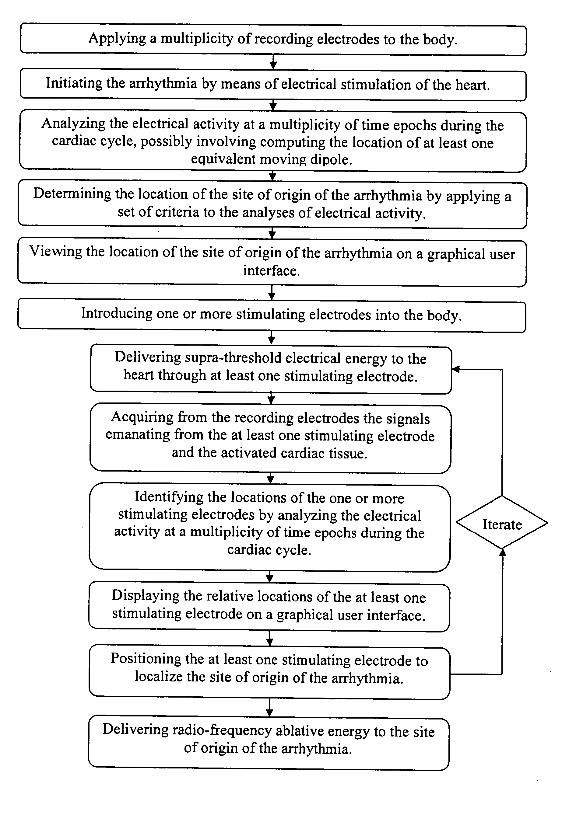
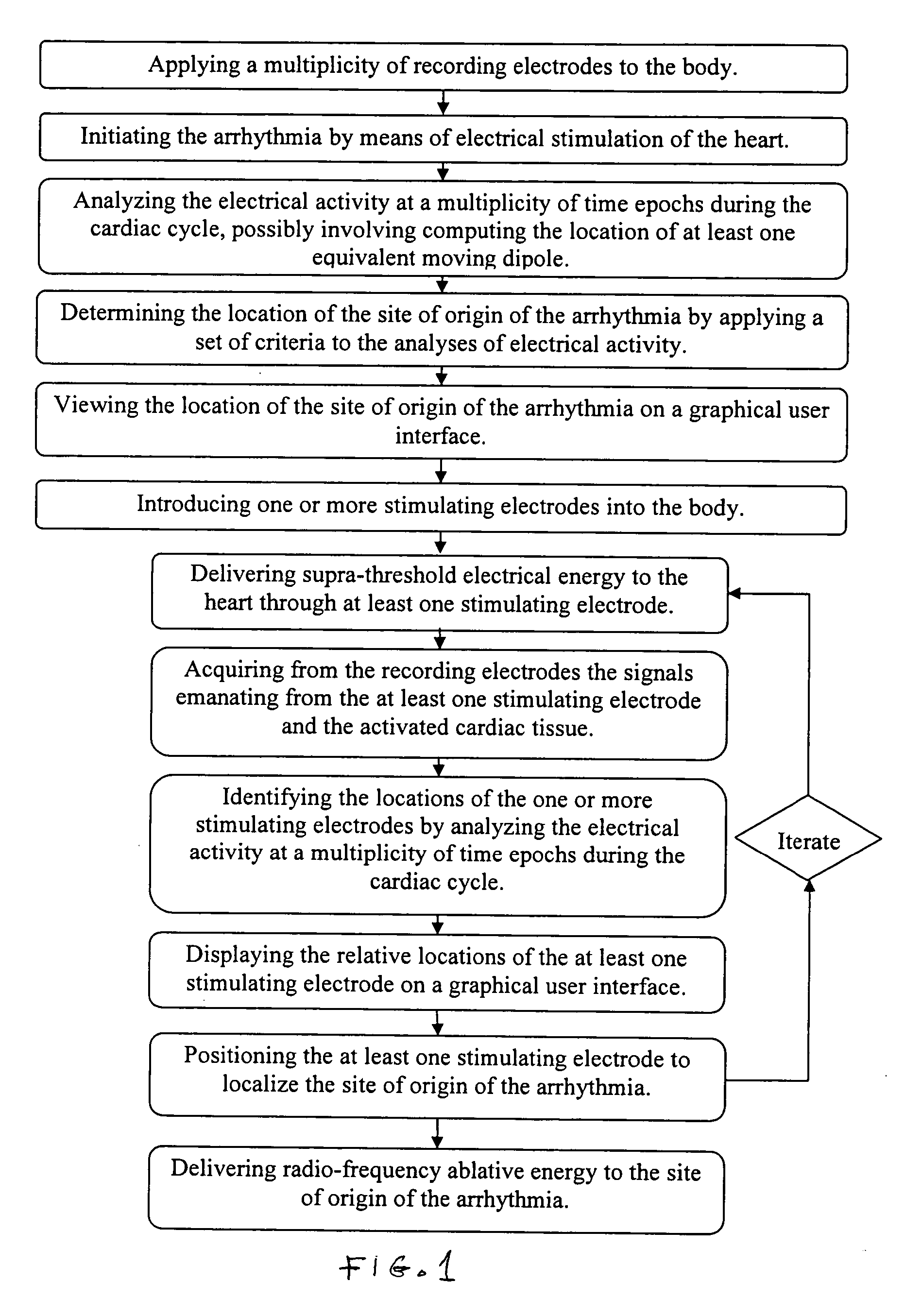
[0029] Previous patents (U.S. Pat. Nos. 6,308,093 and 6,370,412 discussed above) have described a method by which the electrical source of low-frequency electrical activity (such as a slow ventricular tachycardia) within the body can be localized and ablated. However, the ablation catheter tip cannot be accurately guided in the manner described in previous patents in the presence of a rapid arrhythmia (such as in cases of fast ventricular tachycardia, >240 beats per minute).

[0030] In previous methods, low-amplitude sub-threshold bipolar current pulses are delivered from an electrode at the tip of the catheter. The signals at the body surface result solely from the weak electrical fields set up in the body by the bipolar pulses. These surface signals are fitted with a moving dipole model to determine the relative location of the stimulating electrode within a given short time epoch. Thereafter, the processes of delivering electrical energy and determining the relative location of th...


PUM


Abstract

Method and apparatus for guiding ablative therapy of abnormal biological electrical excitation. The excitation from the previous excitatory wave is significant at the beginning of the next excitation. In particular, it is designed for treatment of fast cardiac arrhythmias. Electrical signals are acquired from recording electrodes, and an inverse dipole method is used to identify the site of origin of an arrhythmia. The location of the tip of an ablation catheter is similarly localized from signals acquired from the recording electrodes while electrical pacing energy is delivered to the tip of the catheter close to or in contact with the cardiac tissue. The catheter tip is then guided to the site of origin of the arrhythmia, and ablative radio frequency energy is delivered to its tip to ablate the site.

Description

BACKGROUND OF THE INVENTION [0001] This invention relates to method and apparatus for guided ablative therapy and more particularly to such therapy to treat fast ventricular arrhythmia. [0002] The electrical activity generated in certain organs in the human body is intimately related to the organs' functions. Abnormalities in cardiac and brain electrical conduction processes are principal causes of morbidity and mortality in the developed world. Appropriate treatment of disorders arising from such abnormalities frequently requires a determination of their location. Such localization of the site of origin of an abnormal electrical excitation is typically achieved by painstaking mapping of the electrical activity on the inner surface of the heart or the brain from electrodes or a catheter. Often, this recording must be done while the abnormal biological electrical excitation is ongoing. [0003] Radio frequency catheter ablation procedures have evolved in recent years to become an estab...

Claims


Application Information

Patent Timeline
Patent Type & Authority Applications(United States)
IPC IPC(8): A61B5/02
CPCA61B5/04A61B18/1492A61N1/362A61B2018/00839A61B2018/00351A61B5/24
Inventor COHEN, RICHARD J.BARLEY, MAYA
Owner MASSACHUSETTS INST OF TECH
Why Eureka
Try Eureka