
Method for identifying pre-neoplastic and/or neoplastic states in mammals

a preneoplastic and/or neoplastic state technology, applied in the field of mammals preneoplastic and/or neoplastic states identification, can solve the problems of few diagnostic methods, inconvenient diagnosis, and inability to accurately predict the age of the patient,

Inactive Publication Date: 2007-10-25
BIOSCEPTRE INT
View PDF7 Cites 35 Cited by
  • Summary
  • Abstract
  • Description
  • Claims
  • Application Information

AI Technical Summary

Benefits of technology

"The patent text describes a new method for diagnosing and staging pre-cancerous and cancerous states in mammals, particularly humans. This method involves detecting the expression profile of P2X purinergic receptors in cells and tissue, and comparing it to a predetermined profile of normal cells. The invention provides a new tool for diagnosing and investigating carcinogenesis, as well as a method for identifying specific receptors and their sub-types. The technical effects of the patent include improved diagnosis and treatment of cancer, as well as a better understanding of the physiology and aetiology of carcinogenesis."

Problems solved by technology

Despite the gravity of this condition, diagnostic methods are few and imprecise.
Current methods for assessing prognosis such as digital rectal examination (DRE), ultrasound, prostatic acid phosphatase levels, androgen ablation, prostate specific antigen (PSA) density, PSA velocity, PSA age-specific reference ranges and Gleason histopathological grading, can fail to provide reliable predictive information regarding the clinical outcome of prostate cancer [4].
Unfortunately, serum PSA levels have an incidence of 23% false negative and 36.7% false positive diagnoses [6].
One study has shown that applying an age-specific PSA reference range that increases the upper limit of normal PSA to 4.5 ng / mL results in the failure to detect a substantial number of clinically significant cancers [8].
Given this uncertainty, prostate biopsy is often performed to confirm malignancy but this test also has a highly unsatisfactory 23% incidence of false-negative diagnosis [9].
Unfortunately, prostate cancer tissue is notoriously heterogeneous and a vital diagnostic feature may easily be missed in the section being examined.
To further complicate the situation, there have been no randomised and controlled trials to examine the outcomes of surgery and radiotherapy [2].
The consequences to the patient of these decisions are serious.
Radical prostatectomy for instance, often results in incontinence, impotence, bladder neck stricture and depression [12].

Method used


Image

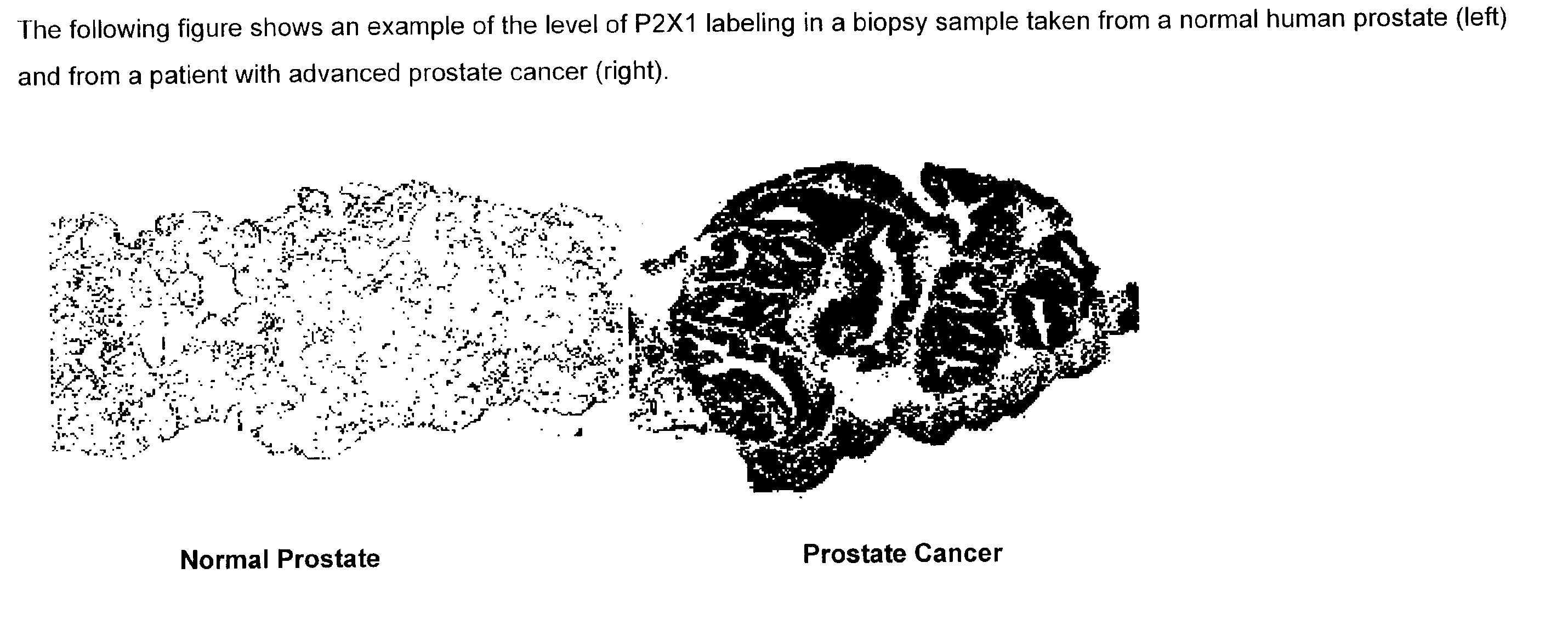
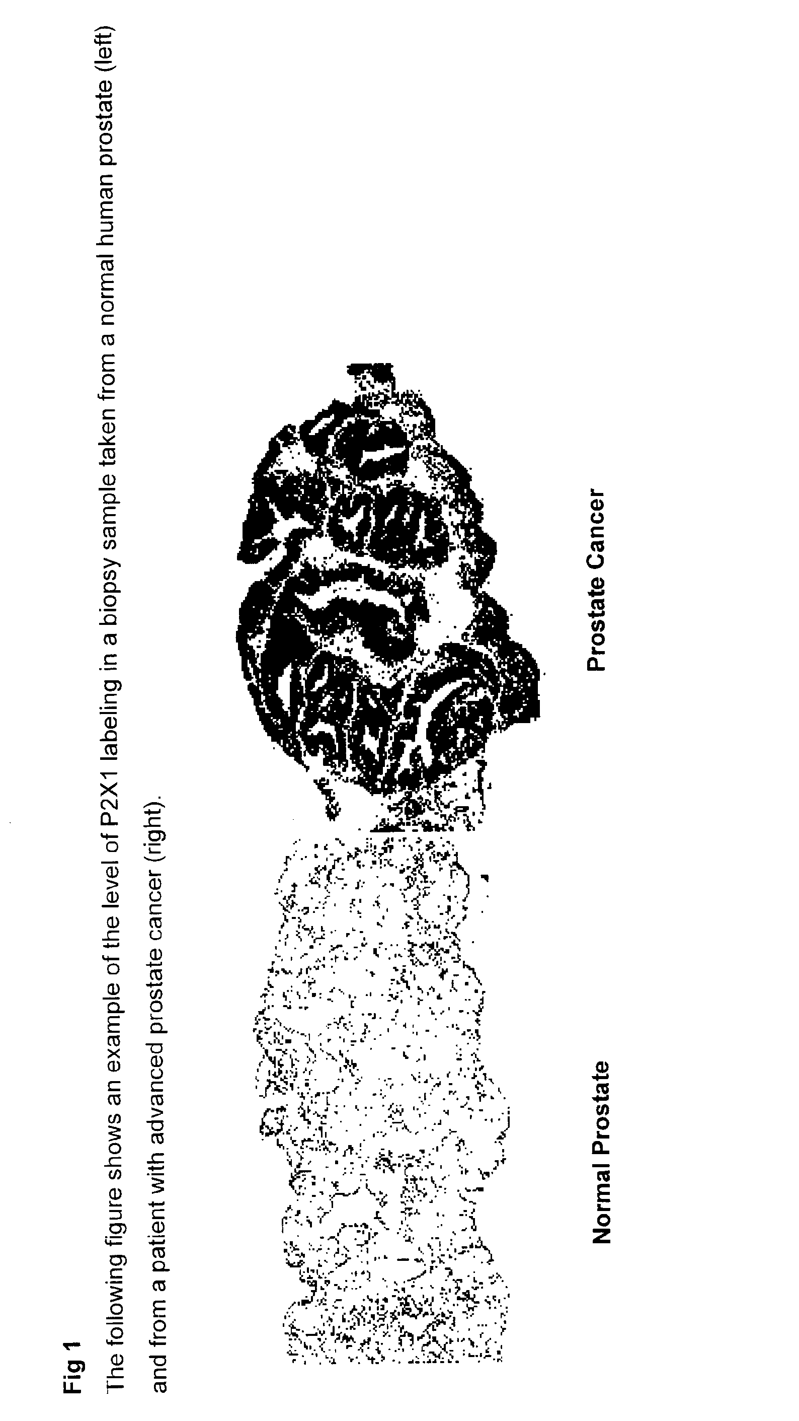
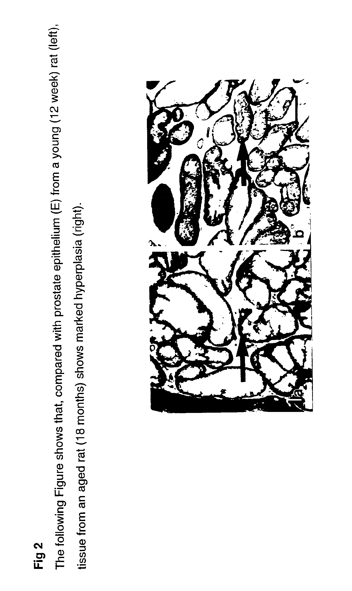
  • Method for identifying pre-neoplastic and/or neoplastic states in mammals
  • Method for identifying pre-neoplastic and/or neoplastic states in mammals
  • Method for identifying pre-neoplastic and/or neoplastic states in mammals

Examples


example 1

Immunohistochemical Procedure

[0042] The immunohistochemical method used in this study was adapted from Barclay [31]. Sections with a thickness of 8 μm were cut from unfixed, frozen tissue using a Reichert Jung 2800 Frigocut cryotome. Sections were air dried at room temperature for 1 hour, fixed for 12 hours in acetone at −20° C. and air dried at room temperature for 1 hour prior to antibody labelling. They were then incubated at room temperature with one of either rabbit or sheep anti-P2X1, P2X2, P2X3, P2X4, P2X5, P2X6 or P2X7 antibody. After washing, sections were then incubated in the secondary antibody; a 1:30 dilution of HRP-conjugated goat anti-rabbit secondary antibody (Dako) for 30 mins for rabbit primaries and HRP-conjugated goat anti-sheep secondary antibody (Dako) for sheep primaries. Slides were again rinsed and then immersed in 15% diaminobenzidine tetrahydrochloride (DAB—Sigma) for 10 minutes. Sections were rinsed, air dried and mounted in DPX (Merck). Control slides w...

example 2

Antibody Production

[0043] The consensus sequences of the rat P2X1 [32], P2X2 [33], P2X3 [34], rat P2X4 [35], rat P2X5 [36], rat P2X6 [36], rat P2X7 [37], human P2X7 [38], human P2X7 [39], human P2X3, [40], human P2X4 [41] and human P2X5 [42] cloned receptors were examined for suitable epitopes following the approach adopted in Hansen et al. [15]. The non-homologous epitopes corresponding to the segment Lys 199-Cys217 used in rat P2X1 were utilised in rat P2X3, rat P2X6 and rat P2X7. Variations were applied to rat P2X4 which used the sequence Ile235-Gly251 to which was attached a C-terminal Cys residue for cross-linking to a 6 kDa diphtheria toxin domain. The P2X2 epitope was selected from a region within the C1 domain [15], Cys130-Gly153. The rat P2X5 epitope was selected from a region closer to the second transmembrane domain but still extracellular (Lys314-Ile333 to which was added a C-terminal Cys also for conjugation). Although largely homologous with rat P2X4, cross-labelling ...

example 3

Specificity of Antibodies

[0045] Each of the P2X antisera used has been shown to possess similar distributions in many cases but with distinctly different distributions in other cases indicating that the antisera do not lack specificity. Specificity was demonstrated by affinity purification of the sera against the cognate peptides. To further verify antibody specificity, individual antibody such as the antibody to P2X1 was added to cells transfected with the corresponding P2X1 cDNA in the presence and absence of a 10 mM concentration of the P2X epitope. Immunolabelling and confocal imaging of the tranfected Xenopus oocytes demonstrated that the expressed P2X1 is located, as expected, within the cell membrane and the presence of a 10 mM concentration of the cognate peptide as an absorption control resulted in the blocking of P2X1 staining [18].

[0046] Individual specificity of all other antibodies has been similarly demonstrated.


PUM

PropertyMeasurementUnit
thicknessaaaaaaaaaa
temperatureaaaaaaaaaa
volumesaaaaaaaaaa

Abstract

The present invention relates to methods of identifying pre-neoplastic and / or neoplastic states in mammals and in particular to a method for identifying pre-neoplastic and neoplastic cells in tissues and body fluids, based on differential expression of purinergic receptors in these cells.

Description

TECHNICAL FIELD [0001] The present invention relates to methods of identifying pre-neoplastic and / or neoplastic states in mammals and in particular to a method for identifying pre-neoplastic and neoplastic cells in tissues and body fluids, based on differential expression of purinergic receptors in these cells. BACKGROUND [0002] When diagnosing cancer, cellular features in biopsy samples are taken into account such as, the degree of variability of cancer cell size and shape, the proportion of actively dividing cells and invasion into neighbouring structures. Commonly used histological stains are haematoxylin (primary stain) and eosin (counterstain) which differentially label subcellular elements. Other diagnostic methods employ antibodies to particular diagnostic molecules within (via intracellular epitopes) or on the surface of cells or tissues (via extracellular epitopes) which can be made visible for microscopic analysis eg, carcino-embryonic antigen (CEA). Some specific examples...

Claims


Application Information

Patent Timeline
Patent Type & Authority Applications(United States)
IPC IPC(8): C12Q1/68C07K16/00C12N5/06G01N33/53C12Q1/02C12N5/07C12N5/078C12N5/079C12N5/09G01N33/574G01N33/577
CPCG01N33/57492Y10S435/975
Inventor SLATER, MICHAELBARDEN, JULIAN
Owner BIOSCEPTRE INT
Learn More