Image processing method and computer readable medium for image processing

a technology of image processing which is applied in the field of image processing method and computer readable medium for image processing, can solve the problems of difficult to understand the depth and running direction, difficult to set an appropriate orthogonal cross section angle, and difficult to understand the three-dimensional structure inside the human body. to achieve the effect of easy to understand depth information

Active Publication Date: 2008-01-10
ZIOSOFT
View PDF5 Cites 42 Cited by
  • Summary
  • Abstract
  • Description
  • Claims
  • Application Information

AI Technical Summary

Benefits of technology

[0017] The present invention has been made in view of the abode circumstances, and provides an image processing method and a computer read ab...

Problems solved by technology

While, three-dimensional structure inside of a human body is hard to understand simply from the tomographic image of a human body.
However, since it is hard to understand the depth and the running direction of the tubular tissue, it is difficult to set an appropriate orthogonal cross section angle.
That is, to o...

Method used


Image

  • Image processing method and computer readable medium for image processing
  • Image processing method and computer readable medium for image processing
  • Image processing method and computer readable medium for image processing

Examples


Embodiment Construction

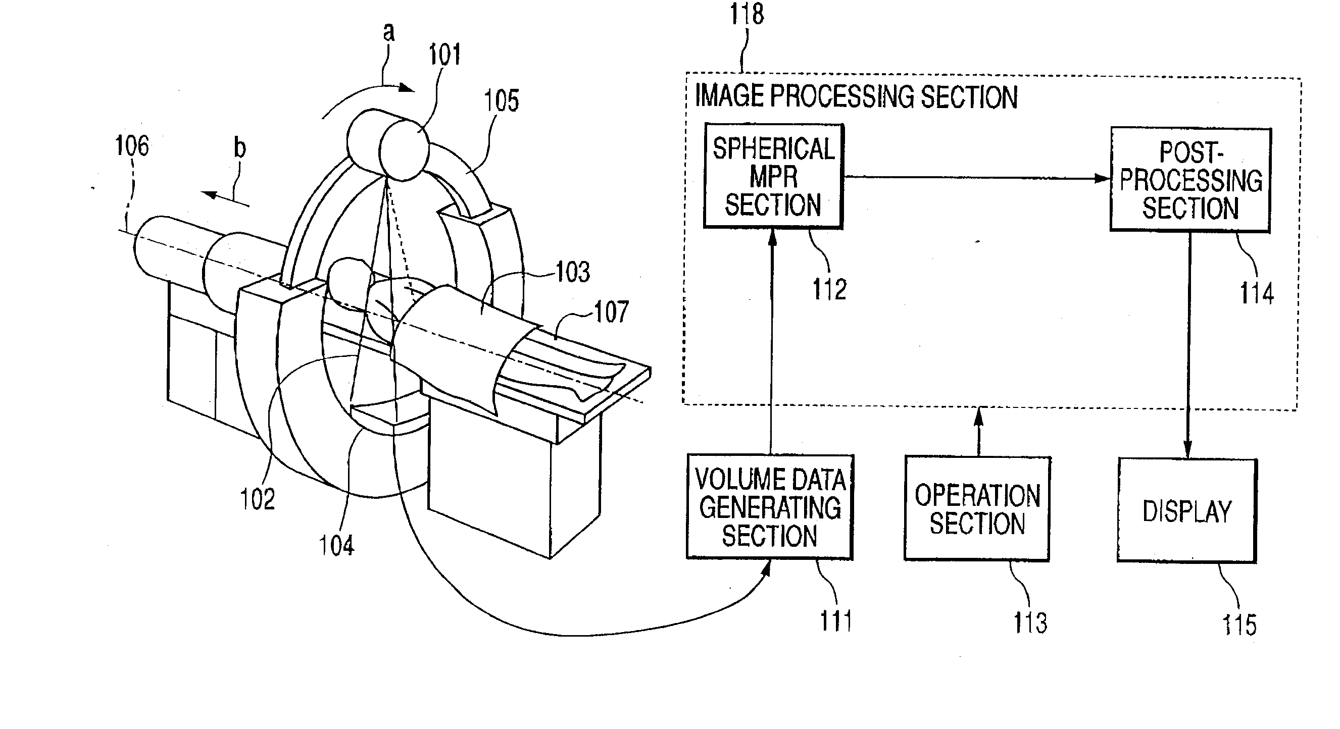
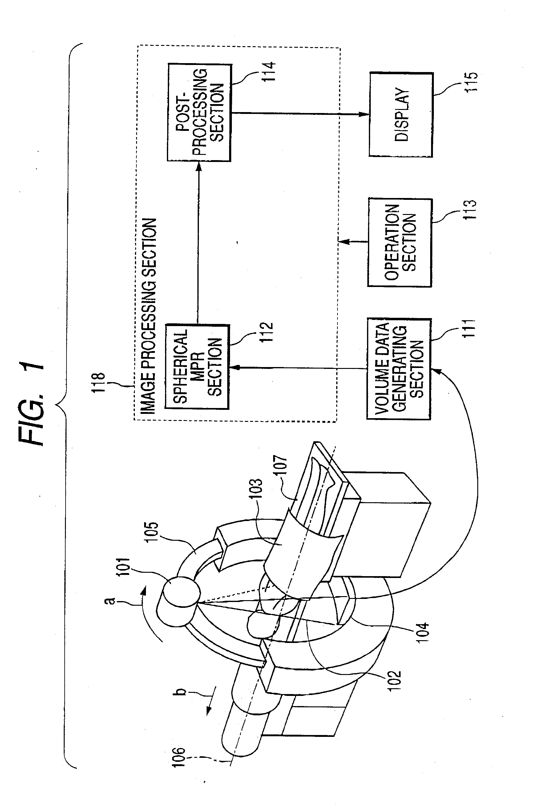
[0064]FIG. 1 schematically shows a computed tomography (CT) apparatus used in an image processing method according to an embodiment of the invention. The computed tomography apparatus is used for visualizing tissues, etc., of a subject. A pyramid-like X-ray beam 2 having edge beams which is represented by dotted lines in FIG. 1 is emitted from an X-ray source 1. The X-ray beam 2 is applied on an X-ray detector 4 after transmitting through the subject, for example, a patient 3. In this embodiment, the x-ray source 1 and the x-ray detector 4 are disposed in a ring-like gantry 5 so as to face each other. The ring-like gantry 5 is supported by a retainer not show in FIG. 1 so as to be rotatable (see the arrow “a”) about a system axis 6 which passes through the center point of the gantry.

[0065] The patient 3 is lying on a table 7 through which the X-rays are transmitted. The table 7 is supported by a retainer which is not shown in FIG. 1 so as to be movable (see the arrow “b”) along the...


PUM


Abstract

An intestine is cut by a cross section of a spherical surface or a combination of a plane and a spherical surface, and voxel values on the cross section are projected onto a projection plane in parallel, whereby a cross-sectional image of the intestine is created. Unlike a CPR image, the image of the spherical portion is projected without being extended. If the radius of the spherical surface is increased, the depth toward a screen can be seen and thus perspective of the inside of the observation object can be understood. On the other hand, if the radius of the spherical surface is lessened, the cut surface of the observation object is displayed as flat MPR and can be observed without distortion. Therefore, the radius of the spherical surface is set appropriately in conjunction with the thickness of the observation object, whereby a precise diagnosis can be conducted.

Description

[0001] This application claims foreign priority based on Japanese Patent application No. 2005-330604, filed Nov. 15, 2005, the content of which is incorporated herein by reference in its entirety. BACKGROUND OF THE INVENTION [0002] 1. Field of the Invention [0003] This invention relates to an image processing method and a computer readable medium for image processing for visualizing volume data. [0004] 2. Description of the Related Art [0005] A revolution is brought about in :the medical field with the advent of a CT (Computed Tomography). apparatus and an MRI (Magnetic Resonance Imaging) apparatus -making it possible to directly observe the internal structure of a human body as the image processing technology using a computer moves forward, and medical diagnosis using the tomographic image of a living body is widely conducted. While, three-dimensional structure inside of a human body is hard to understand simply from the tomographic image of a human body. Further, in recent years, ...

Claims


Application Information

Patent Timeline
IPC IPC(8): G06K9/00A61B6/03G06T15/08
CPCG06T19/00G06T2219/008G06T15/08
Inventor MATSUMOTO, KAZUHIKO
Owner ZIOSOFT
Why Eureka
Try Eureka