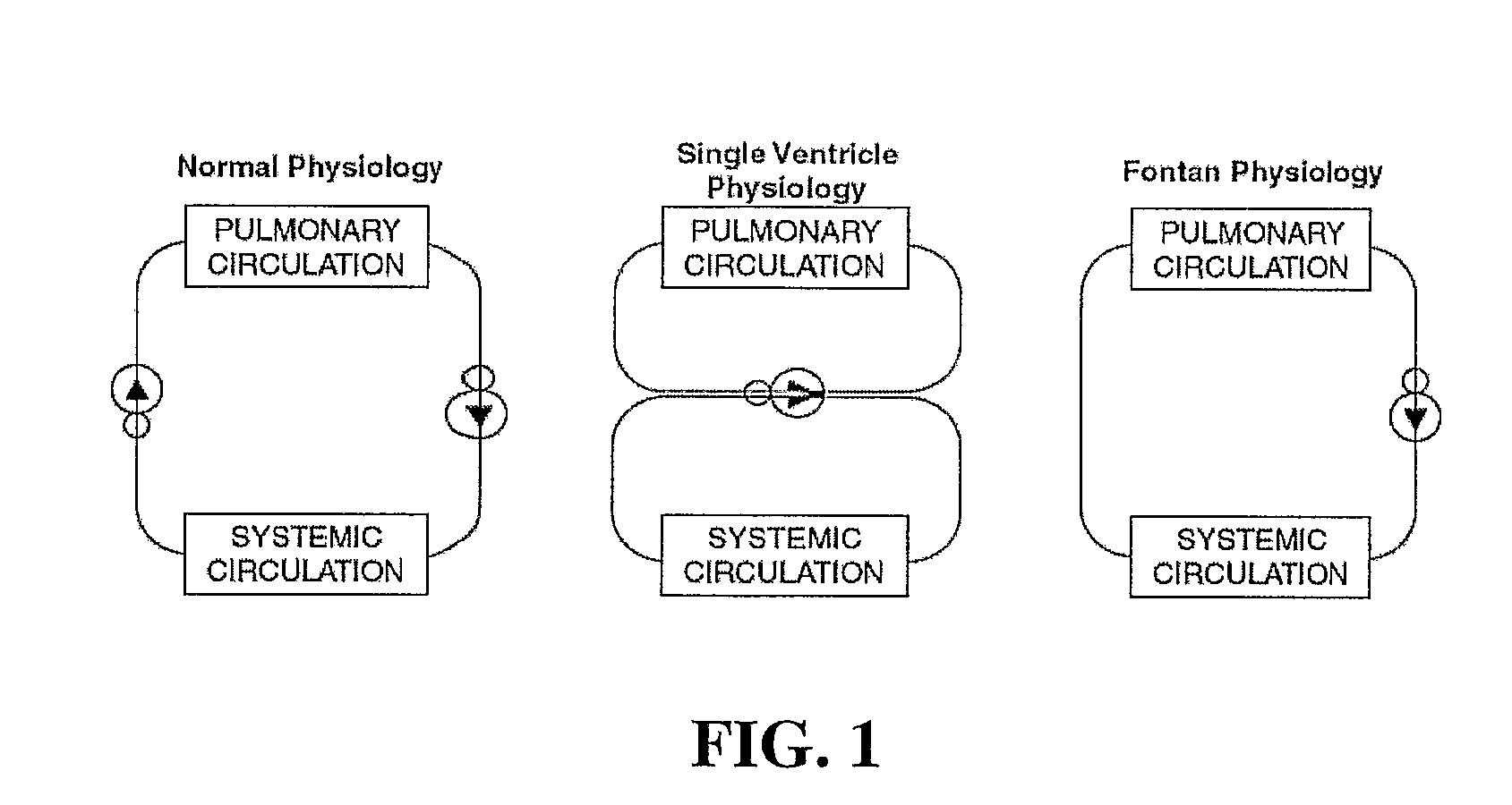
Surgical repairs that separate the pulmonary and systemic circuits, placing them in series with the univentricular pump, termed “Fontan Repairs,” are palliative, and unfortunately not curative.
Operations are routinely staged over many years and survivors often require a lifetime (rather limited) of intensive
medical attention.
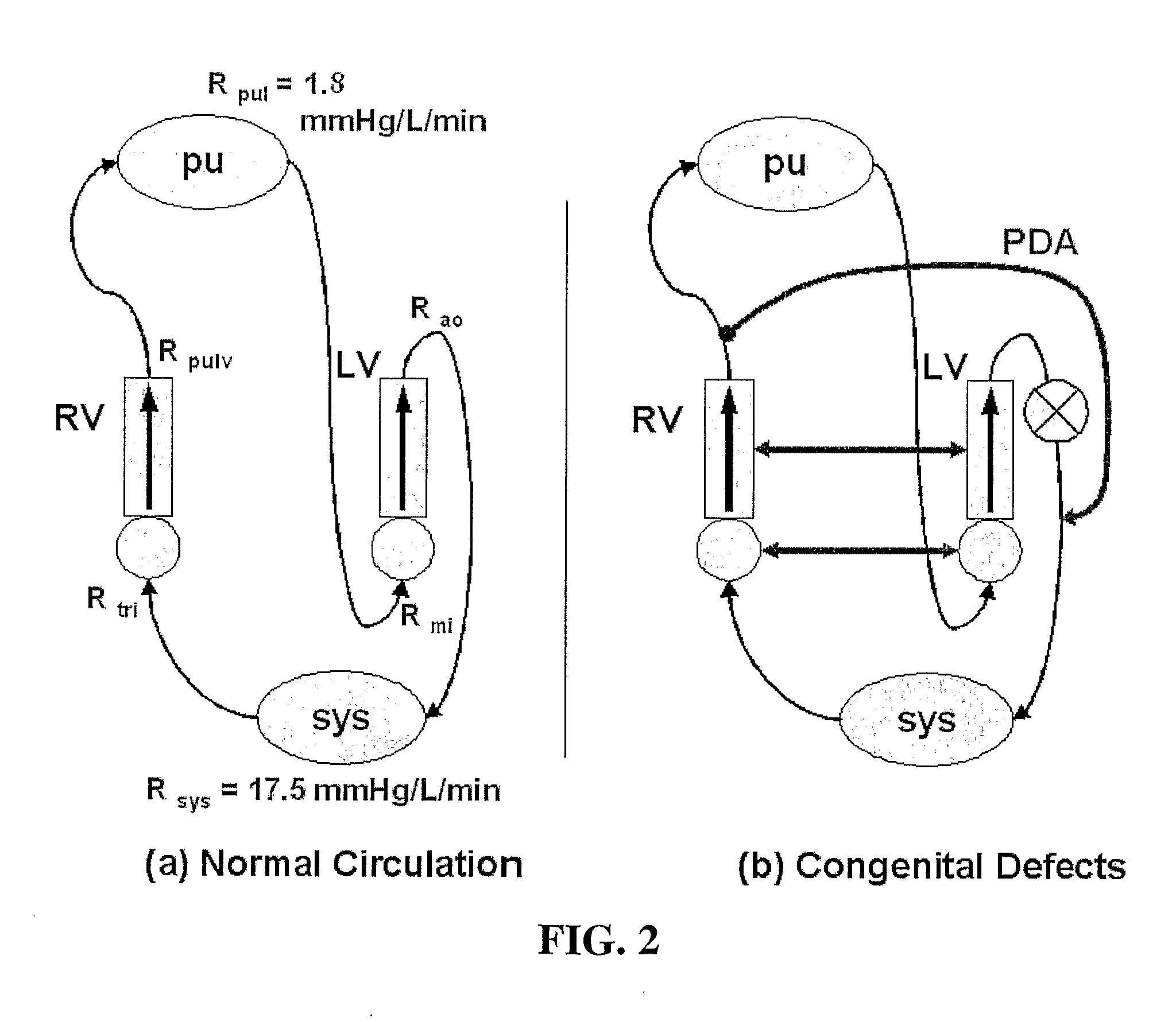
Among all CHDs, particularly challenging are the defects (or combination of defects) observed in about 20% of the CHD cases that effectively lead to a single ventricle (SV)
anatomy.
The
high pressure in the pulmonary circulation and the mixing of oxygenated and deoxygenated blood causes many of problems, which is why the Fontan procedure is performed.
However, ventricular dysfunction, pulmonary
vascular disease, and chronic cyanosis prohibited a normal existence and drastically shortened patient life expectancies with only few patients surviving beyond adolescence.
In such a state, the Fontan operation cannot be performed, or will have a high risk of failure, since the extra energy needed to maintain
lung blood flow is not available.
The elevated pressure in the veins has a few ill effects, including that there may be swelling of the entire body due to fluid from the blood leaking out of the
vein walls, there may be facial puffiness,
fluid accumulation in the
abdomen (
ascites) or chest (
pleural effusion), and sometimes even absorption of nutrients from the intestines is affected.
Indeed, the heart may eventually fail.
The age at which the heart fails and the patient requires a heart transplant depend on many different factors, but a main reason appears to be the excessive
workload placed on the single ventricle.
However, it soon became clear that placing a valve in the caval conduits, rather than being advantageous, resulted in obstruction of the low-pressure VC-to-PA circulation.
Furthermore, the separation of the IVC-to-LPA and SVC-to-RPA tracks, that had been designed to ensure an even
perfusion of the right and left lungs, did not allow for any
adaptation of the LPA / RPA
blood flow ratio, leading to serious complications when one of the pulmonary tracks became obstructed.
Although this procedure was quickly endorsed and has had widespread use in many centers, the long-term follow-up of patients with an AP-connection indicated that they were prone to late complications.
These complications were usually related to a markedly dilated
right atrium appendage, which was suspected to be due to the increased
pressure load imposed on the atrium.
This atrial dilatation was in turn associated with stagnant flows along the dilated right side of the atrium and turbulent flows elsewhere in the connection, resulting in significant fluid energy dissipation.
The
high incidence of right-atrium related complications led many to question the role of the pulsating
right atrium and its actual contribution to the Fontan circulation.
Usually, there are three different stages involved in the completed “Norwood procedure” leading to the
resultant Fontan—each stage with inherent morbidity and associated mortality.
Although most surgeons agree on the staged TCPC as being the current procedure of choice for Fontan repairs, controversies exist about the selection of the
connection type, the type of material to use, the need for fenestration, and the timing of the operations.
On the other hand they provide no growth potential and may lead to conduit
stenosis and thromboembolism.
It is simply that connecting the vena cavae directly to the lungs, bypassing the heart, is a difficult task for the single ventricle due to chronic work and volume overload.
This configuration results in higher pressures in the systemic
bed with severe clinical consequences. FIG. 3 illustrates single ventricle circulation after final stage Fontan
surgery.
Such a region exists as the inlets either present inlet flows at 180 degrees from one another, forcing the flows to collide head on, before separating toward the two outlets, or the inlets do not provide such a
direct path of collision, but even with offset of some amount, the inlet flows nonetheless collide with each other or with a wall sufficiently to present outlet flows with marked
energy loss.
In this mixing region of the connection, there is a lot of swirling of the flows, when the blood analogue from the inlets mix, which causes substantial
energy loss, causing the outlet flow to lose
momentum towards the lungs via the two outlets.
This is known
disadvantage with the current configurations used for Fontan procedures.
Furthermore, the
energy loss in the flow increases the more the vessel diameters in the connection are different.
While, experimentally, the model of FIG. 5 shows good results compared to the others, nutritious blood coming from the
hepatic veins goes primarily to one
lung, increasing the size and functionality of this
lung compared to the other lung, and this is not optimal.
It is evident that current anatomical connections provide non-optimal energy loss in the flow at regions of the connection where there is a high level of
venous flow mixing and disturbance.


