Microencapsulation of Cells in Hydrogels Using Electrostatic Potentials

a technology of electrostatic potential and microencapsulation, which is applied in the direction of skeletal/connective tissue cells, drug compositions, biocide, etc., can solve the problems of difficult to standardize the number of cells contacted with various test compounds, difficult to assess meaningful comparisons between compounds, and difficult to incorporate cell based assays into hts assays

Active Publication Date: 2008-02-07
GEORGIA TECH RES CORP
View PDF75 Cites 44 Cited by
  • Summary
  • Abstract
  • Description
  • Claims
  • Application Information

AI Technical Summary

Benefits of technology

[0012] Still another aspect provides methods of treatment using the disclosed encapsulated cells. In particular aspects, the encapsulated cells are injected directly into pathology sites to repair damaged tissue or to secrete cytokines, growth factors, proteins, or combinations thereof. Because the average diameter of the disclosed encapsulate cells is less than about 200 μm, the encapsulated cells will be minimally damaged by shear forces produced during injection. Microcapsules having a diameter greater than 250 μm tend to block needles used to deliver the microcapsules to a host. Accordingly, the disclosed microcapsules having a diameter of less than about 250 μm, typically less than about 200 μm can be delivered to a host via injection with a standard surgical needle in an amount sufficient to treat the host.

Problems solved by technology

Unfortunately, cell based assays are not easily incorporated into HTS assays because it is difficult to standardize the number of cells contacted with various test compounds.
Without standardizing the number of cells per reaction, meaningful comparisons between compounds are difficult to assess.

Method used


Image

  • Microencapsulation of Cells in Hydrogels Using Electrostatic Potentials
  • Microencapsulation of Cells in Hydrogels Using Electrostatic Potentials
  • Microencapsulation of Cells in Hydrogels Using Electrostatic Potentials

Examples


example 1

Encapsulation of Cells

[0090] Ultrapure alginate compositions containing polysaccharides with ≧60% guluronate or mannuronate resides with average molecular weights > or 6 cells / ml. The solution was then extruded through a 0.18 mm (inner diameter) needle at 10 ml / hr. A 6,000 kV electrostatic potential between the CaCl2 polymerization solution and the needle was used to disrupt the surface tension. Bead size and total cell number per bead were determined by morphometric analysis.

[0091] Cell number and bead size were controlled by varying the distance the cell suspension dropped before hitting the polymerization solution. Additional parameters were modulated according to the following generalized equations to obtain encapsulated cells having a diameter of less than 200 μm and containing a predetermined number of cells. Pi-Po=2⁢ ⁢γRPo=FsAA=4⁢ ⁢π⁢ ⁢R2Fs4⁢ ⁢π⁢ ⁢R2-Po=2⁢ ⁢γRFs=4⁢ ⁢π⁢ ⁢R2⁡(2⁢ ⁢γR+Po)

[0092] Pi=pressure inside droplet

[0093] P0=pressure outside droplet (atmospheric pressure...

example 2

Effect of Alginate Composition on Microencapsulation

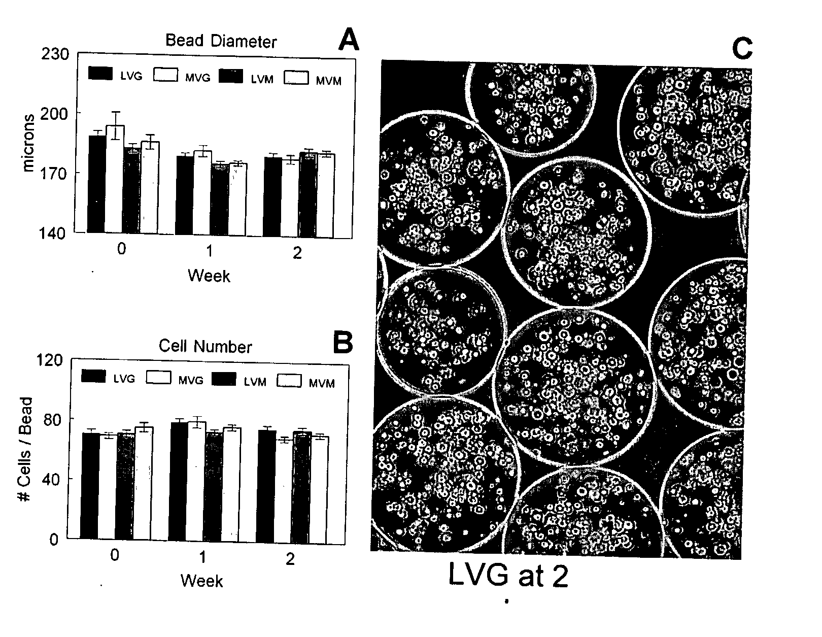
[0103] Alginate is co-polysaccharide composed of guluronate and mannuronate residues. The residue ratio and length of the polymer chains affect the mechanical properties of the alginate hydrogel. Four different alginate formulations (Table 2) were compared based on bead morphometrics and cell viability during two weeks of in vitro culture.

TABLE 1Optimization of Microencapsulation ProcessAlginateBeadCellGelation SolutionConcentrationMorphologyViability100 mM CaCl22.0%Spherical50 mM CaCl2 + 75 mM2.0%Irregular˜50%NaCl20 mM CaCl2 + 120 mM2.0%Irregular˜70%NaCl50 mM CaCl2 + 150 mM2.0%Spherical˜90%Glucose

[0104]

TABLE 2Alginate CompositionsGuluronate ContentMolecular Weight>60%LVGLVM>200,000 g / moleMVGMVM

[0105] The average bead diameter was 176±2 μm to 194±7 μm depending on the alginate composition used (FIG. 1A), but there was not statistically significant difference. In addition, no significant change in bead diameter was measured durin...

example 3

Viability of Encapsulated Cells

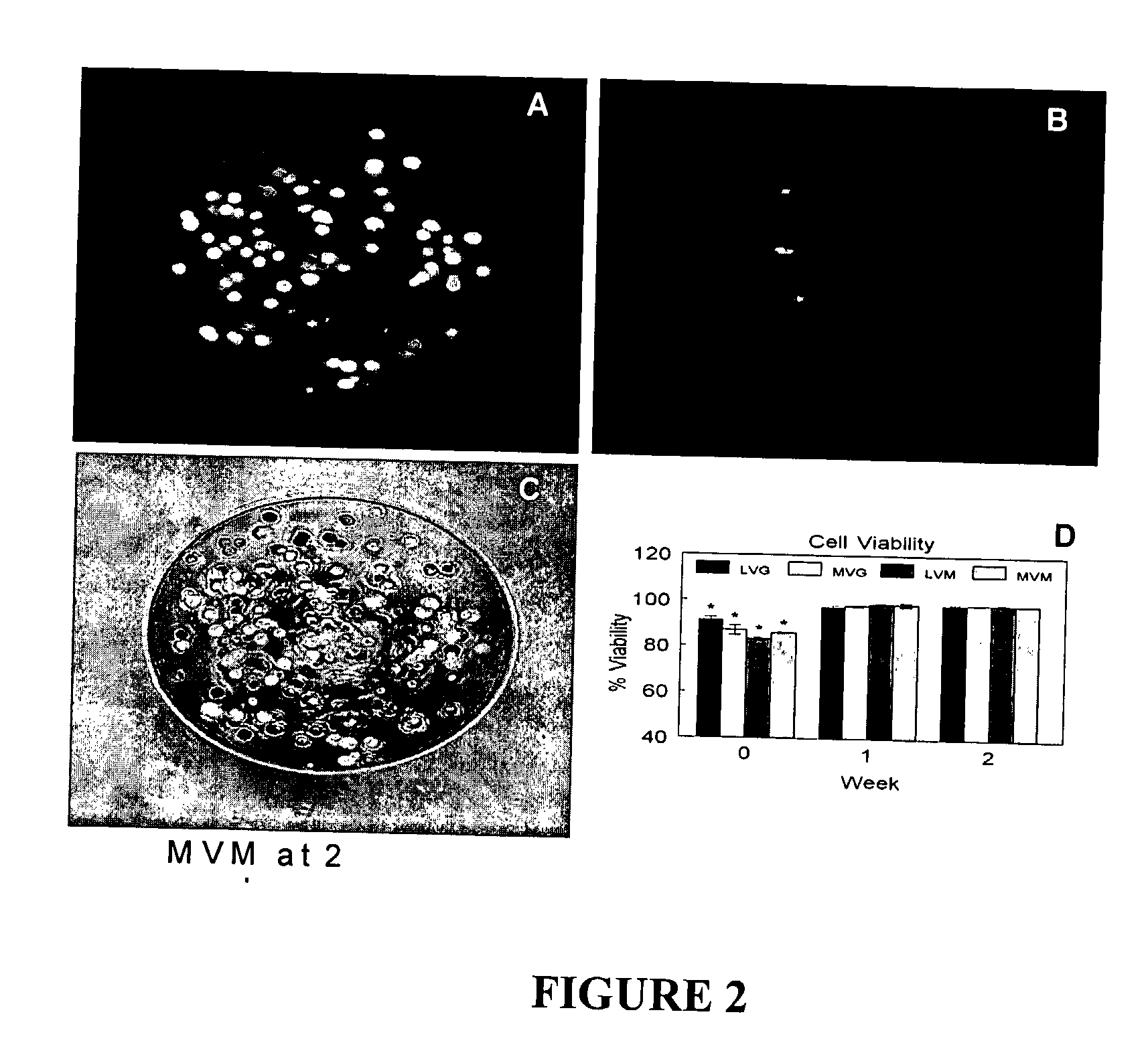
[0106] The viability of the encapsulated chondrocytes was measured by fluorescent confocal microscopy and a live / dead stain consisting of calcein and ethidium homodimer-1. The calcein stains the cytoplasm of the live cells green (FIG. 2A) and the ethidium stains the nucleus of dead cells red (FIG. 2B). The live cells were evenly distributed throughout the beads (FIG. 2C) with an initial viability if 83% to 91% (FIG. 2D). The viability increased after one week in culture to greater than 98% for all alginate compositions and remained constant up to two weeks. No statistically significant differences were observed between the different alginate compositions on cell viability during the in vitro culture.


PUM

PropertyMeasurementUnit
diameteraaaaaaaaaa
diameteraaaaaaaaaa
bead diameteraaaaaaaaaa

Abstract

Compositions and methods for producing encapsulated cells having an average diameter of less than about 200 μm are provided. Methods for using the disclosed encapsulated cells are also provided

Description

CROSS REFERENCE TO RELATED APPLICATION [0001] The application is a 371 application of PCT / US2005 / 036202 filed Oct. 7, 2005, which claims priority to and benefit of U.S. Provisional Patent Application No. 60 / 617,560 filed on Oct. 08, 2004, and where permissible is incorporated by reference in its entirety.STATEMENT REGARDING FEDERALLY SPONSORED RESEARCH OR DEVELOPMENT [0002] Aspects of the work described herein were supported, in part, by Grant No. EEC-9731643 awarded by the National Science Foundation. Therefore, the US government has certain rights in the disclosed subject matter. BACKGROUND [0003] 1. Technical Field [0004] Aspects of the disclosed subject matter are broadly directed to methods and compositions for producing encapsulated cells and methods of using encapsulated cells, for example, in cellular arrays, screening protocols, and methods of treatment. [0005] 2. Related Art [0006] High Throughput Screening (HTS) has been in use for at least the past ten years to screen la...

Claims


Application Information

Patent Timeline
Patent Type & Authority Applications(United States)
IPC IPC(8): A61K45/00A61K9/50A61P43/00C12N11/04C12Q1/02A61K35/12C12N5/077
CPCA61K2035/126C12N2533/74C12N5/0655C12N5/0012A61P17/00A61P19/00A61P21/00A61P9/00
Inventor BOYAN, BARBARA DALEKINNEY, RAMSEY CHRISTIANSCHWARTZ, ZVI
Owner GEORGIA TECH RES CORP
Why Eureka
Try Eureka