Method and apparatus for representing myocardial tissues in different states of damage

Inactive Publication Date: 2008-02-14
SIEMENS HEATHCARE GMBH
View PDF3 Cites 63 Cited by
  • Summary
  • Abstract
  • Description
  • Claims
  • Application Information

AI Technical Summary

Benefits of technology

[0017] The object of the invention is therefore to present an approach with wh

Problems solved by technology

Coronary heart disease results in an inadequate supply of blood to the cardiac muscle and possibly to its necrotization because of narrowing of the coronary arteries.
While revascularization of vital myocardial areas may improve the pumping function of the heart and the patient's prognosis, revascularization of scar tissue (necrotized) does not produce any improvement in results and constitutes ad

Method used


Image

  • Method and apparatus for representing myocardial tissues in different states of damage
  • Method and apparatus for representing myocardial tissues in different states of damage
  • Method and apparatus for representing myocardial tissues in different states of damage

Examples


Embodiment Construction

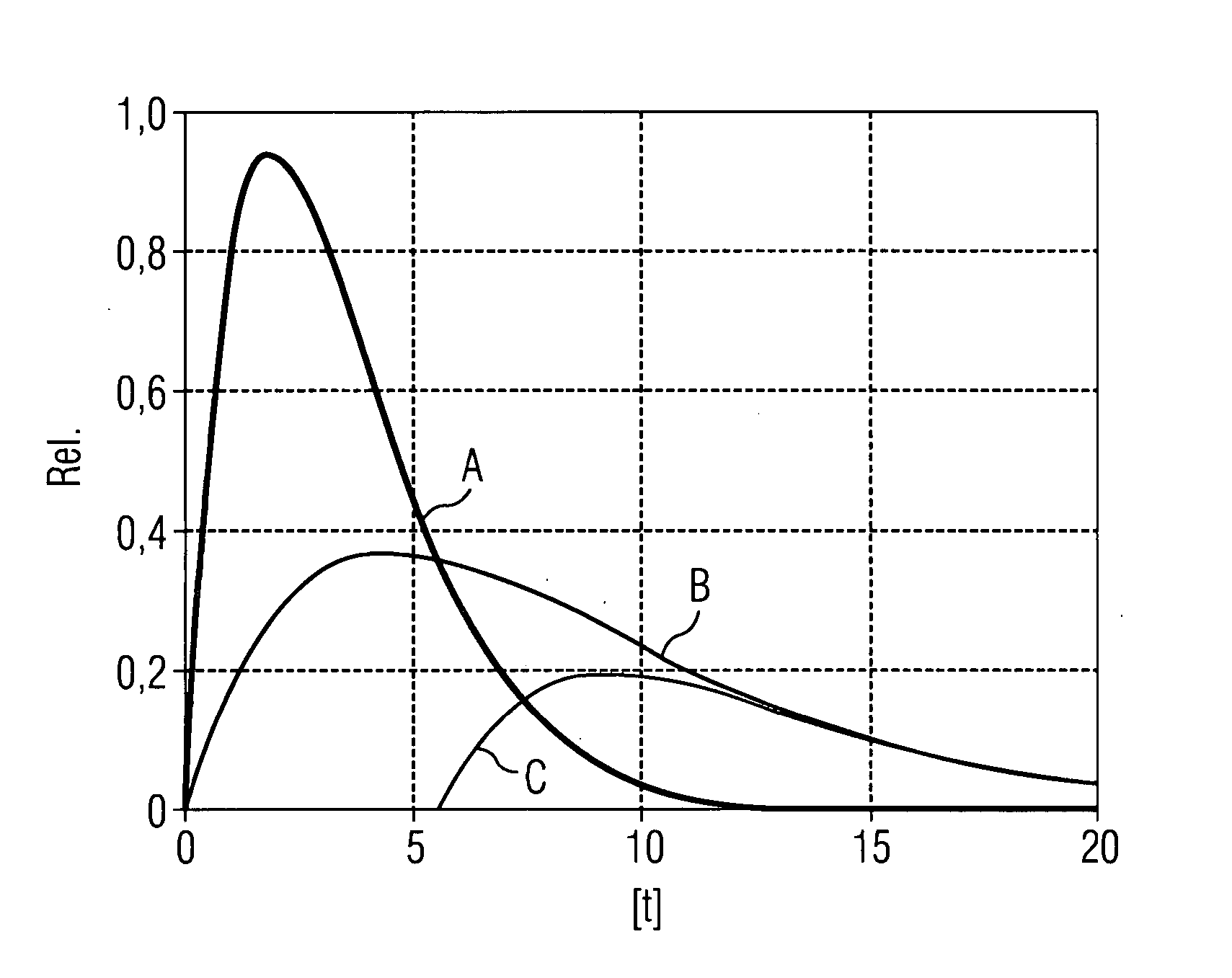
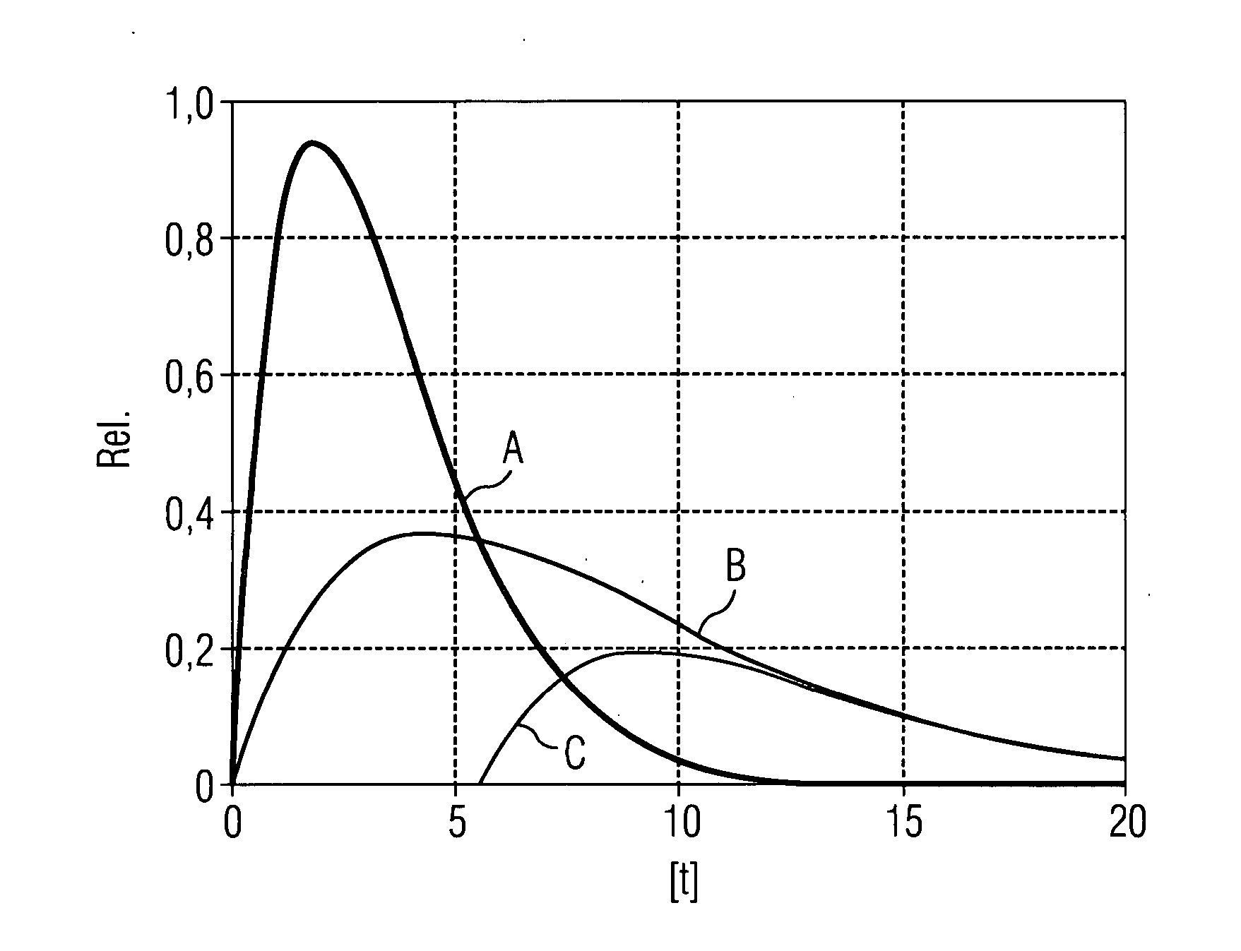
[0068] FIGURE shows a typical contrast agent characteristic for vital and necrotic myocardium and the difference between the two contrast concentrations. The curve A shows the contrast agent characteristic for normally perfused myocardium and less perfused myocardium, while the curve B shows the time-delayed and weaker response of the contrast agent for necrotic myocardium and finally the curve C shows the difference between the two curves A and B, the peak of the curve C representing the maximum value of the difference in contrast agent content to be determined according to the invention, and indicates the point in time of this maximum value at which a late-phase CT scan must be carried out for optimum image results.

[0069] The invention permits the visualization of necrotic myocardial areas using CT scans which are initiated after a particular patient-specific waiting time, determined by the invention, after the administration of contrast agent.

[0070] The present invention thus e...


PUM


Abstract

The invention relates to a method for differentially representing myocardial tissue in different states of damage, comprising the following steps: administering a myocardium-suitable contrast agent to a patient under examination; entering at least one patient-specific parameter affecting the speed of uptake by and elimination from the myocardium of said contrast agent; calculating a point in time after administration of the contrast agent at which a difference between a contrast agent content in necrotic myocardial tissue and a contrast agent content in non-necrotic myocardial tissue attains a maximum value, on the basis of the at least one patient-specific parameter, and carrying out, at the point in time calculated, a late-phase CT scan for accentuation of necrotic myocardial tissue compared to non-necrotic myocardial tissue. The invention likewise relates to apparatus, in particular for carrying out the method. A clean copy of the abstract that incorporates the above amendments is provided herewith on a separate page.

Description

CROSS REFERENCE TO RELATED APPLICATIONS [0001] This application claims priority of German application No. 10 2006 037 284.0 filed Aug. 9, 2006, which is incorporated by reference herein in its entirety. FIELD OF THE INVENTION [0002] The invention relates to a method and apparatus for representing myocardial tissues in different states of damage. BACKGROUND OF THE INVENTION [0003] In the industrialized countries, coronary heart disease (CHD) is the most frequent cause of heart failure. Coronary heart disease results in an inadequate supply of blood to the cardiac muscle and possibly to its necrotization because of narrowing of the coronary arteries. Despite much success with drug therapy, direct treatment of the triggering cause constitutes an important therapeutic concept. For CHD patients, the restoration of adequate perfusion by means of revascularization measures such as thrombolysis, stenting, balloon dilation, a bypass operation, etc. is therefore paramount. However, the succes...

Claims


Application Information

Patent Timeline
IPC IPC(8): A61K49/00A61B5/05
CPCA61B5/0275A61B6/032A61B6/507A61B6/503A61B6/504A61B6/481
Inventor JOHN, MATTHIASLAUTENSCHLAGER, STEFANRAHN, NORBERT
Owner SIEMENS HEATHCARE GMBH
Why Eureka
Try Eureka