Portable endoscope for intubation

a portable, endoscope technology, applied in the field of intubation, can solve the problems of large power requirements, unfavorable use of endoscopes in the battlefield, and the typical medical endoscope is an expensive and bulky instrument, so as to improve compatibility, wide view angle, and limit the resolution of the monitor screen

Inactive Publication Date: 2008-04-17
INTRA LLC
View PDF22 Cites 106 Cited by
  • Summary
  • Abstract
  • Description
  • Claims
  • Application Information

AI Technical Summary

Benefits of technology

[0021]A useful improvement in an intubation procedure is for an endoscope with a wide angle of view. The wide angle is useful for viewing the tip of the intubation tube and the body tissue in front of and around the tube, both located in the immediate proximity of the lens. If the viewing angle is not wide enough it is difficult to make a quick judgment on the exact position of the tip of the endotracheal tube. If the viewing angle is wider than necessary, then a large portion of the screen contains an image of the inner wall of the tube, which has no useful information and waists valuable and limited resolution of the monitor screen. The desired view angle for the intubation endoscope is no less than 80 angular degrees, more typically is no less than 90 degrees and no more than 120 angular degrees, and more typically is no more than 110 angular degrees, while the desired angle typically is 100 degrees. There are certain obstacles that cause difficulties to providing such wide-angle feature. The wide-angle objective lens and the wide-angle illuminating LED must coexist in the very tight quarters of the endoscope head. To improve the compatibility with the most commonly used endotracheal intubation tubes, the outside diameter of the endoscope probe typically does not exceed 6 mm. The end of the probe has a transparent window, sealed inside the probe enclosure. The objective lens and the illuminating LED are placed close to the window to take advantage of their wide-angle geometry and obtain a wide view angle.
[0022]However, the window in the immediate vicinity of the lens and LED creates a direct optical crosstalk between them, which reduces the contrast and can blind the sensor with the specular reflections of the LED light from the window. A wide viewing angle without the artifacts of the optical crosstalk is provided by placing a barrier, such as a thin, opaque light separator, between the illuminating LED and the objective lens. Extending the separator all the way to the inner surface of the window eliminates the undesired crosstalk from the LED. Such separator can be made of black vinyl film, black anodized aluminum foil, or other opaque material.
[0023]Due to a very small space available in the head of the endoscope probe, it is desirable that a single miniature LED provides an amount of light sufficient for performance of the imaging sensor. High sensitivity is one important criteria for selection of the image sensor. The other desired requirements are: compact size, low cost, and placing the light sensing area of the sensor significantly offset from the center of the probe head. One type of the sensor that meets these requirements is a CCD image sensor with frame transfer architecture. A CMOS sensor can also be used, though, in spite of the progress made in the CMOS sensor technology, the best CCD sensors are more sensitive. The frame transfer architecture, where the image sensing area is separate from the image storage area provides improved performance in terms of sensitivity and signal-to-noise ratio due to having largest possible fill factor of the light sensitive area to

Problems solved by technology

This procedure is not a trivial one especially in the inconvenience of emergency situations and with the patients having various irregularities and complications in the throat and neck areas.
However, a typical medical endoscope is an expensive and bulky instrument, utilizing powerful light sources and fiber optic bundles for delivery of light and/or images.
The endoscopes currently known are rather bulky and expensive.
High price, a suitcase-type dimensions at best, and large power requirements preclude use of the endoscopes in the battlefield and the every day typical ambulance

Method used


Image

  • Portable endoscope for intubation
  • Portable endoscope for intubation
  • Portable endoscope for intubation

Examples


Embodiment Construction

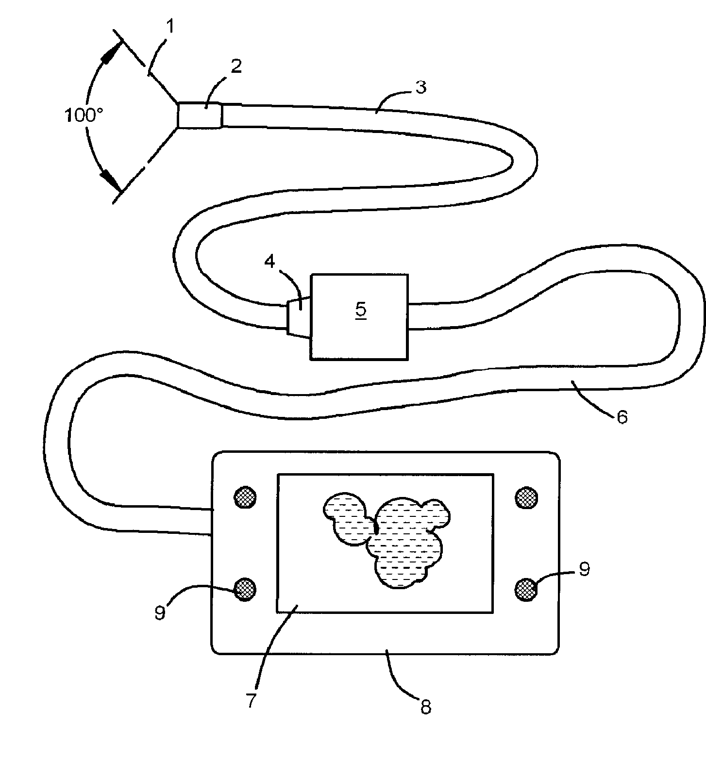
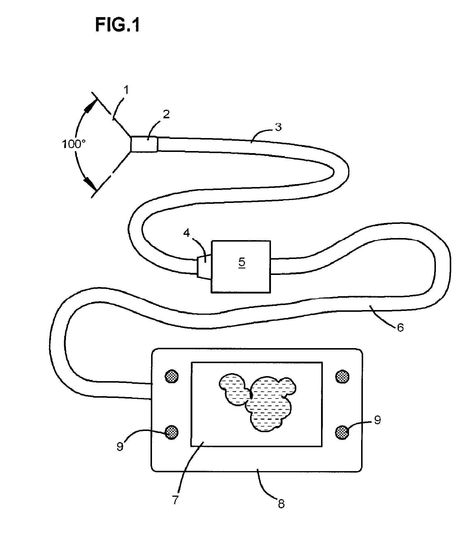
[0031]The FIG. 1 shows the main components of an embodiment of the invention. The flexible probe consists of the imaging head 2 with the viewing angle 1 of 100 degrees, the flexible circuit enclosed into the plastic sleeve 3 and the miniature 14-pin connector 4. The connector 4 is plugged into and electronically communicates with the small in-line control box or enclosure 5 that houses the image sensor controller (not shown). The in-line probe controller box 5 is connected via the second flexible multi-line cable 6 comprising control wiring 42 to the control and display unit 8. The control and display unit 8 has a 3.5″ color TFT LCD video display panel 7, knobs 9 for controlling the power and brightness, battery cell compartment accessible from the back and an electronic hardware positioned inside the enclosure.

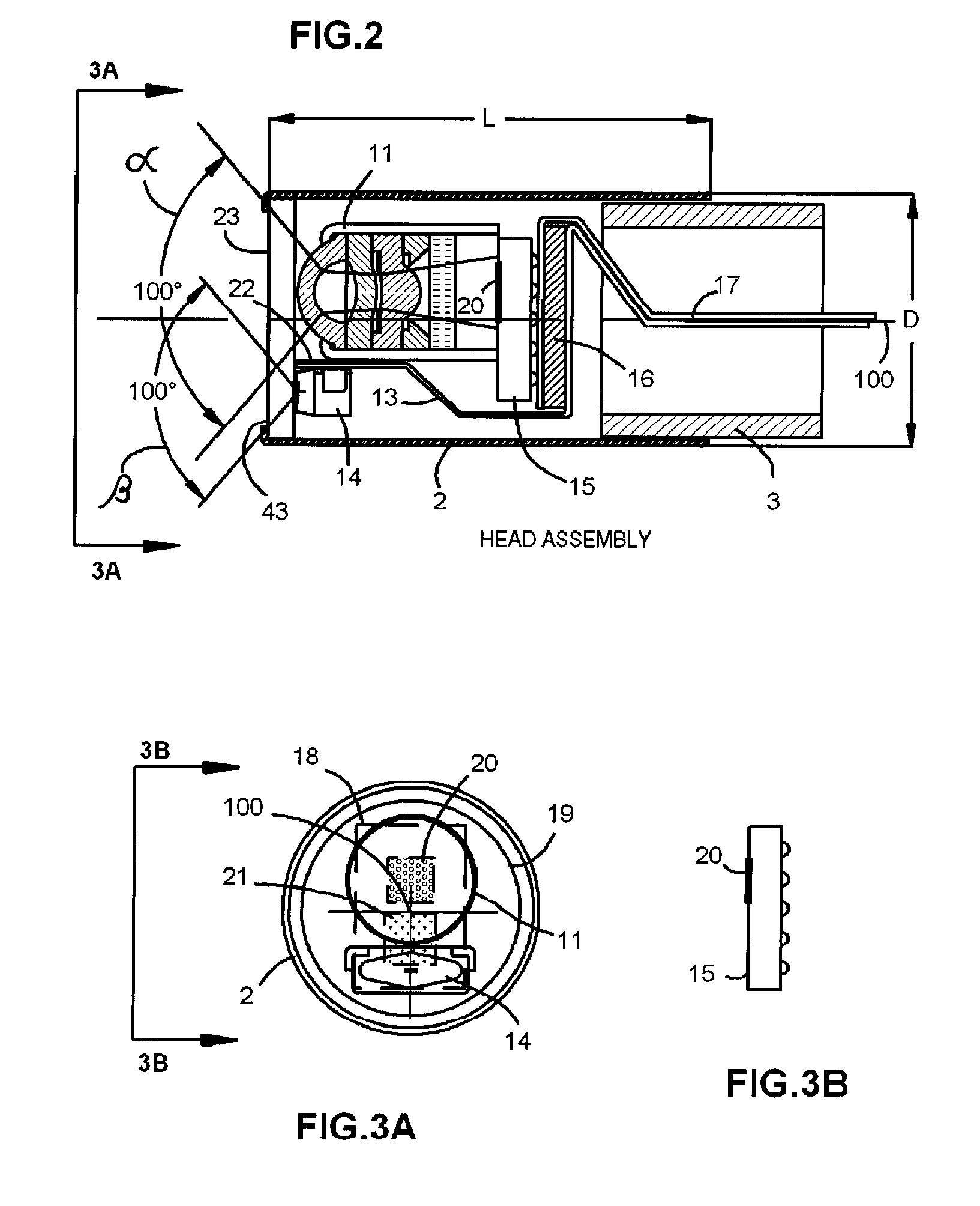
[0032]The longitudinal cross section of the head of the probe, presented on the FIG. 2, shows how the proposed invention resolves the conflicting requirements of the all imag...


PUM


Abstract

A portable, low power, low cost, endoscope having a removable probe with a miniature digital CCD imager, single white LED illuminator and an imaging lens on the distal end and a miniature multi pin connector on the proximal end; both ends connected with a multi trace flexible circuit hidden inside of the protecting flexible sleeve; and a full color LCD monitor and associated image signal processing electronics and powered by the small battery cells.

Description

CROSS-REFERENCE TO RELATED APPLICATIONS[0001]This application claims the benefit of U.S. Provisional Application 60 / 829,763, filed Oct. 17, 2007, the disclosure of which is incorporated herein by reference.FIELD OF THE INVENTION[0002]This invention is concerned with the devices used for assisting a medical practitioner during endotracheal intubation procedure.BACKGROUND OF THE INVENTION[0003]The endotracheal intubation is a commonly used procedure during which a semi rigid plastic tube, called endotracheal tube, is inserted into a patient's trachea through the open mouth. Frequently a laryngoscope is used to view the throat area while the tip of the endotracheal tube advances toward the vocal cords. This procedure is not a trivial one especially in the inconvenience of emergency situations and with the patients having various irregularities and complications in the throat and neck areas. During the last five to ten years many researches and physicians advocated use of endoscopes dur...

Claims


Application Information

Patent Timeline
IPC IPC(8): A61B1/04A61B1/06
CPCA61B1/053A61B1/0676A61B1/0684H04N2005/2255A61B1/00105A61B1/00108H04N5/2253A61B1/267H04N23/54H04N23/555
Inventor LASER, VADIM
Owner INTRA LLC
Why Eureka
Try Eureka