Tissue Marking Devices and Systems

a technology of tissue marking and marking devices, which is applied in the field of implantable and readily detectable medical devices and systems, can solve the problems of compromising future successful localization, requiring image guided localization procedures, and diagnostic imaging has increased the frequency of small lesions throughout the body detection frequency, etc., to achieve the effect of maximizing detectability

Inactive Publication Date: 2008-04-24
MULLEN DAVID
View PDF10 Cites 123 Cited by
  • Summary
  • Abstract
  • Description
  • Claims
  • Application Information

AI Technical Summary

Benefits of technology

[0021] Another aspect of the invention relates to a device that, by nature of...

Problems solved by technology

In addition, the implantation of a small metal “clip” marker following large core biopsy under image guidance may be used when the visualized target has been substantially removed during the diagnostic procedure (thus compromising future successful localization).
The explosive growth of diagnostic imaging has increased the frequency of detection of small lesions throughout the body that cannot be seen or felt by a surgeon charged with the task of removing such a target.
These devices all have the significant limitation of requiring the image guided localization procedure immediately before the surgery.
The need for immediate preoperative localization creates logistical bottlenecks for radiology departments and operating room throughput, as well as additional procedures for patients who have already had a device implanted.
Thus, a device that could be implanted by a radiologist at one time and then independently removed by a surgeon at another time on the day of the needed surgical procedure with no further patient preparation is desirable.
Although some have attempted solutions to these problems, a simple and cost effective approach has not yet been found.
These proposed devices all have one significant drawback in common: they are large and may be expected to be uncomfortable for patients.
This problem may be further compounded when these may need to remain in position for some length of time.
Although these techniques may work in certain circumstances for preoperative wire localization, they have significant limitations for use during operations.
Ultrasound equipment is expensive and requires substantial training to use effectively.
Although some have proposed that surgeons use ultrasound themselves in the operating room, the impediments described above have made this practice uncommon.
Nevertheless, nuclear techniques are cumbersome not only because of the patient preparati...

Method used


Image

  • Tissue Marking Devices and Systems
  • Tissue Marking Devices and Systems
  • Tissue Marking Devices and Systems

Examples


Embodiment Construction

[0040] The subject invention is comprised of a series of implantable sterile and biologically inert metal devices designed to mark the location of the site of a lesion in tissue. These markers are designed to optimize their properties for subsequent metal detection. Such optimization of detection characteristics will be accomplished by creating a device, which, unlike all other existing localization devices, has been specifically designed to provide the maximum metal detection signal and the clearest directional information possible, given the necessities of small size, tissue stability, patient comfort and biocompatibility.

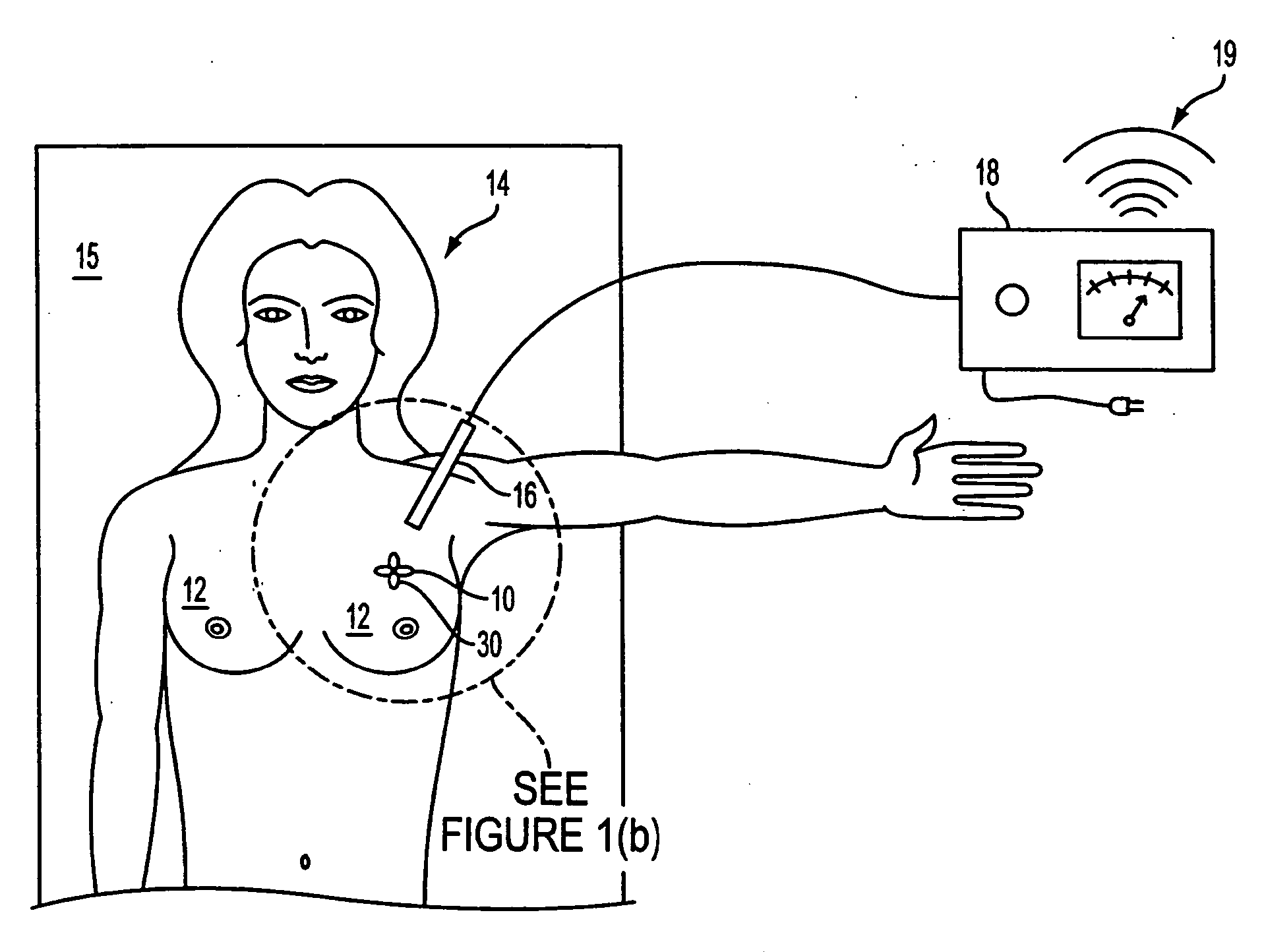
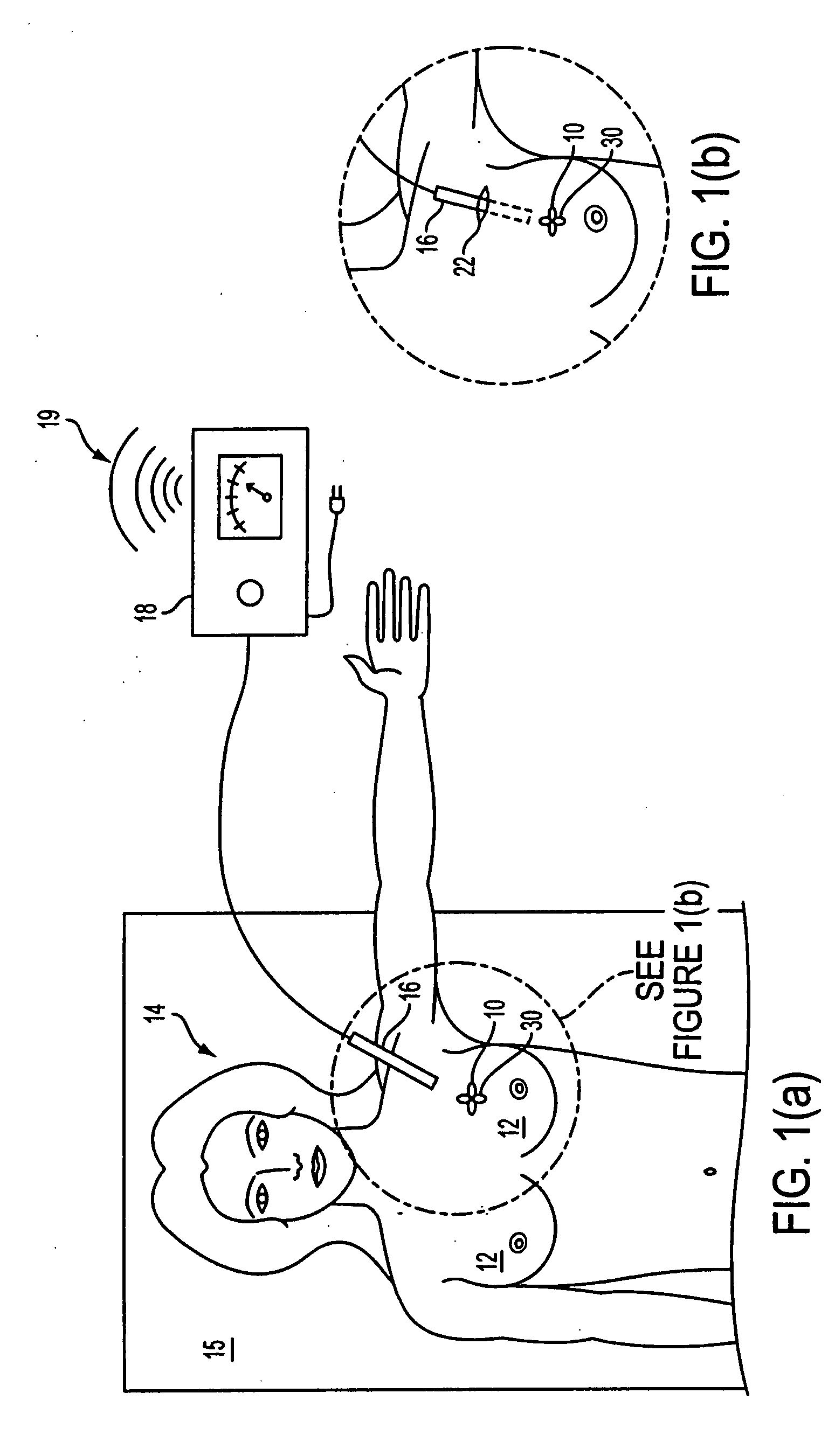
[0041] Turning now to the drawings, FIG. 1(a) shows a marker 10 according to the present invention implanted in a patient's breast 12 with the patient 14 positioned for surgery on a table 15. A metal detection probe 16 is illustrated in communication with a metal detector display 18 with audible output 19. A suitable metal detector and probe is illustrated in Tr...


PUM


Abstract

A marker for marking a site within the body of a mammalian patient is positioned within the tissue of a patient. The marker may be placed in the first instance by a needle or the like or placed where a tissue sample has been removed. The marker has a plurality of loops each at various angles to the other such that when positioned within the patient, one of the loops is positioned orthogonal to a magnetic field of a metal detector. Various shapes of markers may be used, including electron orbital shapes, chains of loops or barbells. Barbs or other anchoring elements may be used to stabilize the marker's position. Normal delivery techniques as needles, catheters or cannulas may readily position the marker within the patent. By having the marker so designed and positioned, at least one of the closed loops is detectable by a metal detection beam of a metal detection device.

Description

FIELD OF THE INVENTION [0001] The present invention relates to implantable and readily detectable medical devices and systems useful for marking a tissue lesion in a subject for later surgical removal. BACKGROUND [0002] The need for accurate preoperative image guided localization of nonpalpable breast lesions has been well described, and the frequency of use for this technique is increasing. Not only mammographically detected lesions require localization, but also lesions that may be found by any other imaging technique such as ultrasound, MRI, nuclear medicine or other technologies not yet described. Such localizations generally require the positioning of a temporary marker, most frequently constructed of a metal anchor on the end of a wire inserted through a needle that has been accurately positioned by image guidance prior to the release of the marker. See, Frank H. A., Hall F. M., Steer M. L., Preoperative Localization of Nonpalpable Breast Lesions Demonstrated by Mammography; N...

Claims


Application Information

Patent Timeline
IPC IPC(8): A61B10/00
CPCA61B19/54A61B2019/5487A61B2019/5454A61B2019/5408A61B90/39A61B2090/3908A61B2090/3954A61B2090/3987
Inventor MULLEN, DAVID
Owner MULLEN DAVID
Why Eureka
Try Eureka