

Method of treating the chemotoxic effects of chemotherapeutic drugs on brain tissue
a chemotherapeutic drug and brain tissue technology, applied in the direction of drug compositions, antibody medical ingredients, peptides, etc., can solve the problems of many side effects, limited clinical effectiveness, and patient undergoing chemotherapy, and achieve the effect of reducing the risk of side effects, and reducing the effect of chemotherapy
- Summary
- Abstract
- Description
- Claims
- Application Information
AI Technical Summary
Benefits of technology
Problems solved by technology
Method used
Image

Examples
example 1
[0109]Eight-week-old male B6C3 mice (25-30 g) were kept under standard conditions. The mice were injected in a single intraperitoneal (i.p.) dose of 20 mg / kg adriamycin (Doxorubicin hydrochloride, Gensia Sicor Phamaceuticals, Inc., Irvine, Calif.), or the same volume of saline as control for 3 hours. This dose and time were based on previous studies in that demonstrated ADR induced cardiomyopathy (Yen et al., J. Clin. Invest. 98:1253-60 (1996); Yen et al. Ach. of Biochem. Biophys. 362:59-66 (1999)).
[0110]To determine whether the neutralization of TNF α in the periphery would mitigate the brain biochemical effects of ADR, anti-mouse TNF α antibody (R&D Systems, Minneapolis, Minn.) was diluted in saline and immediately injected in a single i.p. dose of 40 ng / kg, and anti-TNF α antibody immediately followed by ADR also was injected to separate animals. Controls consisted of anti-TNF α antibody alone, or saline in the same total volume. All results were obtained from at least three sepa...
example 1a
Localization of ADR in Brain Tissue
[0111]Example 1a describes the use of an inverted fluoresence microscope to exhibit the accumulation of ADR in brain tissue.
[0112]Mice were euthanized by the i.p. injection of 65 mg / kg of nembutal (Sodium pentobarbital, Abbott Laboratories, North Chicago, Ill.), and were perfused via cardiac puncture initially with 0.1 M phosphate buffered saline (PBS), pH 7.4, and subsequent fixation with 4% paraformaldehyde. Brain tissues were removed and coronal cryosection at regular intervals of 7 μm thickness was performed. The sections were prepared for immunohistochemistry and detection of ADR. ADR in brain tissue slices was directly visualized using an inverted fluorescence microscope, FIG. 1 (excitation filter 550 nm, and barrier filter 590 nm). Photomicrographs were taken with an Olympus MagnaFire digital camera (Olympus, America, Melville, N.Y.).
[0113]The specific orange-red fluorescence of ADR was observed in several areas outside the blood brain barri...
example 2
TNF α Levels in Serum
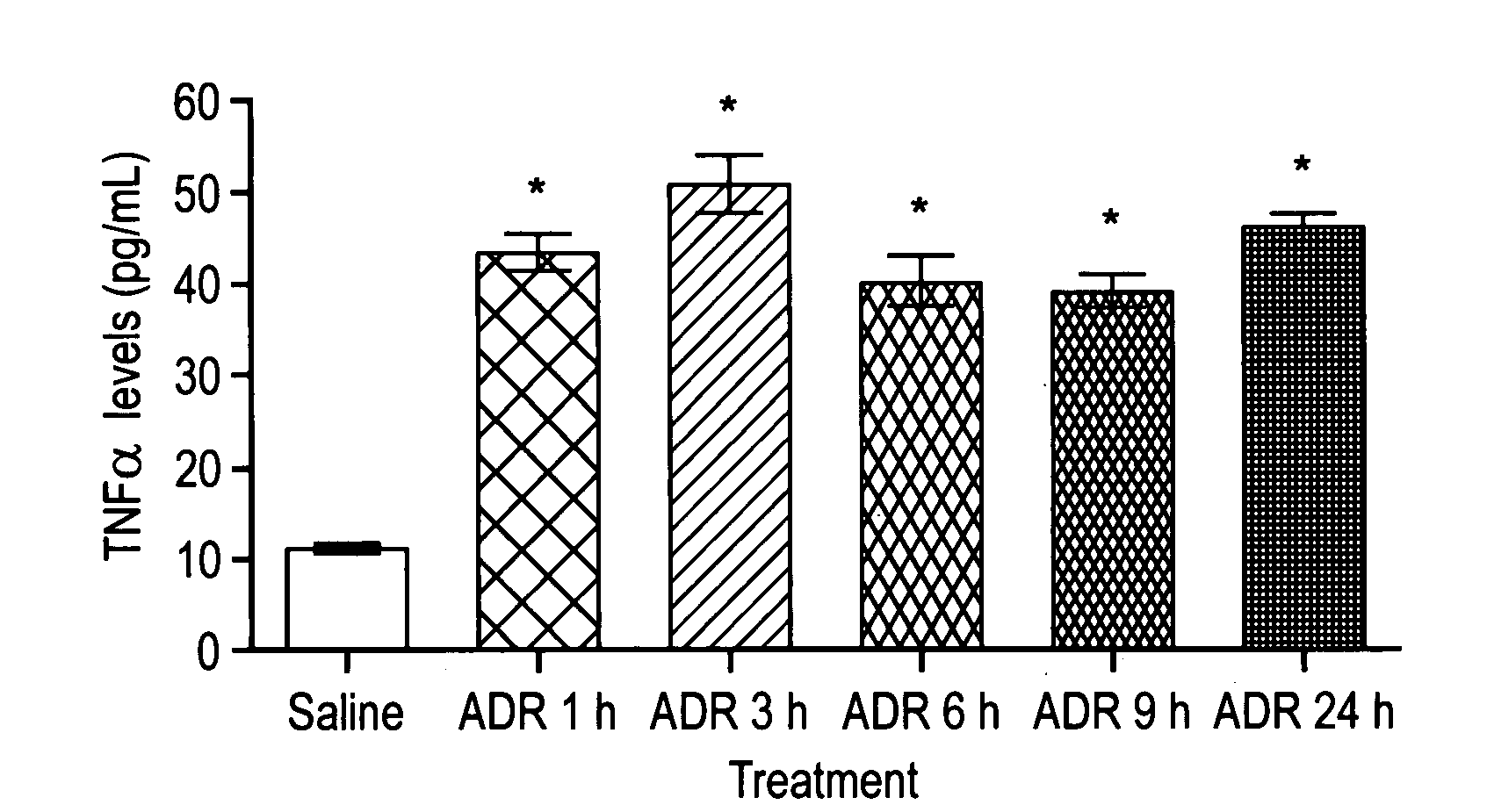
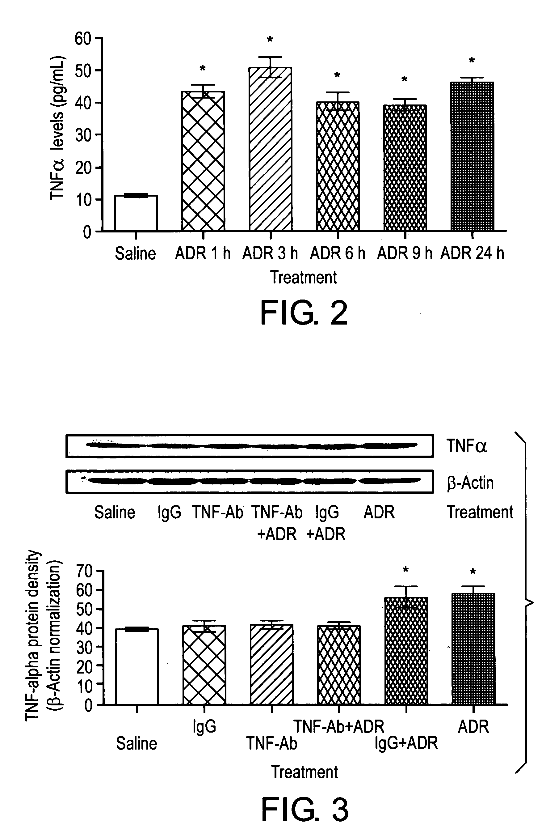
[0114]Example 2 demonstrates that TNF α levels in serum were significantly higher in mice treated with ADR than those from controls.
[0115]Mice were treated with 20 mg / kg ADR or saline as control. Blood samples were collected at 1, 3, 6, 9, 24 hours and allowed to clot at 2-8° C. overnight. Serum samples were used to measure TNF α levels, according to the mouse enzyme-linked immunosorbent assay following the manufacturers' instructions (mouse TNF-α / TNFSF1A immunoassay, R&D Systems, Minneapolis, MN). The TNF α concentration in the sample was calculated from a recombinant mouse TNF α standard curve. The minimum detection limit is typically less than 5.1 pg / mL.
[0116]TNF α levels in serum were significantly higher in mice treated with ADR than those from controls (p<0.001, FIG. 2). The increased TNF α levels were detectable as early as 1 hour and were sustained throughout a 24 hour period after ADR treatment.
PUM
| Property | Measurement | Unit |
|---|---|---|
| Cell angle | aaaaa | aaaaa |
| Chemotherapeutic properties | aaaaa | aaaaa |
Abstract
Description
Claims
Application Information