External and Internal Ultrasound Imaging System

a technology of ultrasound imaging and external ultrasound, applied in the field of medical diagnostic systems and methods, can solve the problems of heavy cabling and equipment, difficult positioning next to the patient, and stiffness of the cabling from the ultrasound system to the proximal connector of the catheter

Inactive Publication Date: 2008-06-19
ST JUDE MEDICAL ATRIAL FIBRILLATION DIV
View PDF99 Cites 114 Cited by
  • Summary
  • Abstract
  • Description
  • Claims
  • Application Information

AI Technical Summary

Benefits of technology

[0004]The various embodiments provide compact, portable ultrasound systems which can operate two or more ultrasound transducers simultaneously, and in certain embodiments include both an intrabody, percutaneous ultrasound imaging probe and an external ultrasound imaging probe suitable for simultaneous examination of an organ from internal and external perspectives.

Problems solved by technology

While ultrasound imaging catheter systems have proven to be invaluable diagnostic tools, the associated cabling and equipment present difficulties for clinicians.
The cabling from the ultrasound system to the catheter's proximal connector is heavy, stiff, and limited in length.
The large cart which normally contains the ultrasound system takes up space in the operating room or catheterization lab, and may be difficult to position next to the patient.
These limitations have made it impractical to perform ultrasound imaging of the heart using transducers positioned internally (e.g., with intracardiac ultrasound catheters) and externally (e.g., a transducer positioned on the chest).
As a result, clinicians have had limited ability to combine and correlate ultrasound images from these two viewing perspectives in order better diagnose and treat heart conditions.

Method used


Image

  • External and Internal Ultrasound Imaging System
  • External and Internal Ultrasound Imaging System
  • External and Internal Ultrasound Imaging System

Examples


Embodiment Construction

[0021]Various embodiments of the present invention will be described in detail with reference to the accompanying drawings. Wherever possible, the same reference numbers will be used throughout the drawings to refer to the same or like parts.

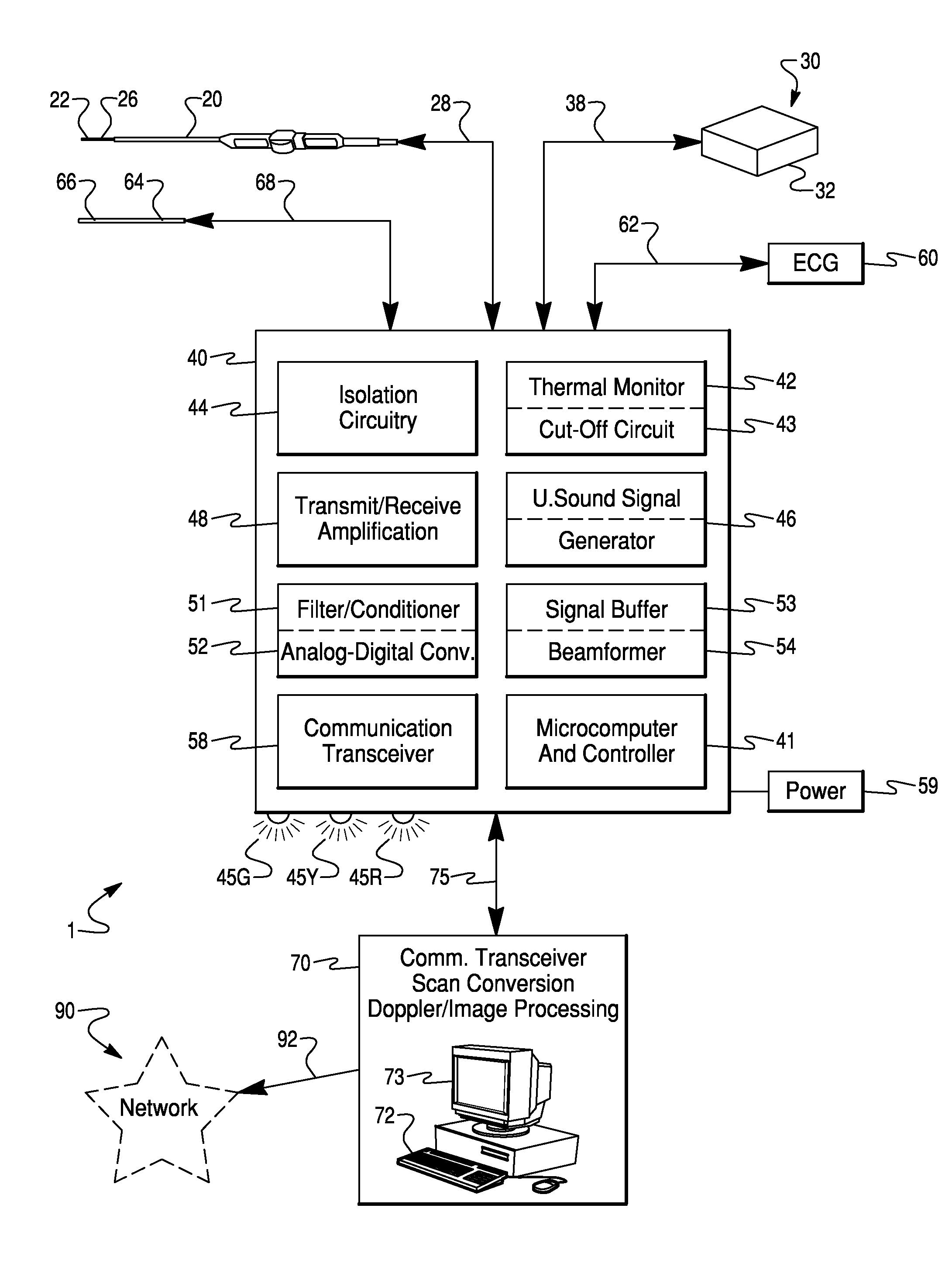
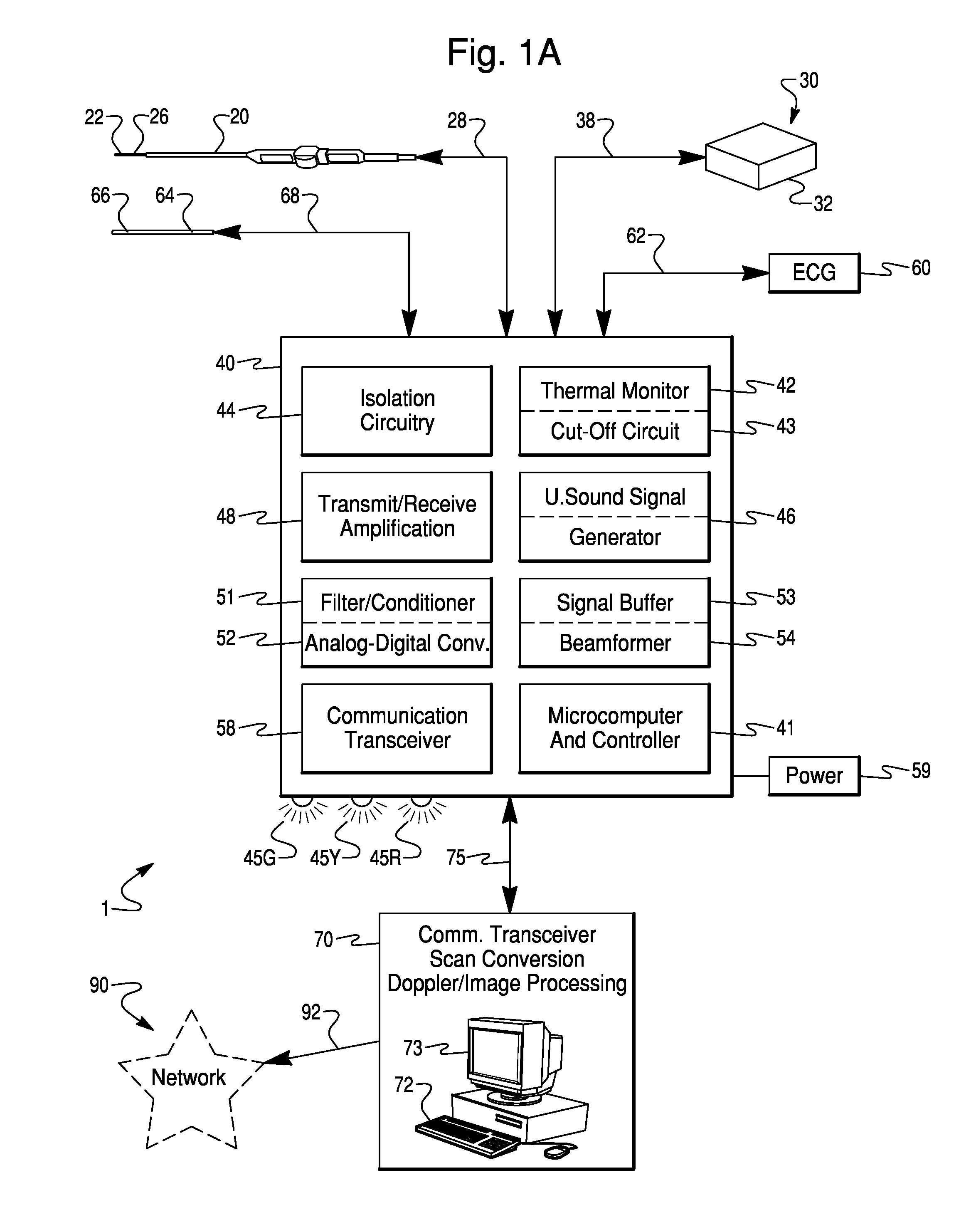
[0022]As used herein, the terms “about” or “approximately” for any numerical values or ranges indicate suitable dimensional tolerances that allow the part or collection of components to function for their intended purposes as described herein. Also, as used herein, the terms “body”“patient”, “host”, and “subject” refer to any human or animal subject and are not intended to limit the systems or methods to human use. Further, embodiments of the invention will be described for use with an intracardiac ultrasound transducer array catheter. However, the embodiments may be applicable to any medical ultrasound transducer.

[0023]The various embodiments provide ultrasound systems and methods that enable simultaneous ultrasound imaging by two or more trans...


PUM


Abstract

An ultrasound imaging system includes a catheter-based ultrasound transducer, an external ultrasound transducer and a compact ultrasound unit including an isolation circuit, an ultrasound signal generator, and a beam former within a single unit. The ultrasound unit may include circuitry and software to enable simultaneous imaging by both transducers. The isolation circuit may limit unintended, leakage current from the ultrasound system through the transducer array of the ultrasound system.

Description

FIELD OF THE INVENTION[0001]The present invention is a medical diagnostic system and method, and more particularly is directed to an ultrasound catheter system having transducer arrays positioned both internal and external to a patient's body.BACKGROUND OF THE INVENTION[0002]Recent advancements in miniaturization of ultrasound technology has enabled the commercialization of catheters including phased array ultrasound imaging transducers small enough to be positioned within a patient's body via intravenous cannulation. By imaging vessels and organs, including the heart, from the inside, such miniature ultrasound transducers have enabled physicians to obtain diagnostic images available by no other means.[0003]While ultrasound imaging catheter systems have proven to be invaluable diagnostic tools, the associated cabling and equipment present difficulties for clinicians. The cabling from the ultrasound system to the catheter's proximal connector is heavy, stiff, and limited in length. T...

Claims


Application Information

Patent Timeline
Patent Type & Authority Applications(United States)
IPC IPC(8): A61B8/00
CPCA61B8/08A61B8/0883A61B8/0891A61B8/12A61B8/4472A61B8/565G01S7/52074G01S7/52082G01S15/899A61B8/56A61B8/5238A61B8/4422A61B8/445A61B8/4477
Inventor JENKINS, DAVID A.BYRD, CHARLES BRYANDALA-KRISHNA, PRAVEEN
Owner ST JUDE MEDICAL ATRIAL FIBRILLATION DIV
Why Eureka
Try Eureka