Endoscopic balloon tissue dissector and retractor

a tissue dissection and endoscopic technology, applied in the field of surgery, can solve problems such as the difficulty of locating and gaining access to internal organs or structures, the difficulty of visualizing adhesions, and the difficulty of anterior and anterolateral access

Inactive Publication Date: 2008-06-19
KAMBIN PARVIZ +1
View PDF30 Cites 33 Cited by
  • Summary
  • Abstract
  • Description
  • Claims
  • Application Information

AI Technical Summary

Benefits of technology

[0017]The image transmitter can also be used for visualization during expansion of the retractor blades. If the blades are independently adjustable, in the spreading step, at least one blade can be moved apart from the other blade independently of movement of the other blade. In this way, tissue can be spread for access to a surgical site with minimal trauma to the patient.

Problems solved by technology

In surgery, adhesions and scar tissue, formed in a patient's thoracic or abdominal cavity as a result of previous surgery, infection or trauma, frequently cause difficulties in locating, and gaining access to, the internal organs or structures that require repair or removal.
For example, adhesions frequently cause problems in anterior and anterolateral access to the spinal column or the content of the abdominal cavity.
However, when a gloved finger is used, it is not possible to visualize the adhesions, and unnecessary bleeding, and tearing, or damage to the normal anatomy, almost inevitably occurs.

Method used


Image

  • Endoscopic balloon tissue dissector and retractor
  • Endoscopic balloon tissue dissector and retractor
  • Endoscopic balloon tissue dissector and retractor

Examples


Embodiment Construction

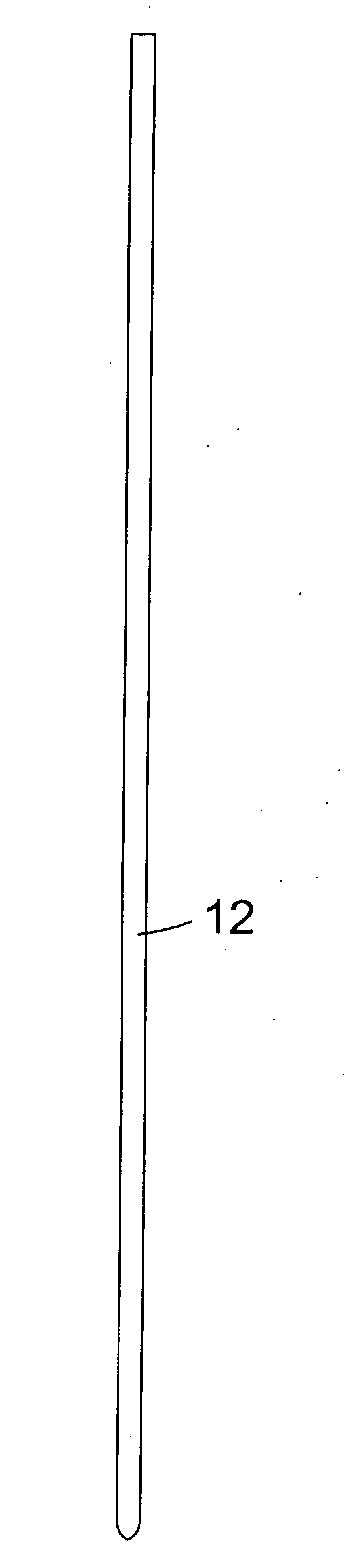
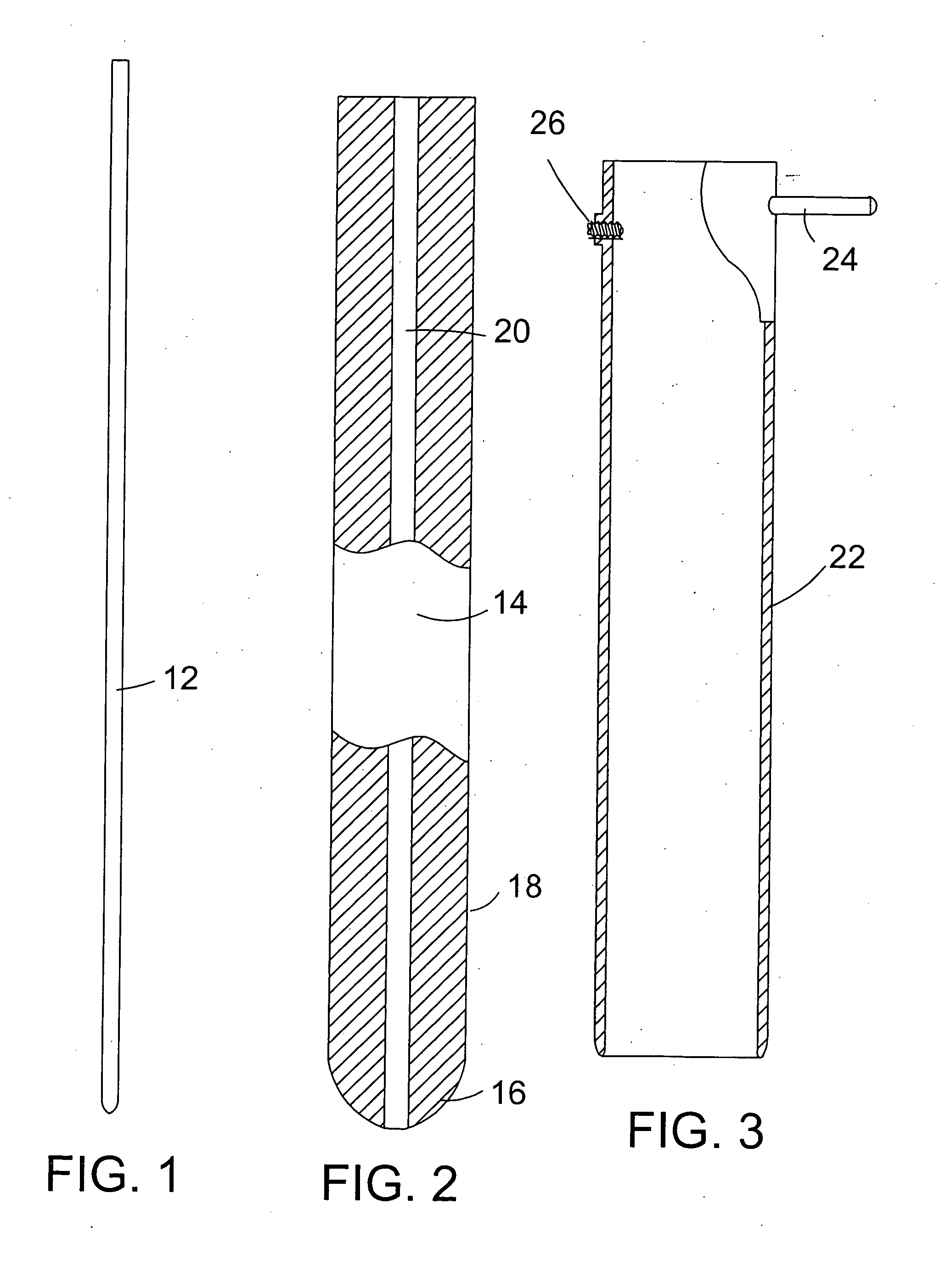
[0031]FIG. 1 shows a guide wire 12, which is typically inserted through an incision in a patient's skin and advanced toward the site at which surgery is to be performed. For example, in the case of spinal surgery performed using a posterior or posterolateral approach, the guide wire can be inserted through the patient's skin, using fluoroscopic guidance to avoid tissue damage. With the guide wire 12 in place, an obturator 14, as shown in FIG. 2, is passed over the guide wire. The obturator is elongated, has a blunt tip 16, a preferably circular, cylindrical exterior wall 18, and a centrally located longitudinal channel 20 for receiving the guide wire 12. As an alternative, a solid obturator can be used without a guide wire, especially in cases where the depth to which the obturator is inserted is small.

[0032]With the obturator 14 in place, a cannula 22, as shown in FIG. 3 can be passed over the exterior of the obturator. The cannula can be manipulated by a handle 24, which can also ...


PUM


Abstract

An instrument for endoscopic surgery comprises a retractor having independently spreadable retractor blades mounted at the distal end of a tube, through which a balloon dissector can be passed. The balloon is transparent, and the balloon dissector is provided with an image transmitter for visualization of tissue dissection.

Description

FIELD OF THE INVENTION[0001]This invention relates to surgery, and particularly to instruments for dissection and retraction of tissue in endoscopic surgery.BACKGROUND OF THE INVENTION[0002]In surgery, adhesions and scar tissue, formed in a patient's thoracic or abdominal cavity as a result of previous surgery, infection or trauma, frequently cause difficulties in locating, and gaining access to, the internal organs or structures that require repair or removal. For example, adhesions frequently cause problems in anterior and anterolateral access to the spinal column or the content of the abdominal cavity.[0003]Typically, a surgeon releases adhesions manually, using a gloved finger. However, when a gloved finger is used, it is not possible to visualize the adhesions, and unnecessary bleeding, and tearing, or damage to the normal anatomy, almost inevitably occurs. In addition, access to certain organs requires the identification and separation of two muscle groups through their anatom...

Claims


Application Information

Patent Timeline
Patent Type & Authority Applications(United States)
IPC IPC(8): A61B17/02
CPCA61B2017/22038A61B2017/320048A61B2017/347A61B2017/00907A61B17/32A61B17/34A61B2017/00685A61B17/0218
Inventor KAMBIN, PARVIZKAMBIN, SHEILA P.
Owner KAMBIN PARVIZ
Why Eureka
Try Eureka