
Functionalized Magnetic Nanoparticles and Methods of Use Thereof

a magnetic nanoparticle and functional technology, applied in the field of magnetic nanoparticles, can solve problems such as the inability to distinguish between diseased tissues and normal tissues

Inactive Publication Date: 2008-08-28
RGT UNIV OF CALIFORNIA
View PDF19 Cites 67 Cited by
  • Summary
  • Abstract
  • Description
  • Claims
  • Application Information

AI Technical Summary

Benefits of technology

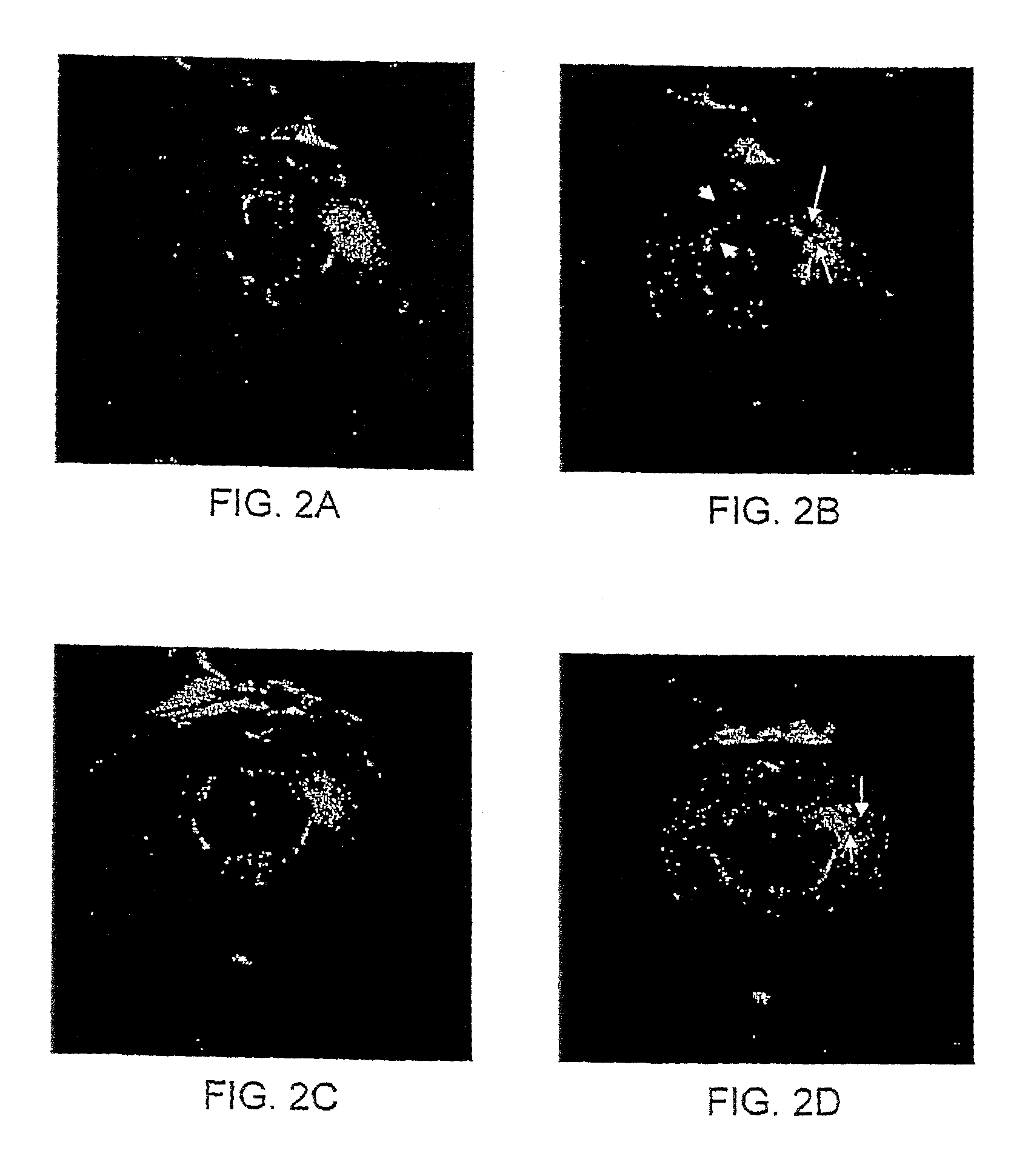
[0008]FIGS. 2A-D depict magnetic resonance images of brains of kainic acid-treated rats zero hour after injection with AMT-MNP (FIG. 2A); 6 hours after injection with AMT-MNP (FIG. 2B); zero hour after injection with non-functionalized MNP (FIG. 2C); and 6 hours after injection with non-functionalized MNP (FIG. 2D).

Problems solved by technology

A major challenge in imaging is the ability to distinguish between diseased tissues and normal tissue.

Method used


Image

  • Functionalized Magnetic Nanoparticles and Methods of Use Thereof
  • Functionalized Magnetic Nanoparticles and Methods of Use Thereof
  • Functionalized Magnetic Nanoparticles and Methods of Use Thereof

Examples


example 1

Preparation of Functionalized Magnetic Nanoparticles

Nanoparticle Preparation

[0108]200 mg of human serum albumin (USA) are dissolved in 2.0 ml water containing magnetic nanoparticles (MNP; e.g., magnetite particles). The pH of the solution is raised to 8.4 under constant stirring by dropwise addition of 0.01 M and 0.1 M solution of NaOH. Under constant stirring desolvatation of the 10% HSA solution is performed by dropwise addition of 8.0 ml ethanol. After addition of ethanol, 235 μl of an 8% glutaraldehyde solution are added. After 24 h, the resulting nanoparticles are purified by threefold centrifugation (16.100 g, 8 min) and redispersion in water. Redispersion is performed in an ultrasonication bath. HSA-MNP synthesized using this method have an average diameter of about 60 nm to about 990 nm, depending on the pH of the preparation and addition of non-conjugated or conjugated MNP. AMT-MNP nanoparticles have an average diameter of approximately 20 Dill, and a size range of from abo...

example 2

In Vivo Characterization of Functionalized MNP

[0132]Non-functionalized MNP and AMT-conjugated MNP were administered to a kainic acid (KA) model of epilepsy. The data demonstrated that AMT-MNP display affinity for epileptic tissues.

[0133]Two Lewis rats (90 days old) were injected in the right hippocampus with 1 μl KA solution. The rats developed status epilepticus immediately post-injection with KA. Status epilepticus stopped approximately 48 hours post-injection. On day 3 post KA injection, baseline MRI were obtained, using T2 sequences (TR=6000 ms; TE=50 ms; slice thickness=1.5 mm; interstices distance=0.25 mm). After the baseline MRI, the first rat was injected (i.v.) with AMT-MNP (300 μmol / kg) and the second rat was injected (i.v.) with non-functionalized MNP (300 μmol / kg). MRI were repeated 6 hours after each rat was injected with the MNP.

[0134]FIG. 2A shows baseline MRI of the first rat; FIG. 2B shows the areas of (negative) enhancement in the CA1 (upper arrowhead) and dentate ...


PUM

PropertyMeasurementUnit
Therapeuticaaaaaaaaaa
Affinityaaaaaaaaaa
Disorderaaaaaaaaaa

Abstract

The present invention provides functionalized magnetic nanoparticles comprising a functional group, which functionalized magnetic nanoparticles exhibit differential binding to a tissue, indulging brain tissue, bone, and vascular tissues. The present invention further provides compositions, including pharmaceutical compostions, comprising a subject functionalized magnetic nanoparticle. The present invention further provides diagnostic and research methods including use of subject functionized magnetic nanoparticles. The present invention further provides a magnetic resonance imaging (MRI)-visible drug delivery system; drugs using MRI, as well as tissue-specific drug delivery.

Description

CROSS-REFERENCE[0001]This application claims the benefit of U.S. Provisional Patent Application No. 60 / 664,046, filed Mar. 21, 2005, which application is incorporated herein by reference in its entirety.FIELD OF THE INVENTION[0002]The present invention is in the field of magnetic nanoparticles, and their use in imaging, e.g., magnetic resonance imaging, of tissues.BACKGROUND OF THE INVENTION[0003]Nanoparticles are very small particles typically ranging in size from as small as one nanometer to as large as several hundred nanometers in diameter. Their small size allows nanoparticles to be exploited to produce a variety of products such as dyes and pigments; aesthetic or functional coatings; tools for biological discovery, medical imaging, and therapeutics; magnetic recording media; quantum dots; and even uniform and nanosize semiconductors.[0004]Use of magnetic nanoparticles has been proposed for various biomedical purposes, including magnetic resonance imaging, hyperthermic treatmen...

Claims


Application Information

Patent Timeline
IPC IPC(8): A61K49/18A61K9/14A61K39/395A61P25/28A61K9/00A61K38/02
CPCA61K47/48176A61K47/4823A61K47/48238A61K47/48861B82Y5/00A61K49/1854A61K49/1863A61K49/1869A61K47/48884A61K47/58A61K47/61A61K47/62A61K47/6923A61K47/6929A61P19/00A61P21/04A61P25/00A61P25/08A61P25/14A61P25/16A61P25/18A61P25/28A61P25/30A61P25/36A61P29/00A61P35/00A61P9/00A61K9/14A61B5/055A61K49/06
Inventor AKHTARI, MASSOUDENGEL, JEROME
Owner RGT UNIV OF CALIFORNIA
Learn More