
Combined radiation therapy and magnetic resonance unit

a combined radiation therapy and magnetic resonance technology, applied in radiation therapy, medical science, radiation therapy, etc., can solve the problems of limited solution, tumor growth or shrinkage, and the irradiation target in the body can move, so as to reduce the adverse effects of radiation therapy on healthy tissu

Active Publication Date: 2008-08-28
SIEMENS HEALTHCARE GMBH
View PDF21 Cites 113 Cited by
  • Summary
  • Abstract
  • Description
  • Claims
  • Application Information

AI Technical Summary

Benefits of technology

The invention is a combined radiation therapy and magnetic resonance unit that allows for high-quality image monitoring during radiation therapy. The unit includes a magnetic resonance diagnosis part with a main magnet and a radiation therapy part located inside the diagnosis part. The radiation therapy part can be an electron accelerator, which can easily generate a therapy beam. The unit also includes a beam deflection arrangement, such as an electromagnet, to direct the electron beam inward into the interior of the unit. The X-ray beam generated by the unit can be directed and homogenized using a collimator. The collimator can also be adjusted to change the cross-section of the X-ray beam. The invention allows for independent operation of the radiation therapy part and the magnetic resonance diagnosis part, reducing adverse effects on healthy tissue during radiation therapy.

Problems solved by technology

During irradiation the problem often arises that the irradiation target in the body can move.
On the other hand, in the period between radiation treatment planning and actual radiation treatment a tumor may have grown or have already shrunk.
These, however, provide only a limited solution to the problem.
In the case of ultrasound imaging the necessary penetration depth is lacking for many applications.
In X-ray imaging the X-ray sensors can be disrupted or damaged by the gamma radiation of the accelerator.
Furthermore, the quality of the tissue images is often unsatisfactory.
These positioning aids and fixing devices are, however, expensive and in most cases they are also uncomfortable for the patient.
In addition, they conceal the risk of irradiation errors because as a rule no further check of the actual position of the irradiation center is carried out during irradiation.
A division of the magnet, a rotatable magnet and shielding of the radiation source represent elaborate technical solutions and increase the cost.

Method used


Image

  • Combined radiation therapy and magnetic resonance unit
  • Combined radiation therapy and magnetic resonance unit
  • Combined radiation therapy and magnetic resonance unit

Examples


Embodiment Construction

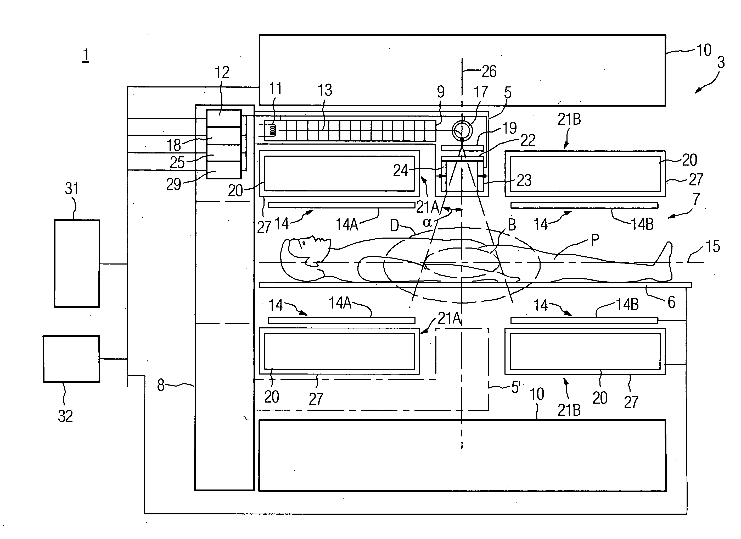
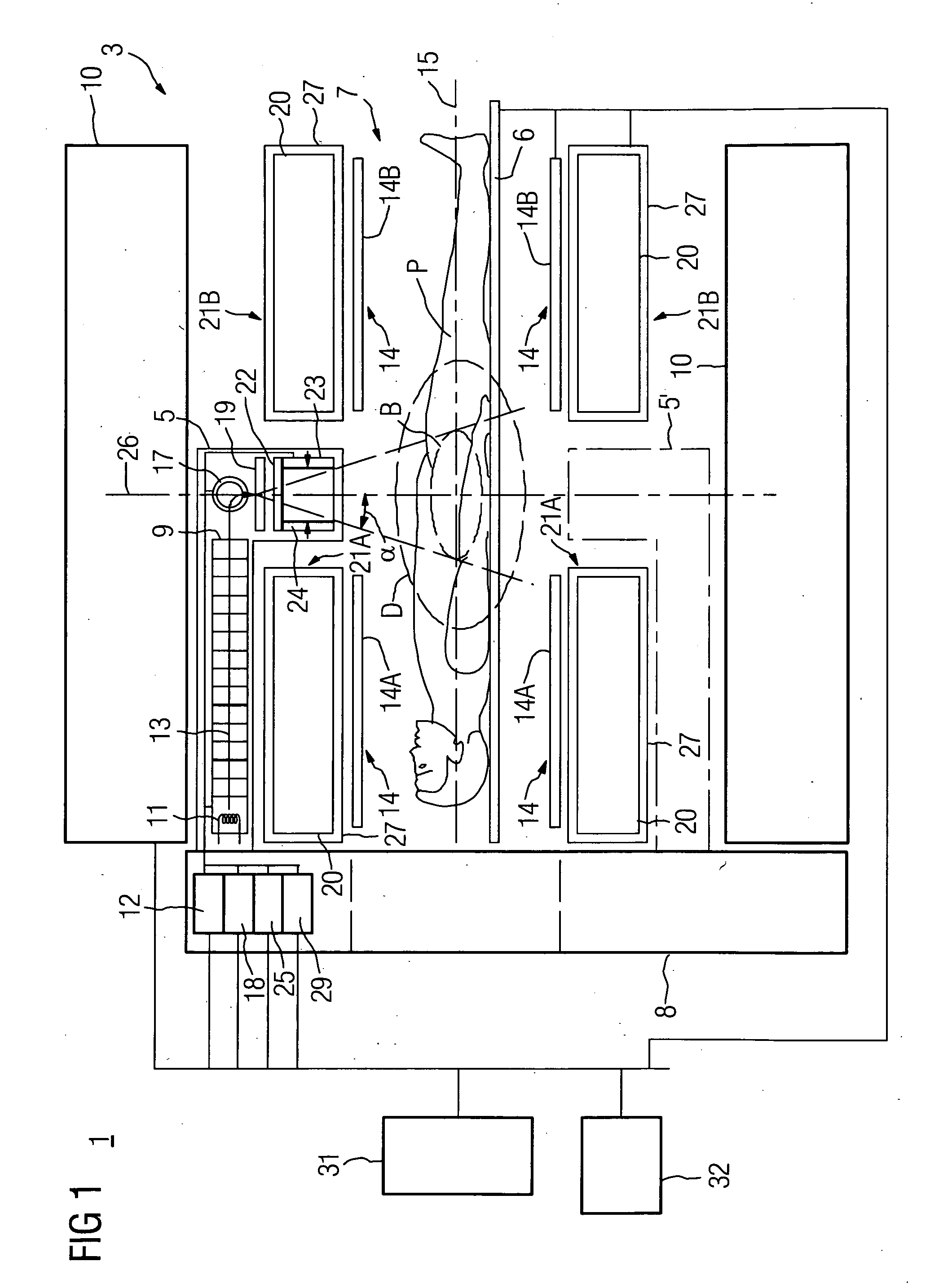
[0030]FIG. 1 shows a schematic representation (not to scale) of a combined radiation therapy and magnetic resonance unit 1 with a magnetic resonance diagnosis part 3 and a radiation therapy part 5. The magnetic resonance diagnosis part 3 comprises a main magnet 10, a gradient coil system comprising two in this case symmetrical partial gradient coils 21A,21B, high-frequency coils 14, for example two parts of a body coil 14A,14B, and a patient bed 6. All these components of the magnetic resonance part are connected to a control unit 31 and an operating and display console 32.

[0031]In the example presented, both the main magnet 10 and the partial gradient coils 21A,21B are essentially shaped like a hollow cylinder and arranged coaxially around the horizontal axis 15. The inner shell of the main magnet 10 limits in radial direction (facing away vertically from the axis 15) a cylinder-shaped interior 7, in which the radiation therapy part 5, the gradient system, high-frequency coils 14 a...


PUM


Abstract

The invention relates to a combined radiation therapy and magnetic resonance unit. For this purpose, in accordance with the invention a combined radiation therapy and magnetic resonance unit is provided comprising a magnetic resonance diagnosis part with an interior, which is limited in radial direction by a main magnet, and a radiation therapy part for the irradiation of an irradiation area within the interior, wherein at least parts of the radiation therapy part, which comprise a beam deflection arrangement, are arranged within the interior.

Description

CROSS REFERENCE TO RELATED APPLICATIONS[0001]This application claims priority of German application No. 10 2007 010 132.7 filed Feb. 28, 2007 and claims priority of German application No. 10 2008 007 245.1 filed Feb. 1, 2008, which are incorporated by reference herein in their entirety.FIELD OF THE INVENTION [0002]The invention relates to a combined radiation therapy and magnetic resonance unit.BACKGROUND OF THE INVENTION [0003]Generally in radiation therapy the aim is to irradiate a target within the human body in order to combat diseases, in particular cancer. For this purpose a high dose of radiation is specifically generated in an irradiation center (isocenter) of an irradiation apparatus. During irradiation the problem often arises that the irradiation target in the body can move. For instance, a tumor in the abdomen can move during breathing. On the other hand, in the period between radiation treatment planning and actual radiation treatment a tumor may have grown or have alre...

Claims


Application Information

Patent Timeline
Patent Type & Authority Applications(United States)
IPC IPC(8): A61B5/055A61N5/00
CPCA61N5/1049A61N2005/1055G01R33/4808G01R33/385G01R33/4215G01R33/3806
Inventor AMIES, CHRISTOPHER JUDEBEASLEY, PAULCELI, JUAN CARLOSHEID, OLIVERHERNANDEZ-GUERRA, FRANCISCO MIGUELKRUIP, MARCEL JAN MARIEVESTER, MARKUS
Owner SIEMENS HEALTHCARE GMBH
Learn More