Method and apparatus for non-invasive ultrasonic fetal heart rate monitoring

a non-invasive, ultrasonic technology, applied in the direction of ultrasonic/sonic/infrasonic image/data processing, mechanical vibration separation, application, etc., can solve the problems of loss of heart rate information, relative cumbersome array of singles, experienced by hospital staff members, etc., and achieve the effect of less interferen

Inactive Publication Date: 2008-08-28
GENERAL ELECTRIC CO
View PDF2 Cites 29 Cited by
  • Summary
  • Abstract
  • Description
  • Claims
  • Application Information

AI Technical Summary

Benefits of technology

[0013]In accordance with the disclosed embodiment, each ultrasonic transducer array patch comprises a multiplicity of subelements that are switchably reconfigurable to form elements having different shapes, e.g., annular rings. Each subelement comprises a plurality of interconnected capacitive micromachined ultrasonic transducer (cMUT) cells that are not switchably disconnectable. The use of cMUT patches will provide the ability to interrogate a three-dimensional space electronically (i.e. without mechanical beam steering) with ultrasound, using a transducer device that is thin and lightweight enough to stick to the patient's skin like an EKG electrode.
[0014]Thus the present invention applies cMUT technology to the monitoring of fetal heart rate in a labor and delivery or ICU setting. The ultrasound device can track the fetal heart in three-dimensional space as it moves due to the mother's motion or the motion of the unborn child within the womb. This will allow the use of a narrower beam and a smaller sample volume than is used by prior art devices, so that less interference will be present in the Doppler signal that is processed to derive the fetal heart rate. While this tracking is taking place, the fetal heart rate can be derived by standard means from the Doppler shift of the ultrasound reflected from the fetal heart.

Problems solved by technology

One challenge in clinical applications is ensuring that the ultrasonic transducers are properly positioned relative to the fetal heart.
Known instrumentation uses a relatively cumbersome array of single-element ultrasound transducers designed to cover the entire maternal abdomen.
Unfortunately, as the result of fetal or maternal motion, the alignment of these transducers is such that the transducers fail to capture some echoes from the fetal heart and there will be a loss of heart rate information.
The resulting frustration experienced by hospital staff members because of this inconvenience is apparently to the degree that the instrument is often ignored and the mother and fetus do not receive the benefit of monitoring or the assessment of fetal distress.
This is contrary to good practice, since the mother's motion is important in causing the onset of heavy labor.

Method used


Image

  • Method and apparatus for non-invasive ultrasonic fetal heart rate monitoring
  • Method and apparatus for non-invasive ultrasonic fetal heart rate monitoring
  • Method and apparatus for non-invasive ultrasonic fetal heart rate monitoring

Examples


Embodiment Construction

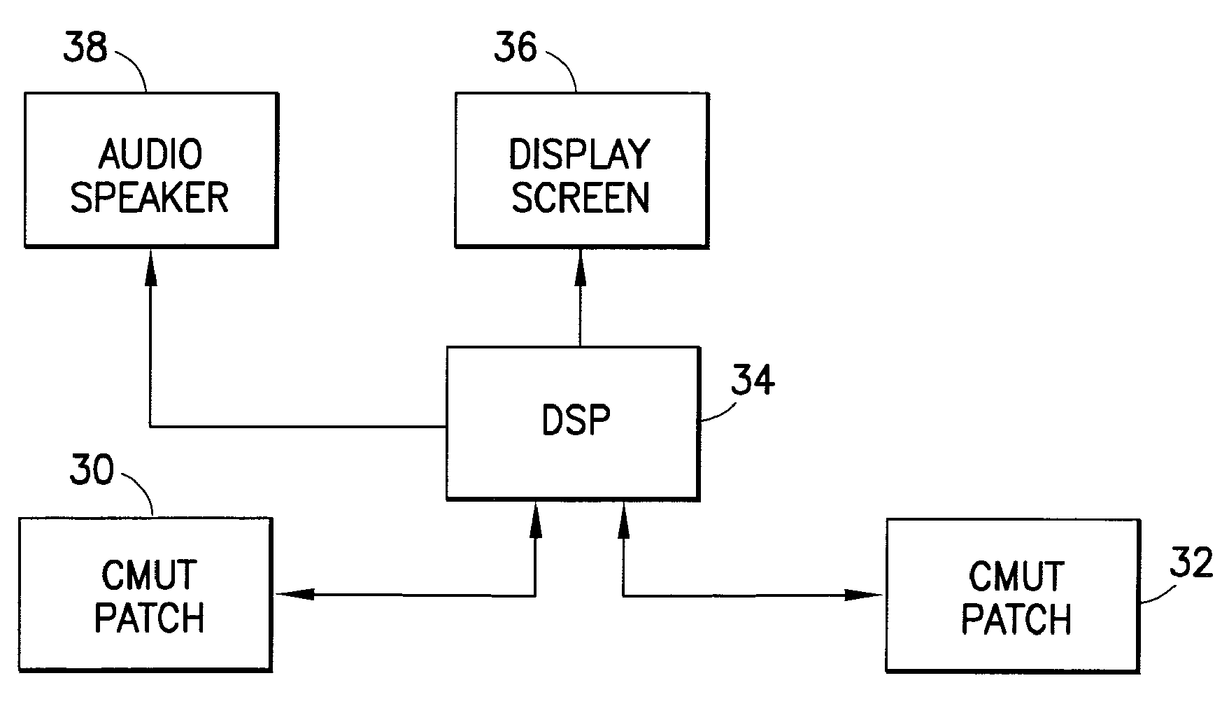
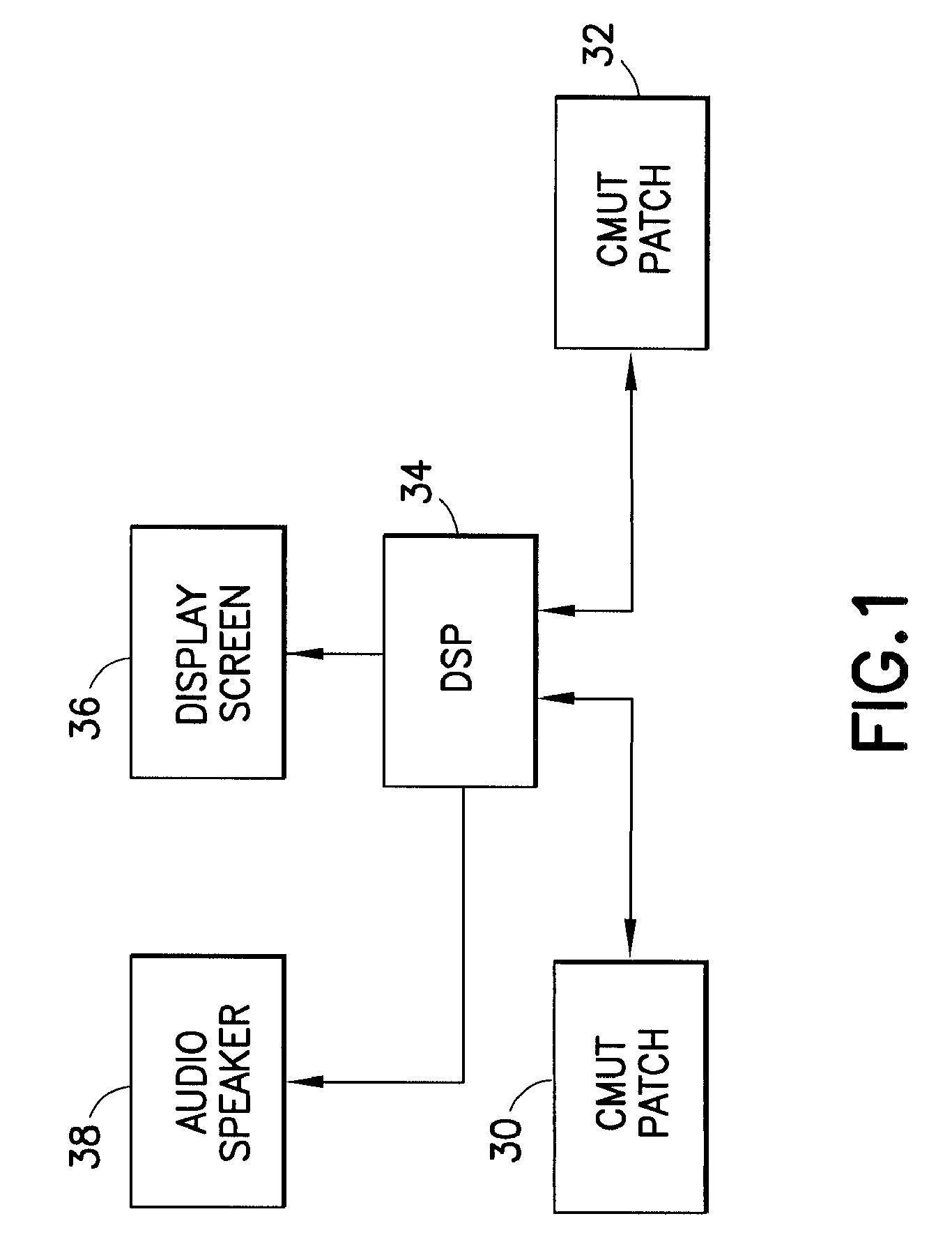
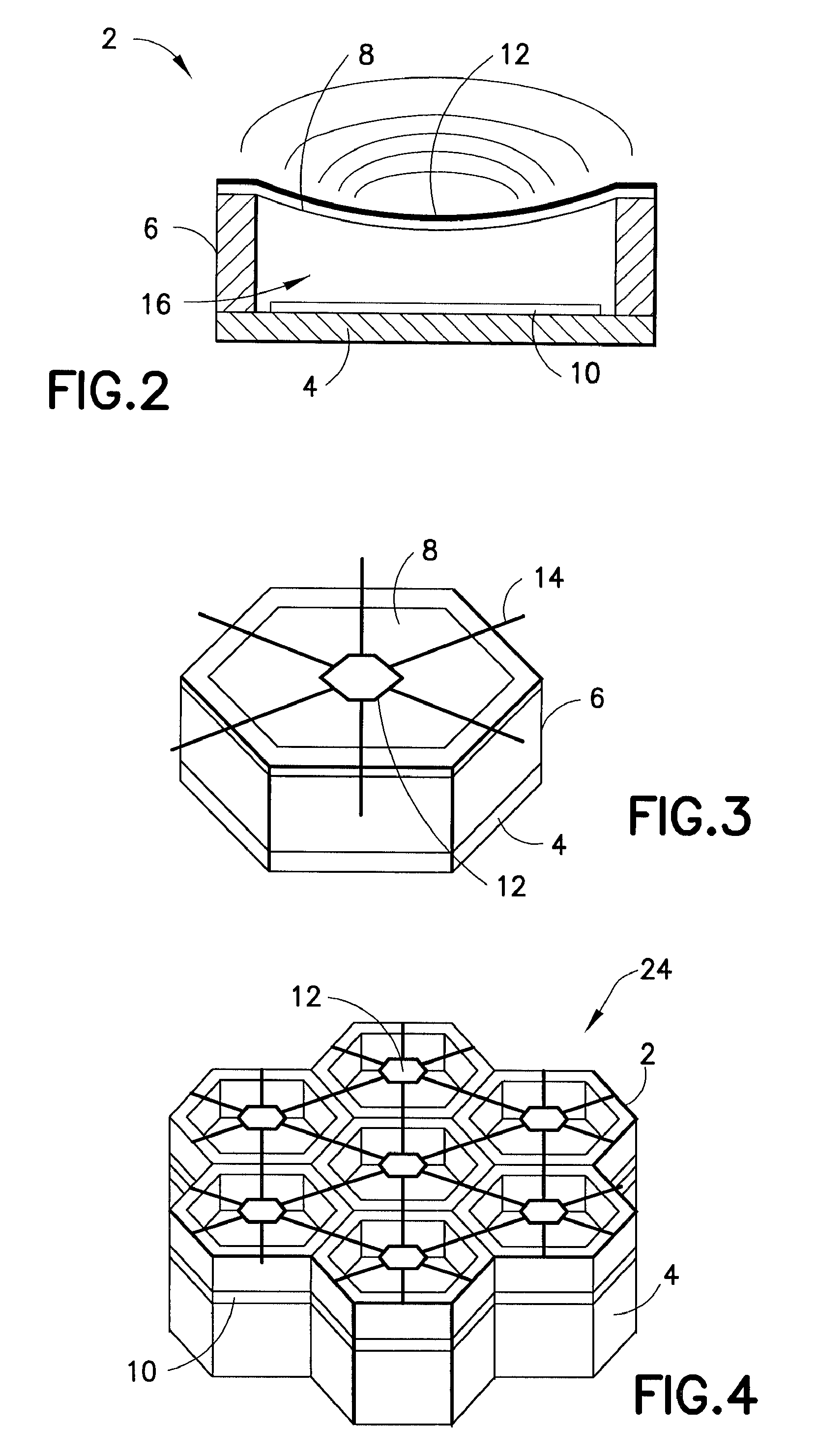
[0031]In accordance with one embodiment of the present invention, one or more cMUT patches are adhered to the mother's abdomen. Each cMUT patch comprises a two-dimensional ultrasound transducer array that is steerable in three-dimensional space. The cMUT patches are electrically connected to a bedside instrument.

[0032]When the instrument is powered on, a search is performed to acquire the Doppler signal of the fetal heart. An initial coarse search, using wide beams and long range gates, will be followed by higher-resolution searches that will successively locate the fetal heart to a greater precision. Once the fetal heart has been located precisely, its position will be tracked using multiple sample volumes surrounding the heart's location. The sample volume that is directly over the heart's location will produce a Doppler waveform that can be processed to extract the fetal heart rate.

[0033]Based on acoustic data from the cMUT patch probes, a processor incorporated in the bedside in...


PUM


Abstract

A continuous, non-invasive fetal heart rate measurement is produced using one or more ultrasonic transducer array patches that are adhered or attached to the mother. Each ultrasound transducer array is operated in an autonomous mode by a digital signal processor to obtain data from which fetal heart rate information can be derived. Each ultrasonic transducer array patch comprises a multiplicity of subelements that are switchably reconfigurable to form elements having different shapes, e.g., annular rings. Each subelement comprises a plurality of interconnected cMUT cells that are not switchably disconnectable. The use of cMUT patches will provide the ability to interrogate a three-dimensional space electronically (i.e. without mechanical beam steering) with ultrasound, using a transducer device that is thin and lightweight enough to stick to the patient's skin like an EKG electrode. The ultrasound device can track the fetal heart in three-dimensional space as it moves due to the mother's motion or the motion of the unborn child within the womb.

Description

CROSS REFERENCE TO RELATED APPLICATIONS[0001]This application is a continuation of U.S. patent application Ser. No. 10 / 838,799, entitled “METHOD AND APPARATUS FOR NON-INVASIVE ULTRASONIC FETAL HEART RATE MONITORING”, filed May 4, 2004, which is herein incorporated by reference.BACKGROUND[0002]The present invention generally relates to methods and apparatus for determining the heart rate of a subject, and particularly relates to methods and apparatuses for determining the beat-to-beat heart rate of a fetus.[0003]Fetal monitoring (i.e., monitoring of the fetal condition during gestation and at birth) usually comprises monitoring uterine activity and the fetal beat-to-beat heart rate. The fetal heart rate, which provides an indication of whether the fetus is sufficiently supplied with oxygen, is preferably calculated from beat to beat.[0004]To obtain a signal indicative of the fetal heart rate prior to rupture of the membranes, a noninvasive monitoring technique must be used. The most ...

Claims


Application Information

Patent Timeline
Patent Type & Authority Applications(United States)
IPC IPC(8): A61B8/02A61B8/00A61B8/08B06B1/02
CPCA61B8/02A61B8/08B06B1/0292A61B8/4483A61B8/488A61B8/0866A61B8/4236
Inventor HOCTOR, RALPH T.THOMENIUS, KAI E.
Owner GENERAL ELECTRIC CO
Why Eureka
Try Eureka