
Method and a dual-array transducer probe for real time mechanical imaging of prostate
Patent Information
- Authority / Receiving Office
- US · United States
- Current Assignee / Owner
- Publication Date
- 2008-09-11
- Estimated Expiration
- Not applicable · inactive patent
Smart Images
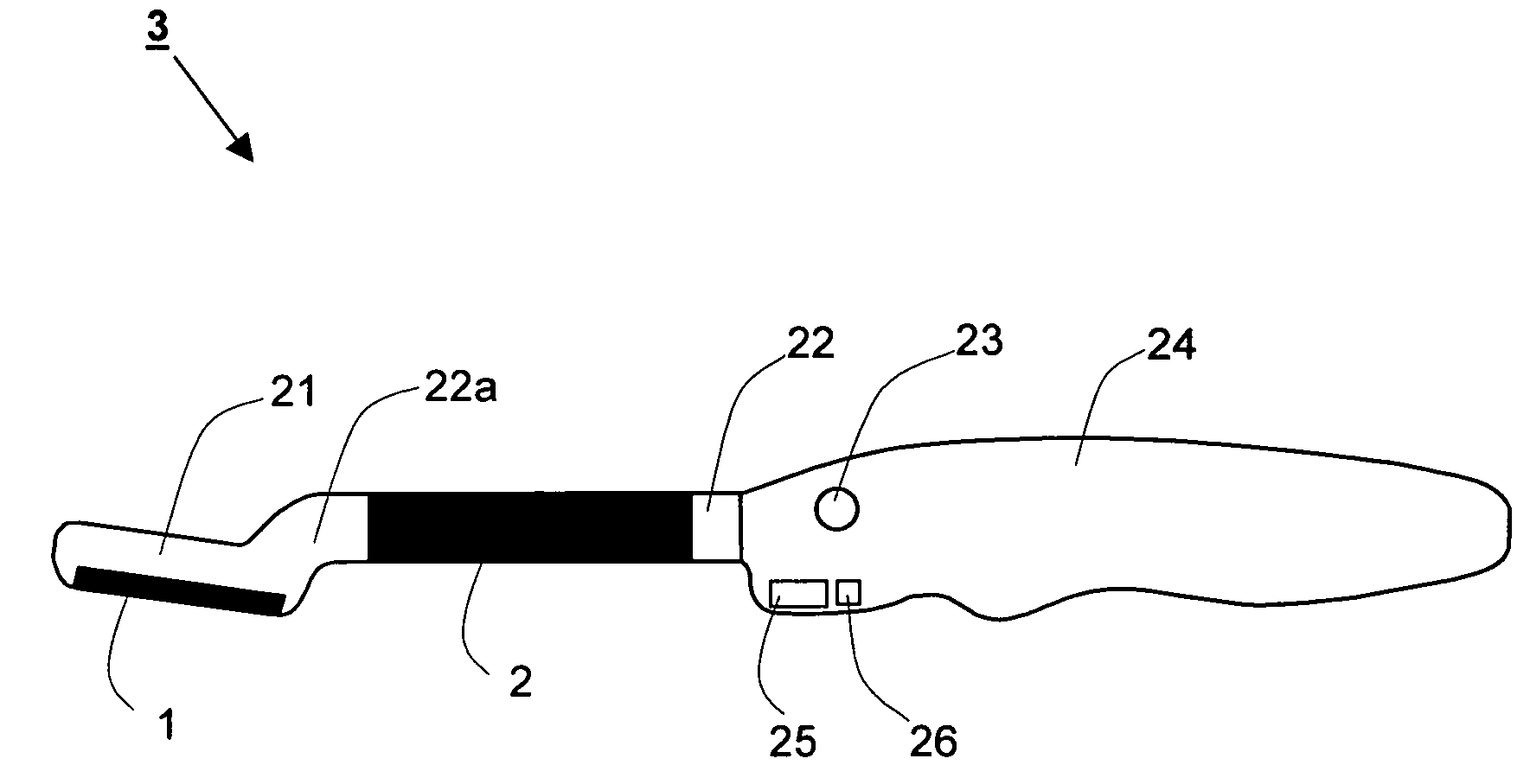
Figure 1 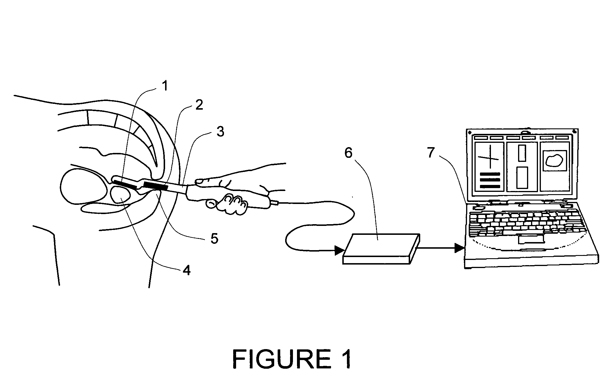
Figure 2 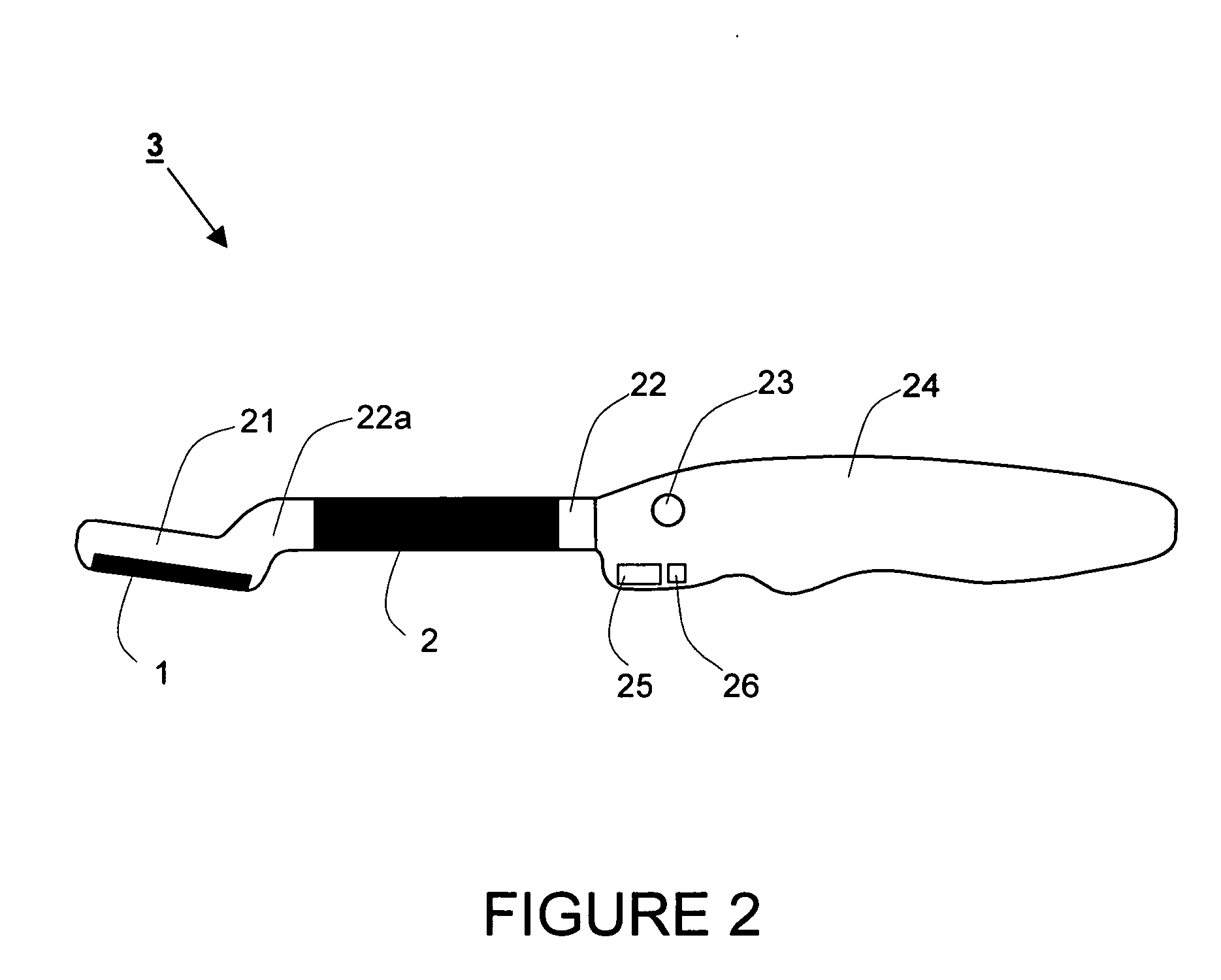
Figure 3
Abstract
Description
CROSS-REFERENCE DATA
[0001] This application is a continuation-in-part of a co-pending U.S. patent application Ser. No. 11 / 123,999 filed May 6, 2005 and entitled “Method and device for real time mechanical imaging of prostate”.STATEMENT REGARDING FEDERALLY SPONSORED RESEARCH OR DEVELOPMENT
[0002] This invention was made with government support under SBIR Grant 2 R44 CA82620-02A1 awarded by the National Institutes of Health, National Cancer Institute. The government has certain rights in this invention.BACKGROUND OF THE INVENTION
[0003] The present invention relates generally to medical devices. More specifically, it relates to a mechanical imaging system and process for examining, mapping, and diagnosing diseases of a palpable organ such as a prostate gland in a male patient, especially the prostate cancer. It is also applicable more generally to mechanical imaging of palpable tissues, including but not limited to, through natural body openings in a human being, i.e. mouth, ear(s), rectum...
Examples
Embodiment Construction
[0052]A detailed description of the present invention follows with reference to accompanying drawings in which like elements are indicated by like reference letters and numerals.
[0053]Referring now to the drawings, FIG. 1 is a schematic view of a preferred embodiment of a device for generating a mechanical image of a three-dimensional prostate volume from a plurality of data frames corresponding to a scan of the prostate. The device comprises the following major elements:[0054]a dual-array transrectal probe 3 with incorporated head pressure sensor array 1 for receiving pressure response data for the prostate 4 and shaft pressure sensor array 2 for receiving supplemental pressure response data for a sphincter area 5,[0055]electronic unit 6, and[0056]a processing and displaying means 7, which may be for example incorporated into a compact personal computer.
[0057]The prostate examination is performed using the following general steps. The patient is instructed to take off all clothes b...