
System and method for providing noninvasive diagnosis of compartment syndrome using exemplary laser speckle imaging procedure

a laser speckle imaging and compartment syndrome technology, applied in the field of system and method for providing noninvasive diagnosis of compartment syndrome using exemplary laser speckle imaging procedure, can solve the problems of delayed diagnosis and intervention with disastrous outcomes, major cause of morbidity and limb loss, permanent damage to muscles and nerves, etc., and achieves low cost, simple interface, and high or low risk of compartment syndrome.

Inactive Publication Date: 2008-09-25
THE GENERAL HOSPITAL CORP
View PDF99 Cites 9 Cited by
  • Summary
  • Abstract
  • Description
  • Claims
  • Application Information

AI Technical Summary

Benefits of technology

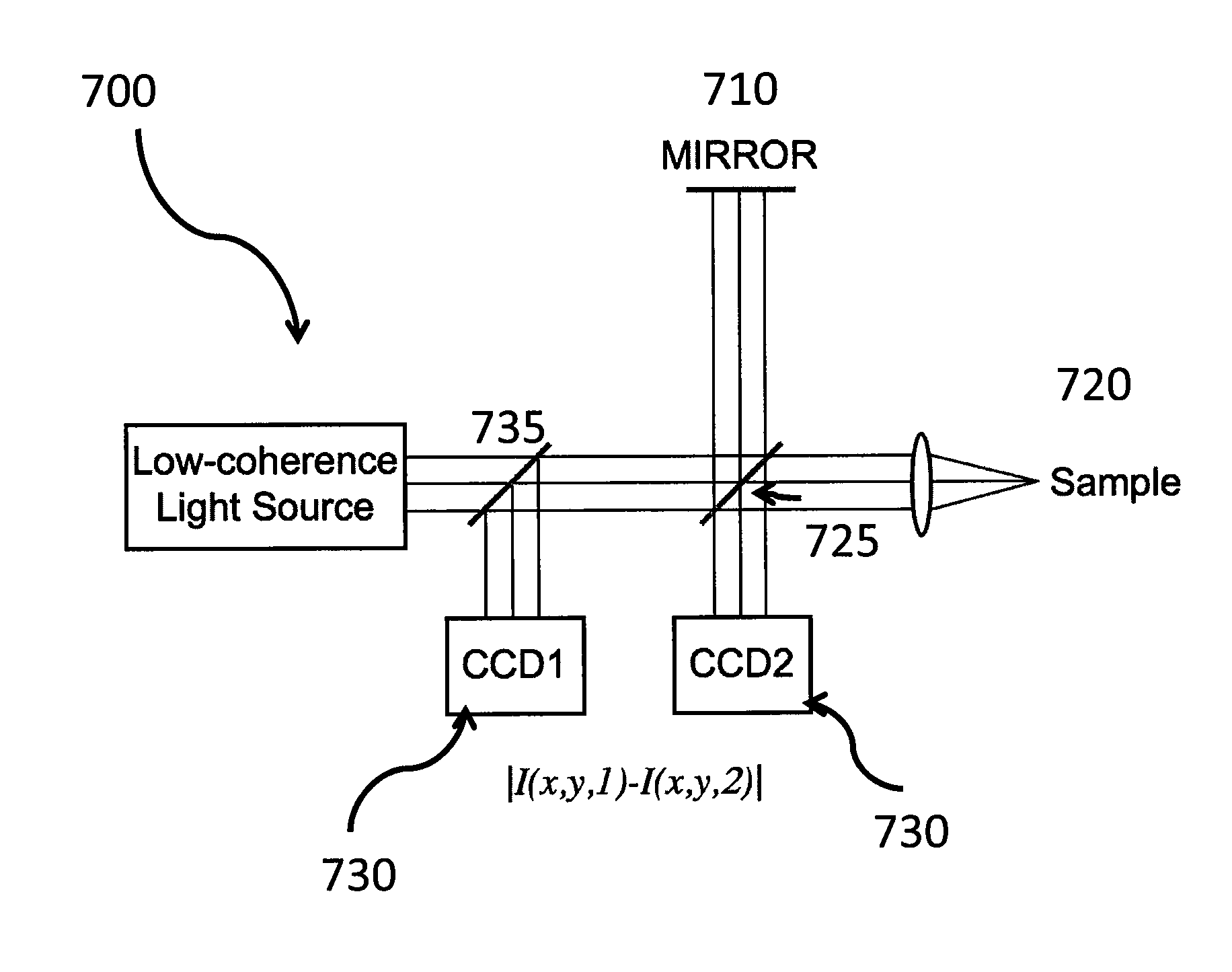
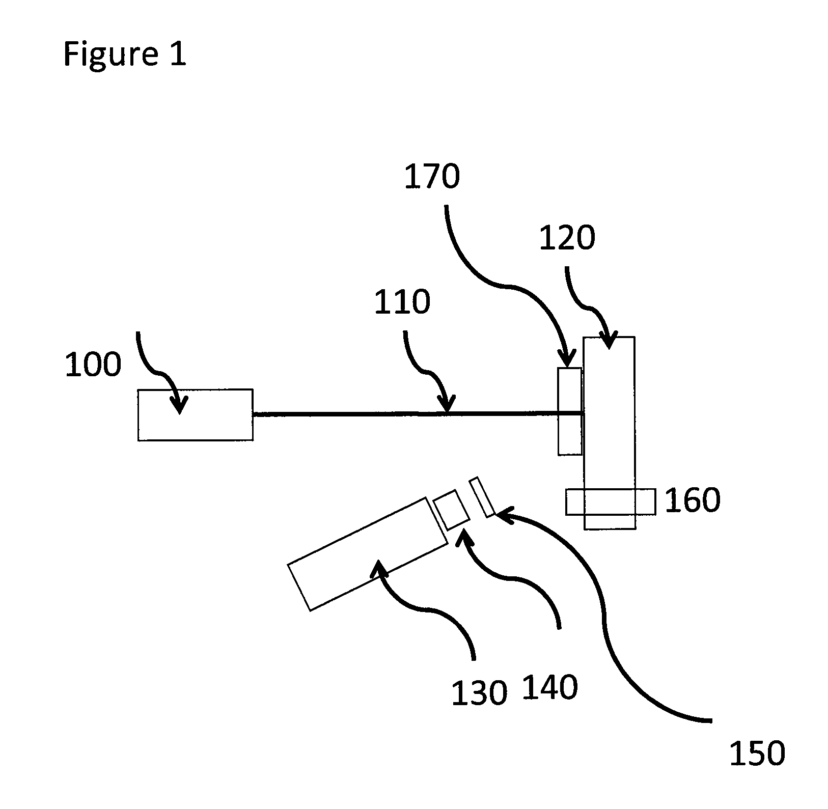
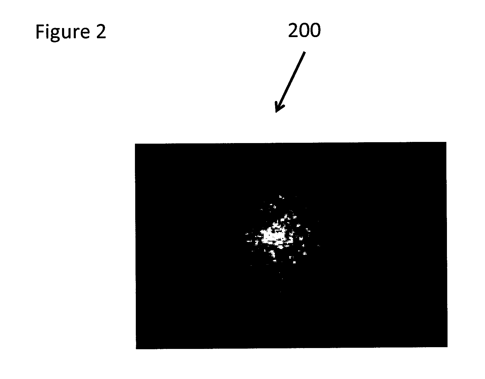
[0009]Since the early stages of compartment syndrome can change muscle capillary blood flow, according to one exemplary embodiment of the present invention, it has been believed that the detection of capillary blood flow within fascial compartments can provide an index for predicting compartment syndrome. Using such exemplary embodiment, a noninvasive method and system [e.g., termed Laser Speckle Imaging (LSI)] can be provided for measuring a depth-dependent tissue perfusion of skin. With the exemplary LSI methods and systems, coherent light can illuminate the tissue, may multiply scattered within the tissue, and can be remitted to form a speckle pattern that is imaged at the surface and analyzed spatially and temporally.
[0010]The exemplary LSI methods and systems may be capable of measuring blood flow since speckle formed from light that has traversed vascular tissue is temporally modulated, where the modulation frequency can dependent on blood flow rate. Due to optical diffusion, spatial analysis of the speckle pattern allows measurement of blood flow distributions at different depths within tissue. Exemplary advantages of these exemplary LSI methods and systems for measuring tissue perfusion can include noninvasiveness, have the potential for portability, and may be relatively low cost. Further, according to another exemplary embodiment of the present invention, it is possible to utilize a hand held device with a simple interface that can indicate high or low risk of compartment syndrome, and which may not need significant training to interpret.
[0011]One difficulty with utilizing the LSI method and system for measuring internal blood flow can be that the skin blood flow also can modulate the laser speckle pattern, making it difficult to measure the components of the laser speckle modulation that may be due to motion or flow in a fascial compartment or internal body cavity such as the abdominal cavity, peritoneum or pleural cavity. One exemplary method for obviating external (e.g., skin) flow can be to apply a tourniquet to the skin or external member in a manner such that the external flow is substantially diminished, so that the LSI measurement primarily only reflects the motion or flow of the internal member, cavity, or fascial compartment. Another exemplary embodiment of system and method according to the present invention can be provided that may apply local pressure at the measurement site, thereby substantially terminating blood flow or motion in the measurement area. Thus, with such exemplary embodiment, it possible to measure the subsurface motion or flow using laser speckle pattern modulation measurement of the internal cavity or fascial compartment below the skin.

Problems solved by technology

Extremity compartment syndrome is believed to be a major cause of morbidity and limb loss following civilian and military trauma.
Cessation of capillary blood flow results in edema which further increases interstitial pressure, eventually leading to ischemia and permanent damage to the muscles and nerves.
Unfortunately, the unreliability of clinical symptoms—particularly in multiply injured patients—and the lack of accurate diagnostic techniques often lead to delayed diagnosis and interventions with disastrous outcomes.
Presently, the only accepted method for measuring compartment pressure is invasive and requires insertion of the device into a body cavity or fascial compartment.

Method used


Image

  • System and method for providing noninvasive diagnosis of compartment syndrome using exemplary laser speckle imaging procedure
  • System and method for providing noninvasive diagnosis of compartment syndrome using exemplary laser speckle imaging procedure
  • System and method for providing noninvasive diagnosis of compartment syndrome using exemplary laser speckle imaging procedure

Examples


Embodiment Construction

[0005]Exemplary objects of the present invention may include, but not limited to the detection of blood within compartments, detecting motion and blood flow below the skin, and validating (e.g., in humans at risk of compartment syndrome.

[0006]Detection of motion and blood flow within compartments The exemplary embodiments of the methods and systems according to the present invention described herein can be utilized to measure blood flow in fascial or abdominal compartments. A further exemplary embodiment can quantitatively determine the distributions of blood flow in compartments. An additional exemplary embodiment determines the presence, absence, or degree of capillary blood flow in compartments. Another exemplary embodiment can determine the pressure in fascial or abdominal compartments by measuring blood flow or Brownian motion or a combination thereof.

[0007]Detection of motion and blood flow below the skin An exemplary embodiment of the system and method according to the presen...


PUM


Abstract

Exemplary systems and methods can be provided for providing information associated with tissue. For example, it is possible to illuminate the tissue with at least one electromagnetic radiation which is a coherent light and / or a partially coherent light. The electromagnetic radiation reflected from the tissue can be received and speckle patterns may be formed associated with the electromagnetic radiation. In addition, changes can be analyzed in the speckle patterns at time intervals sufficient to measure motion of or within a fascial compartment of the tissue. For example, it is also possible that the electromagnetic radiation is an interfered radiation from a sample and a reference. Further, the speckle patterns can be measured at different depths within the sample by moving the reference.

Description

CROSS-REFERENCE TO RELATED APPLICATION(S)[0001]The present invention claims priority from U.S. patent application Ser. No. 60 / 895,642 filed on Mar. 19, 2007, the entire disclosure of which incorporated herein by reference.FIELD OF THE INVENTION[0002]The present invention relates to system and method which can utilize a noninvasive optical device capable of detecting extremity compartment syndrome.BACKGROUND INFORMATION[0003]Extremity compartment syndrome is believed to be a major cause of morbidity and limb loss following civilian and military trauma. In modern warfare, the combination of new explosive devices and more effective protection of the torso makes injuries to the extremities a primary concern. Abdominal compartment syndrome is also a concern in patients with trauma. In the early phases of compartment syndrome, an increased interstitial pressure within enclosed fascial compartments likely decreases capillary arteriovenous pressure gradients, slowing capillary blood flow. C...

Claims


Application Information

Patent Timeline
IPC IPC(8): A61B5/02
CPCA61B5/0059A61B5/02007A61B5/02028G01N2021/479A61B5/103A61B5/4878A61B2562/0242A61B5/0261
Inventor TEARNEY, GUILLERMO J.VELMAHOS, GEORGEBOUMA, BRETT E.
Owner THE GENERAL HOSPITAL CORP
Learn More