[0007]In reviewing the obstacles of providing safe access to the
peritoneal cavity, it becomes clear that over- and under-
insertion of invasive
instrumentation is a major issue. During
catheter placement in the
peritoneal cavity, for example, even with manual tenting of the
abdomen and an optically-guided trocar, damage to major abdominal vessels, e.g., the
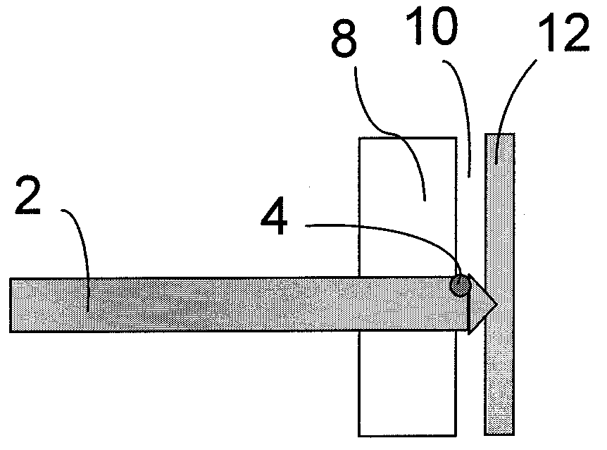
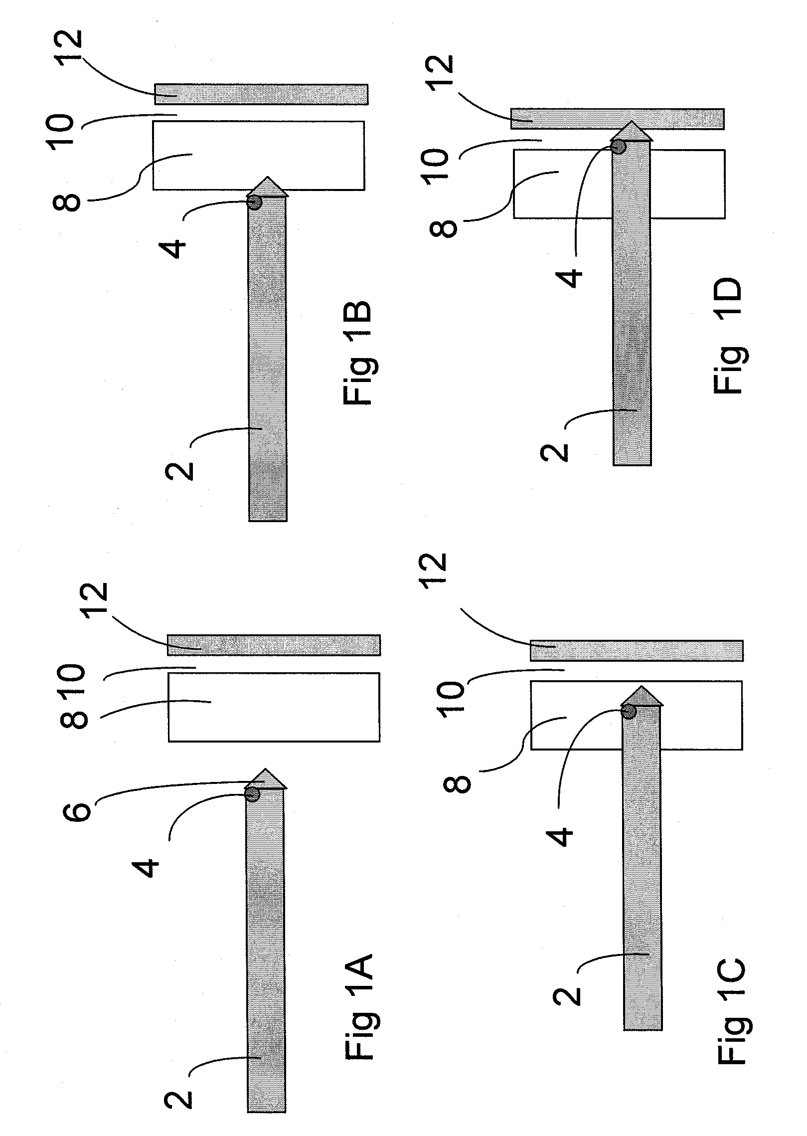
aorta or iliac arteries, and bowel has been reported. One aspect of the invention is a method and device for safe access of the peritoneal cavity. The improved safety of the current invention is based, in part, on the ability of the access
system to detect and report entry into the peritoneal cavity. Additional safety may be provided by the ability to tent the
abdomen in a focused manner directly at the site of trocar insertion, by the use of a blunt-tipped trocar and / or by the use of a force-gauge or force-
limiter to help guide the level of insertion force (which is frequently excessive or inadequate). In one embodiment, additional sensing capabilities may be incorporated as well to optimize the desired intervention or therapy to be delivered.
[0008]The tenting mechanism of the current invention may involve capturing the tissue around the site of insertion (via superficial puncture, suction, use of adhesives, etc.) at one or more sites and then applying an upward force during cavity entry. This tissue capture, in one embodiment, is fully, or nearly fully, circumferential to the
access site to provide optimal tenting directly adjacent to the site of puncture. In addition, the tissue capture mechanism may, after application to the
abdominal skin, allow for single-handed application of abducting force while the trocar or entry device is driven into the cavity. The tenting device may also consist of multiple components such that the grasping component may be detached for the low-profile tissue capture element such that remains at the site of cavity entry. The tenting may also be reversible and allow for immediate removal of the entire device once access has been obtained. In this embodiment, the tissue puncture may be released, the suction may be deactivated, the
adhesive may be dissolved, etc., once the trocar has been inserted into the cavity and the at-risk organs have been spared.
[0009]A
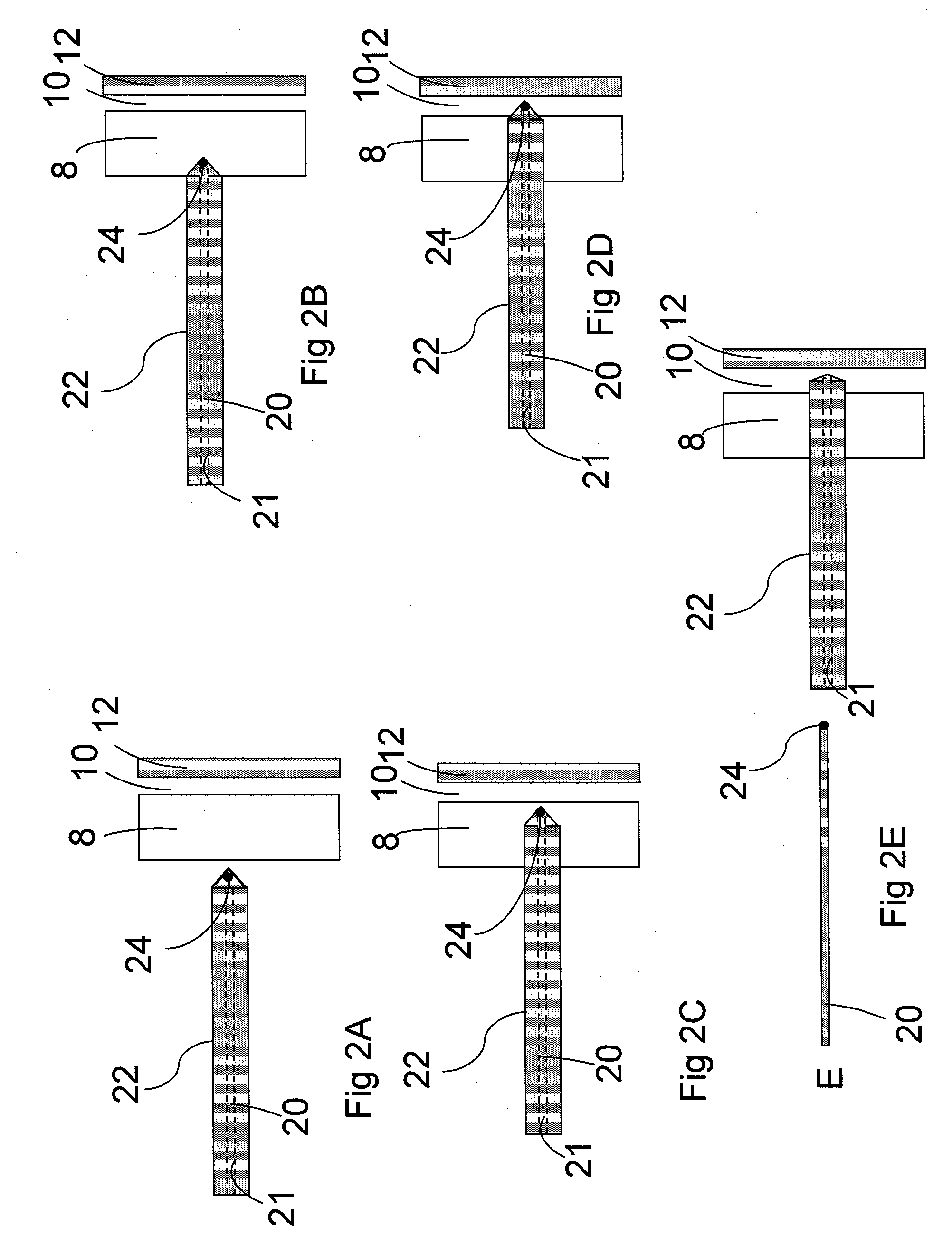
force gauge or force limiting mechanism may also be employed along with the above-mentioned feature or on its own in safely accessing the peritoneal, or any other, cavity. This component provides feedback to the user and prevents application of excessive force during cavity entrance. In one embodiment, the device alerts the user to both inadequate and excessive pressures via tactile, visual, auditory, or other stimulus. The device may also be capable of alerting the user to slightly inadequate or slightly excessive forces application. In the peritoneal embodiment, for example, the blunt-tipped trocar may be driven by a
handle or other component which signals the amplitude of the force along the axis of the
insertion device. This
signal may be as simple as a circuit which is closed with appropriate pressure and not with excessive or inadequate pressure. In another embodiment utilizing the asymmetric peritoneal
insertion device, the feedback to the user may be based on rotation. With asymmetric blunt trocars, safe insertion requires forward motion while rotating the trocar itself. In one example of this embodiment, the spring-loaded or shape-memory component of the
handle will allow the interdigitating elements of the trocar to engage and permit application of rotational forces only when the appropriate force is applied. Too much force will overshoot the appropriate engagement site and inadequate force will undershoot the engagement site.
[0010]This
force gauge feature, however, could utilize any mechanism to report the appropriate force range and to prevent over-insertion of the penetrating element. The feature could also be used in the accessing of other body cavities, e.g.,
bone marrow biopsies,
lumbar punctures, orthopedic screwing / plating or other manipulations of bone,
thoracentesis,
paracentesis, etc.
[0011]Some embodiments may include a
force gauge or force-
limiter that utilizes the tenting
handle to engage or disengage the rotational forces, as in threaded and / or asymmetric blunt trocar. In this embodiment the penetrating element may only advance when the appropriate force is applied in abducting the tenting handle. This safety feature will help ensure that the appropriate tenting force is applied while the penetrating element is advanced.

