
Methods and kits for detecting prostate cancer biomarkers

a prostate cancer and biomarker technology, applied in the field of biomarkers associated with prostate cancer, can solve the problems of time and effort of a trained clinician, a false positive rate of almost 75%, and difficult early diagnosis of prostate cancer

Inactive Publication Date: 2008-10-16
LIFE TECH CORP
View PDF4 Cites 71 Cited by
  • Summary
  • Abstract
  • Description
  • Claims
  • Application Information

AI Technical Summary

Benefits of technology

[0030]In some embodiments a biomarker detection panel can further comprise antibodies such as but not limited to one or more of antibodies to ACCP, BCL2, PSA (total), PSA (free), CXCR4, PTGER2, IL-6, IL-8, PAP, or PSMA. In some preferred embodiments, biomarker detection panel comprises antibodies to ACCP and/or IL-6.
[0031]In some preferred embodiments of biomarker detection panels, at least one of the autoantibody capture molecules is selected from Table 10. In some exemplary embodiments, a biomarker detection panel of the invention comprises one or more autoantibody detection sets of Table 5, Table 6, Table 7, Table 8, or Table 9.
[0032]In some preferred embodiments, the biomarker detection panel is provided bound to one or more solid or semi-solid supports, such as, for example, a gel or matrix, beads, particles, fibers, rods, filaments, or a filter, strip, sheet, membrane, plate (for example, a multiwell plate), dish, chip or array. In some preferred embodiments, at least 50% of the human proteins bound to the solid support are test antigens of the biomarker detection panel. In some preferred embodiments

Problems solved by technology

Accurate, early diagnosis of prostate cancer has proven challenging however, as the current diagnostic test for prostate cancer relies on detection of prostate-specific antigen (PSA) levels, an indicator that also correlates with benign prostate hypertrophy (BPH), a non-life threatening condition that does not increase cancer risk.
While the PSA test has a fairly good sensitivity (80%), it suffers from a false positive rate that approaches 75%.
However, the reliability of the test depends on its being performed in conjunction with an attentive digital rectal exam (DRE), which means the time and effort of a trained clinician are required, as well as the willingness of the patient to undergo DRE.
Such biomarkers will always be “unavailable” for convenient in-vitro blood-based diagnostic tests.

Method used


Image

  • Methods and kits for detecting prostate cancer biomarkers
  • Methods and kits for detecting prostate cancer biomarkers
  • Methods and kits for detecting prostate cancer biomarkers

Examples


example 1

Test Protein Array for Diagnostic Autoantigens

[0201]Biomarker detection panels were sought having at least as high a sensitivity as the standard PSA test (80%), with higher specificity.

Experimental Design

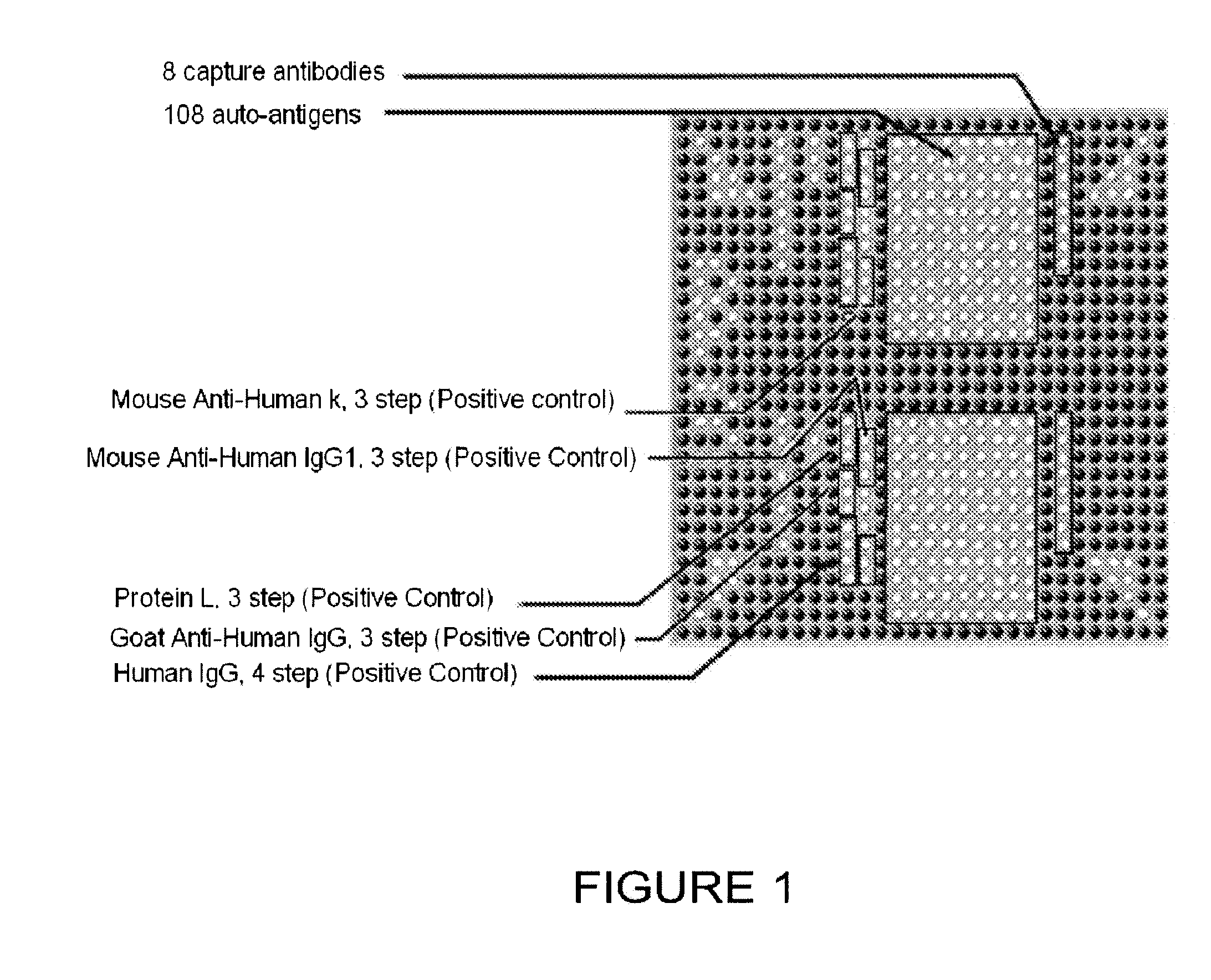
[0202]A protein array was fabricated by spotting proteins on a planar nitrocellulose substrate. The overall design of the array is depicted in FIG. 1, which shows half of an array used to test for the diagnostic utility of the 108 antigens and 8 antibodies listed in Table 1. The antigens and antibodies were selected based on biological experiments, knowledge of biological pathways altered by cancer progression (including immunological pathways), analogy with other cancers, and literature searching to compile a list of proteins that were overexpressed, inappropriately expressed, or differentially modified or degraded in prostate cancer cells when compared with normal prostate cells.

[0203]Table 1 provides in the first column (“Marker”) the term used throughout this application for the...

example 2

Identification of Autoantigens Present in Prostate Cancer Sera on ProtoArrav™ Human Protein Array

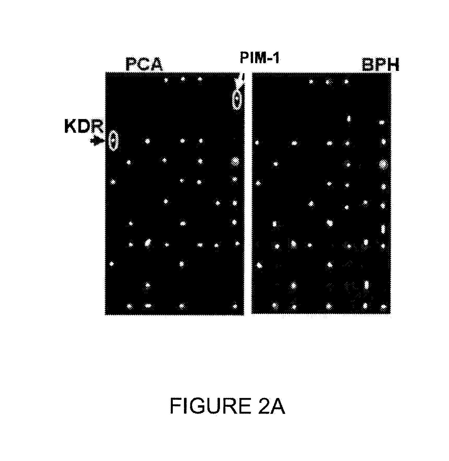
[0215]The human Protoarray™ high content protein microarray from Invitrogen (Carlsbad, Calif.) was screened with sera using the methods provided in Example 1. A combination of single-patient sample and pooled-patient samples were utilized with these arrays. A total of 32 patient samples were screened (16 prostate cancer and 16 BPH) as well as series of pooled-patient samples representing high, medium, and low PSA values. All of this data was analyzed together to generate a list of approximately 98 candidate prostate cancer biomarkers (Table 11a).

[0216]The high density Protoarray™ microarray data was normalized using a Quantile Normalization method for all chips used. After normalization, the diagnostic value of individual markers was estimated by calculating all possible order M-statistics and their associated p-values. The order with the lowest p-value was selected for each marker, the ...

example 3

Autoantibodies Differentiating Prostate Cancer from Benign Prostatic Hyperplasia in Patients

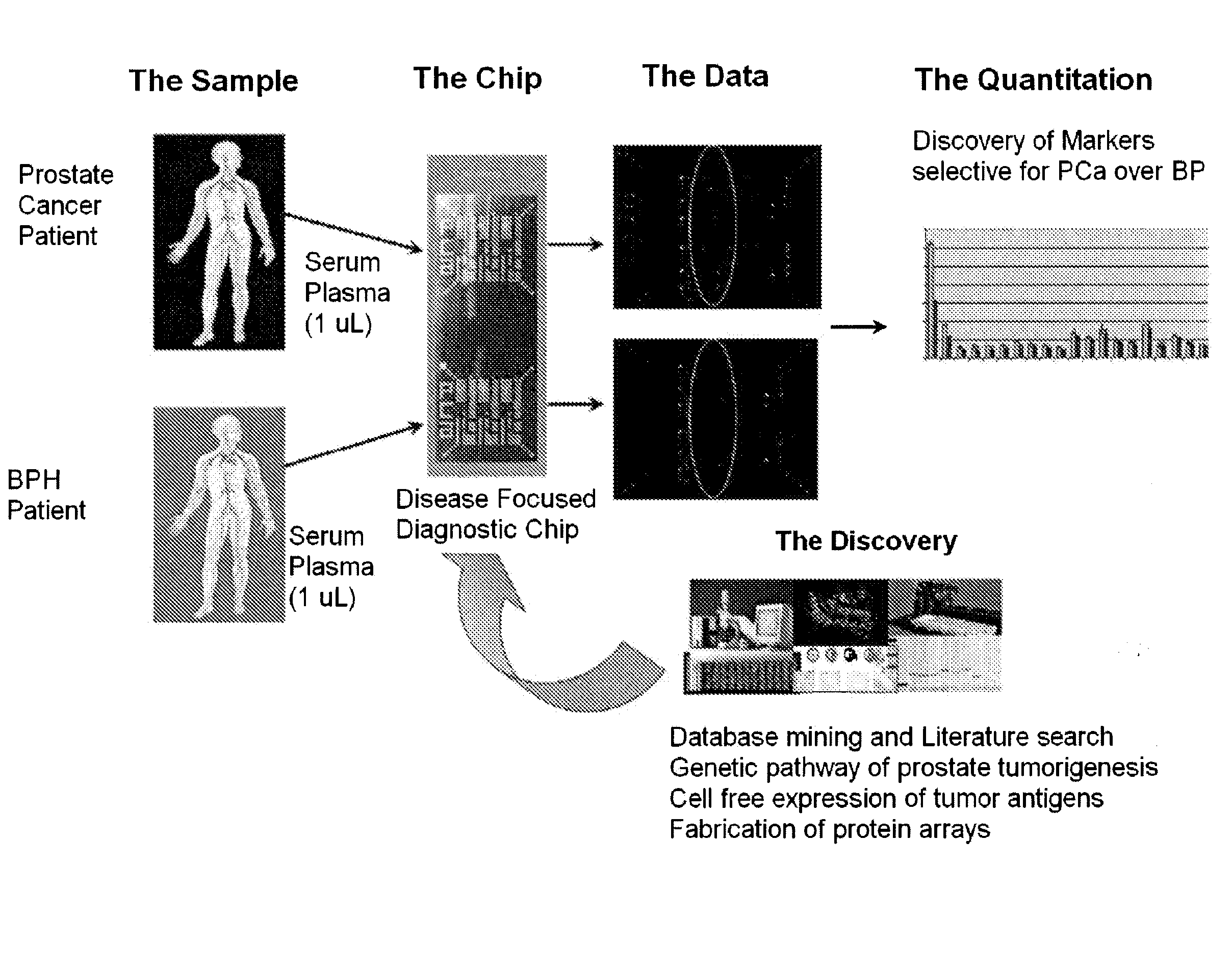
[0218]Autoantibody profiling using a protein microarray chip containing 96 proteins thought to be associated with prostate cancer development was conducted using sera from 32 patients with prostate cancer and 32 patients with benign prostatic hyperplasia. The goal was to find biomarkers that are stable in blood, easily measured using approximately 1 μL of serum (or plasma), and that can differentiate true prostate cancer from the closely-related benign prostatic hyperplasia, the major weakness in the current, clinically used, PSA-based prostate cancer diagnostic test.

[0219]The scheme for testing chips with human sera from individuals with prostate cancer and BPH is provided in FIG. 5. Serum samples from individuals having prostate cancer and BPH were collected and contacted with a chip containing the possible target antigens. The resulting binding of target antigens to autoantibodies was quan...


PUM


Abstract

Provided herein are novel autoantibody biomarkers, and panels for detecting autoantibody biomarkers for prostate cancer, and methods and kits for detecting these biomarkers in the serum of individuals suspected of having prostate cancer.

Description

CROSS REFERENCE TO RELATED APPLICATIONS[0001]This application claims benefit of U.S. Provisional Application No. 60 / 865,621, filed Nov. 13, 2006, which is incorporated by reference in its entirety herein to the extent that there is no consistency with the present disclosure.BACKGROUND[0002]The invention generally relates to biomarkers associated with prostate cancer, and methods and compositions for the detection, diagnosis, prognosis, and monitoring of the progression of prostate cancer.[0003]Prostate cancer (also referred to herein as “PCa”) is the most prevalent form of cancer and the second most common cause of cancer death in American men (Jemal et al. (2007) “Cancer statistics,” CA Cancer J. Clin. 57(1):43-66). When prostate cancer is diagnosed in its early stages, however, the prognosis is very good, with a ten year survival rate of greater than 85%. Current treatment modalities include radiation therapy, surgery, and androgen deprivation therapy. Treatment of prostate cancer...

Claims


Application Information

Patent Timeline
IPC IPC(8): G01N33/53G01N33/566B01J19/00
CPCG01N33/564G01N33/57434C12Y207/10001C12Y207/11001G01N2333/91205
Inventor LOVE, BRADLEYROGERS, JEFFREYBEECHEM, JOSEPHWANG, LILIN
Owner LIFE TECH CORP
Learn More