Add-On For Invasive Probe

a technology of invasive probes and add-ons, which is applied in the field of electrodes, can solve the problems of relative largeness, and achieve the effect of simple and cheaper

Inactive Publication Date: 2008-10-16
HADANI RON
View PDF23 Cites 41 Cited by
  • Summary
  • Abstract
  • Description
  • Claims
  • Application Information

AI Technical Summary

Benefits of technology

[0016]An aspect of some embodiments of the present invention relates to a compact electrode carrier for mounting on an endoscope or other elongate invasive probe, such as a trans-esophageal ultrasound probe, without covering the distal end of the probe. The electrode carrier occupies a small portion of the axial extent of the endoscope or probe. Optionally, the electrode carrier occupies less than 25% or even less than 15% of the surface area of the portion of the probe suitable for insertion into the patient. In an exemplary embodiment of the invention, the electrode carrier has an area of less than 1500 square millimeters or even less than 800 square millimeters. In some cases, using a compact electrode carrier is simpler and cheaper than using a large electrode carrier sheath or sheet which covers most (or a large portion) of the endoscope. Optionally, the electrode carrier comprises a substrate (e.g., an isolating material) with one or more electrodes mounted thereon.
[0017]The electrode is optionally very thin (flat), so as not to substantially affect the cross-section area of the probe. Optionally, the attachment of the electrode increases the cross-section diameter of the probe by less than 2 millimeters, or even less than one millimeter. In an exemplary embodiment of the invention, the electrode increases the cross-section diameter of the probe by less than half a millimeter.
[0024]In some embodiments of the invention, the electrode carrier includes a substrate which surrounds the entire circumference of the probe. Alternatively, the electrode carrier covers only a portion of the circumference of the probe. In some embodiments of the invention, the electrode carrier includes an adhesive which is used to connect the electrode carrier to the probe or endoscope. Optionally, the electrode carrier is removably attached to the endoscope or probe, so that it is easily removed after completion of a medical procedure in which the one or more electrodes on the electrode carrier are used. The adhesive is optionally a temporary adhesive which allows complete removal of the electrode, without damaging the probe and / or without leaving remnants on the probe.
[0025]In some embodiments of the invention, the electrode carrier includes a protective layer, which separates the probe from the electrode (or electrodes), in order to prevent damage to the probe from the electrical currents and / or heat in the electrode.
[0027]An aspect of some embodiments of the present invention relates to a compact electrode carrier with an adhesive substrate for mounting on an endoscope or other elongate invasive probe. Using an adhesive for attachment of the electrode carrier to the probe, instead of mechanical attachment makes the attachment of the electrode carrier simpler and usable with a large range of probes.
[0028]An aspect of some embodiments of the present invention relates to a single-electrode carrier for mounting on an endoscope or other elongate invasive probe. The use of a single electrode, rather than a plurality of electrodes, may simplify the electrode carrier.

Problems solved by technology

These sheaths are relatively large and may interfere with the insertion of the endoscope into the patient and / or its use therein.

Method used


Image

  • Add-On For Invasive Probe
  • Add-On For Invasive Probe
  • Add-On For Invasive Probe

Examples


Embodiment Construction

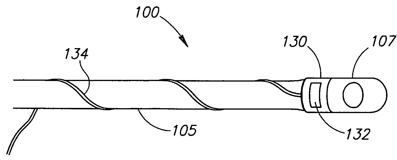
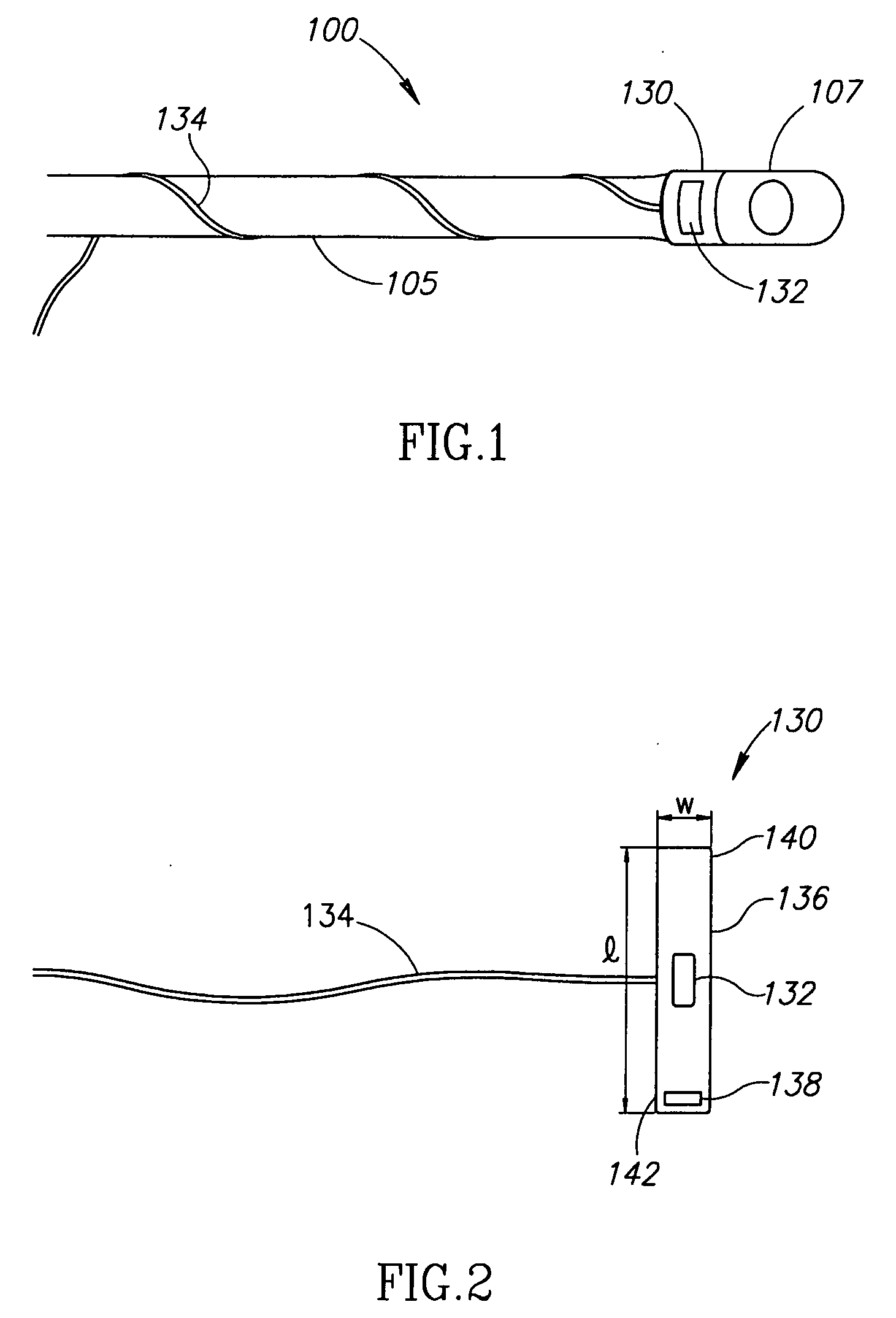
[0071]FIG. 1 is a schematic side view of an ultrasound probe 100 with an electrode carrier band 130 mounted thereon, in accordance with an exemplary embodiment of the present invention. Probe 100 includes an echocardiography sensor 107 and an elongate insertion tube 105. Probe 100 is optionally used for transesophageal echocardiography. By adding electrode carrier band 130 with an electrode 132 thereon to probe 100, probe 100 can be used for transesophageal cardioversion or cardiac pacing during a same procedure as the echocardiography.

[0072]FIG. 2 is a schematic illustration of electrode carrier band 130, in accordance with an exemplary embodiment of the invention. Carrier band 130 includes an electrode 132 and a wire 134 (or a group of wires), which electrically connects the electrode to a power generator, sensor, controller or other apparatus, at a proximal end of probe 100. Electrode 132 is optionally mounted on a substrate 136, which serves to attach the electrode to probe 100....


PUM


Abstract

An invasive probe assembly including an invasive probe having a surface area and a tool carrier removably mounted on the invasive probe, including a substrate having an area smaller than 25% of the surface area of the probe and at least one tool mounted on the substrate. The layout of the substrate or of one or more of the at least one tool is optionally substantially different in different sectors of the circumference of the probe.

Description

RELATED APPLICATIONS[0001]This application claims the benefit under 119(e) of U.S. provisional patent application 60 / 632,739, titled “Add on Electrode for Endoscope”, filed Dec. 1, 2004, U.S. provisional patent application 60 / 653,135, titled “Endoscopic Sheath with Illumination System”, filed Feb. 16, 2005, and U.S. provisional patent application 60 / 669,007, filed Apr. 7, 2005, titled “Emergency Electrode on Medical Tube”. The disclosures of all of these applications are incorporated herein by reference.FIELD OF THE INVENTION[0002]The present invention relates to apparatus for minimally invasive procedures and particularly to electrodes to be used in such procedures.BACKGROUND OF THE INVENTION[0003]Endoscopes and other invasive probes are used to view internal tissue of humans, and for many other tasks.[0004]U.S. Pat. No. 4,354,502 to Colley et al., the disclosure of which is incorporated herein by reference, describes a catheter having an ultrasound transducer and an ECG electrode ...

Claims


Application Information

Patent Timeline
Patent Type & Authority Applications(United States)
IPC IPC(8): A61B5/042
CPCA61B1/00105A61B1/00135A61B1/00142A61B1/05A61B2018/1495A61N1/05A61N1/0517A61N1/3621A61N1/3625A61N1/3918
Inventor HADANI, RON
Owner HADANI RON
Why Eureka
Try Eureka