Method and apparatus for entry of specimens into a microfluidic device

a microfluidic device and specimen technology, applied in the field of microfluidic devices, can solve the problems of affecting the movement of liquid and analytical, introducing difficult problems for small samples, and affecting the accuracy of liquid/fluent solid measurements, etc., to achieve accurate and repeatable assays

Inactive Publication Date: 2008-10-23
SIEMENS HEALTHCARE DIAGNOSTICS INC
View PDF106 Cites 56 Cited by
  • Summary
  • Abstract
  • Description
  • Claims
  • Application Information

AI Technical Summary

Benefits of technology

[0009]The invention relates in particular to entry ports adapted to supply small samples of 0.1 to 20 μL to microfluidic chips, thereby making possible accurate and repeatable assays of the analytes of interest in such samples. Such entry ports provide access for small samples and transfer of the samples uniformly into an inlet chamber while purging air from the microfluidic chip without trapping air bubbles in the chamber. Uniform distribut

Problems solved by technology

However, smaller samples introduce difficult problems.
As the sample is moved into a well, it is important that the liquid is uniformly distributed such that all the air in the well is expelled, since air will adversely affect the movement of liquid and the analytical results.
Also, there are other problems associated with the initial introduction of the sample to the microfluidic device.
Specimens on the surface will cause carry-over and contamination between analyses.
Air in the port will cause under-filling and, consequently, under estimation of the analytical results.
Air bubbles in the inlet port o

Method used


Image

  • Method and apparatus for entry of specimens into a microfluidic device
  • Method and apparatus for entry of specimens into a microfluidic device
  • Method and apparatus for entry of specimens into a microfluidic device

Examples


example 1

[0054]In a test chip similar to that of FIG. 5c, the geometry of inlet port opening was varied to demonstrate that the shape of the opening was not critical to filling the inlet chamber. The results of these tests are given in the following table:

DepthWidthLengthFluidGeometrymmmmmmSampleForceFill timeRectangle0.030.1501.0WholeCapillarybloodCylinder0.1000.1001.0WholeCapillarybloodRectangle0.030.1502.0WholeCapillarybloodRectangle0.030.1502.0UrineCapillaryRectangle0.030.1502.0UrinePositivewithpressureadapterRectangle0.030.1502.0WholePositivewithbloodpressureadapterRectangle0.030.1502.0WholeNegativewithbloodpressureadapter

[0055]Using a capillary as the inlet port, the inlet chamber was filled in the less than 2 seconds with and without an adapter at the inlet. The fill time was dependent on the fluid used as well as the surface energy of the capillary and the length, width or shape of the capillary.

example 2

[0056]Using a test chip similar to that of Example 1, the pressure and volumes used to add fluid to the inlet chamber via the port opening were varied. The inlet chamber volume was 5 μL and a metering loop having a volume of 0.3 μL received liquid when the inlet chamber was filled. The experiment was performed with blood and urine.

Volume(μL)Sample delivery devicePressureObservation5Capillary without plungerTargetMetering occurs4Capillary without plungerTargetMetering occurs6Capillary without plungerTargetMetering occurs &excess overflows5Capillary with plungerHighMetering occurs4Capillary with plungerHighMetering occurs6Capillary with plungerHighMetering occurs &excess overflows5Capillary with plungerLowMetering occurs4Capillary with plungerLowMetering occurs6Capillary with plungerLowMetering occurs &excess overflows

[0057]Pressure applied either by capillary action or by use of a plunger allowed acceptable filling over a wide range of sample volumes 4-6 μL. In the case of an over fi...

example 3

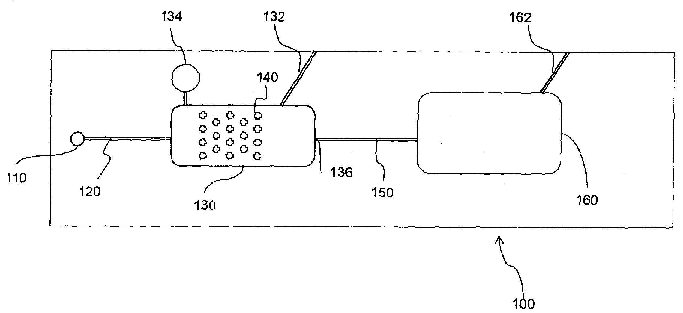
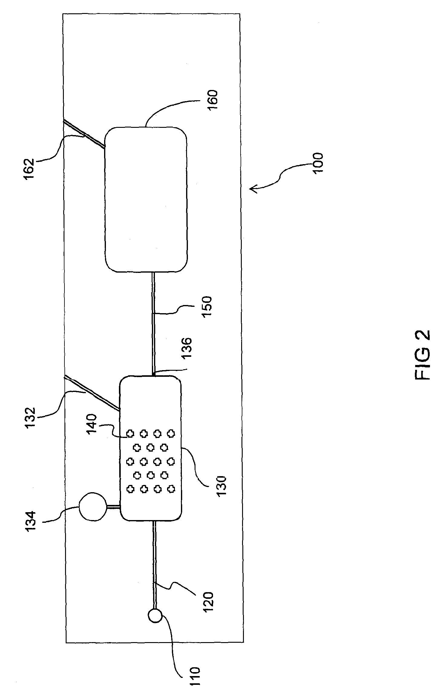
[0058]The microfluidic device of FIGS. 3a-c and 4 was used to measure the glucose content of blood. Whole blood pretreated with heparin was incubated at 250° C. to degrade glucose naturally occurring in the blood sample. The blood was spiked with 0, 50, 100, 200, 400, and 600 mg / μL of glucose as assayed on the YSI glucose instrument (YSI Instruments Inc.). A glucose reagent (chromagenic glucose) reagent as described in Bell U.S. Pat. No. 5,360,595 was coated on a nylon membrane disposed on a plastic substrate. A sample of the reagent was placed in chamber 44 and the bottom of the device covered with Excel Sealplate (Excel Scientific Inc.).

[0059]Samples of blood containing one of the concentrations of glucose were introduced into inlet port 40 using a 2 μL capillary with plunger (Drummond Aqua). Since the inlet port is sealed when the sample is dispensed, a positive pressure is established which forces the sample into the inlet passageway 42 and then into the reagent area 44. The sam...


PUM

PropertyMeasurementUnit
Forceaaaaaaaaaa
Pressureaaaaaaaaaa
Flow rateaaaaaaaaaa

Abstract

A microfluidic device for analyzing biological samples is provided with a sample inlet section including an inlet port, a capillary passageway communication with the inlet port and with an inlet chamber. The inlet chamber includes means for uniformly distributing the sample liquid across the inlet chamber and purging the air initially contained therein.

Description

[0001]This is a continuation in-part of U.S. Ser. No. 10 / 608,671, filed Jun. 27, 2003.BACKGROUND OF THE INVENTION[0002]This invention relates to microfluidic devices, particularly those that are used for analysis of biological samples. Microfluidic devices are intended to be used for rapid analysis, thus avoiding the delay inherent in sending biological samples to a central laboratory. Such devices are intended to accept very small samples of blood, urine, and the like. The samples are brought into contact with reagents capable of indicating the presence and quantity of analytes found in the sample.[0003]Many devices have been suggested for carrying out analysis near the patient, some of which will be discussed below. In general, such devices use only small samples, typically 0.1 to 200 μL. With the development of microfluidic devices the samples have become smaller, which is a desirable feature of their use. However, smaller samples introduce difficult problems. In microfluidic dev...

Claims


Application Information

Patent Timeline
IPC IPC(8): G01N27/26
CPCB01L3/5025B01L3/502723B01L3/50273B01L2200/027B01L2200/0684B01L2300/0806B01L2300/0816B01L2400/0406B01L2400/0409B01L2400/0478B01L2400/0487B01L2400/0688B01L2400/086
Inventor PUGIA, MICHAEL J.PROFITT, JAMES A.BLANKENSTEIN, GERTPETERS, RALF-PETER
Owner SIEMENS HEALTHCARE DIAGNOSTICS INC
Why Eureka
Try Eureka