Method for Diagnosis of Functional Lung Illnesses

a functional lung disease and lung disease technology, applied in the field of functional lung disease diagnosis, can solve the problems of reduced lung capacity, at least hindered exchange, and least delayed exchange, and achieve the effect of precise acquisition of the effect of therapies, fast, inexpensive, and radiation-fr

Active Publication Date: 2008-10-30
SIEMENS HEALTHCARE GMBH
View PDF8 Cites 15 Cited by
  • Summary
  • Abstract
  • Description
  • Claims
  • Application Information

AI Technical Summary

Benefits of technology

[0007]An object of the present invention is to provide a method that allows a fast, inexpensive, radiation-free and better spatially-resolved detection of functional defects of the lung as well as a precise acquisition of the effect of therapies.

Problems solved by technology

In the case of certain other illnesses, however, parts of the lung are filled with air without an exchange with the fresh inhaled air ensuing, or the exchange is at least hindered.
With asthma, a narrowing of the air passages leads to an at least delayed, but mostly insufficient exhalation of the air.
This leads not only to a correspondingly reduced capacity of the lung but also over longer periods of time to a permanent damage of the lung tissue (emphysema).
Unfortunately, the suitable isotopes are very rare or extremely difficult to provide (for example He3 forms only 0.01% of naturally-occurring helium and, due to its low density, escapes forever after the release into the atmosphere).
Alternative isotopes provide only a relatively low contrast due to the low resonance frequency and the poor degree of hyper-polarization.
Moreover, the apparatus expenditure in the MR scanner given the use of hyper-polarized gases is not in consequential.
Due to the low resonance frequency relative to protons, a dedicated RF system is required, which incurs additional costs.
Moreover, an HPG MR measurement can be repeated only in a limited manner since these gases supply no contribution to the oxygen saturation and therefore can be tolerated to a limited degree in patients, in particular those with limited lung function.

Method used


Image

  • Method for Diagnosis of Functional Lung Illnesses
  • Method for Diagnosis of Functional Lung Illnesses
  • Method for Diagnosis of Functional Lung Illnesses

Examples


Embodiment Construction

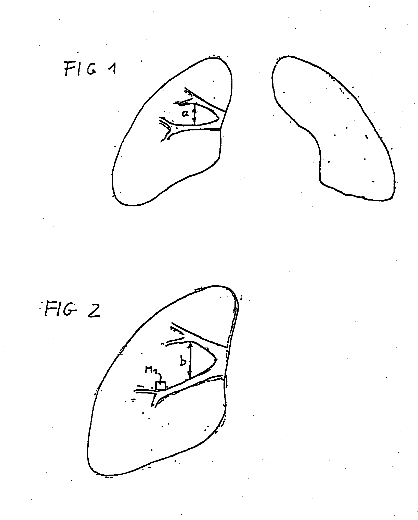
[0029]The lung is shown at maximum exhalation in FIG. 1 and at maximum inhalation in FIG. 2. To produce these images the patient is positioned in an MR scanner such that the lung lies in the FOV. An MR exposure is generated at each of various phase points of the respiration, in the shown exemplary embodiment at maximum inhalation and maximum exhalation. The change of the proton density dependent on the phase point of the respiration (or the time) provides a measure for the homogeneity and thus also for possible functional lung illnesses.

[0030]For this purpose the 2D image of the lung can be subdivided in a computer into macroscopic areas, for example 100 squares. For each square the computer then determines the signal difference in comparison to the air at inhalation and exhalation. The computer also segments the blood vessels of the lung and associates the segments with these vessels.

[0031]In order to compare the same lung region in each of the inhaled state and exhaled state, thus...


PUM


Abstract

In a method for diagnosing functional lung illnesses, image exposures of the lungs are obtained at various phase points in time of the respiration of a subject, such as at maximum inhalation and maximum expiration, and the image exposures are segmented and at least two of the image exposures are compared on a segment-by-segment basis to identify a change in tissue density between the compared segments, as an indicator of lung functioning.

Description

BACKGROUND OF THE INVENTION[0001]1. Field of the Invention[0002]The present invention relate to methods for diagnosing functional long illnesses, particularly with the use of images of the lungs of a subject obtained with a medical imaging modality.[0003]2. Description of the Prior Art[0004]For the diagnosis and therapy of lung illnesses it is necessary to record spatially resolved and time-resolved functional parameters of the lungs using measurement technology. In healthy people the lung is nearly homogeneous, meaning that macroscopically the lung is uniformly filled with fresh air upon inhalation and uniformly emptied upon exhalation. The very thin lung tissue is permeated by a few large and many small blood vessels and bronchi. In a series of different illnesses of the lung, specific areas do not participate in the exchange with fresh air. Such areas can, for example, be filled with mucus, for example in the case pneumonia lung inflammation. Such areas can already be detected in...

Claims


Application Information

Patent Timeline
Patent Type & Authority Applications(United States)
IPC IPC(8): A61B5/08A61B5/05A61B5/055A61B6/03
CPCA61B5/055A61B5/08A61B6/507A61B6/03A61B5/411
Inventor KUTH, RAINERRUPPRECHT, SABINERUPPRECHT, THOMASZAPKE, MAREN
Owner SIEMENS HEALTHCARE GMBH
Why Eureka
Try Eureka