Deformable phantom apparatus

a phantom apparatus and phantom technology, applied in the field of phantom apparatus, can solve the problems of not imparting a physiologically correct movement of the organs during breathing, prone to errors and inaccuracy, and the dose to be received by the patient is estimated, so as to reduce the difficulty and disadvantages, accurately and reproducibly simulate

Inactive Publication Date: 2008-12-04
MCGILL UNIV
View PDF10 Cites 93 Cited by
  • Summary
  • Abstract
  • Description
  • Claims
  • Application Information

AI Technical Summary

Benefits of technology

[0012]The present invention reduces the difficulties and disadvantages of the aforementioned designs and provides an apparatus which can more accurately and more reproducibly simulate the motion of a patient's anatomy during breathing. Applicants have discovered a deformable phantom apparatus which simulates movement of a patient's anatomy, e.g. an organ, during breathing. Surprisingly, the Applicants have found that the deformable phantom apparatus is highly reproducible and simulates anatomical motion anthropomorphically and in a very close manner to the in vivo situation. The deformable phantom apparatus can find application in four-dimensional (4-D) radiation therapy and aid the management of the respiratory motion in quality assurance for dynamic imaging, treatment planning and dose delivery in radiation therapy.
[0033]Advantageously, the deformable member of the phantom apparatus of the present invention can be made of simple and cheap materials such as natural sponge and rubber. The deformable member and the phantom apparatus can be easily and cheaply constructed and used in many methods that deal with the investigation and minimization of the impact of respiratory motion such as: motion-encompassing methods, respiratory gating techniques, and real-time tumour-tracking techniques. One possible application is in 4D radiation therapy. Other possible applications exist in radiology whenever time dependent imaging is required on anatomically realistic representations of the human body.
[0035]Advantageously, the inclusion of a deformable mass in the deformable member can realistically simulate the presence and movement of a tumour or lesion during breathing. In the case of the lung, tumours inside the lung are known to move significantly due to respiratory movement. By means of the deformable mass being incorporated within the deformable member, the complex 3D trajectory of a real-life tumour can be simulated and tracked. Simulation of hysteresis in the tumour motion can be achieved by regulating the flow of fluid in and out of the deformable member using valves, for example, or by dynamically blocking and unblocking a number of openings in mouth of the cover portion of the deformable member. A compensation may then be incorporated into the radiotherapy treatment to accommodate this movement.

Problems solved by technology

However, this technique is affected by motion artefacts on the resultant images which makes localization of the tumour, and therefore the estimation of the dose to be received by the patient, prone to errors and inaccuracies.
However, this mechanism may not impart a physiologically correct movement of the organs during breathing.

Method used


Image

  • Deformable phantom apparatus
  • Deformable phantom apparatus
  • Deformable phantom apparatus

Examples


Embodiment Construction

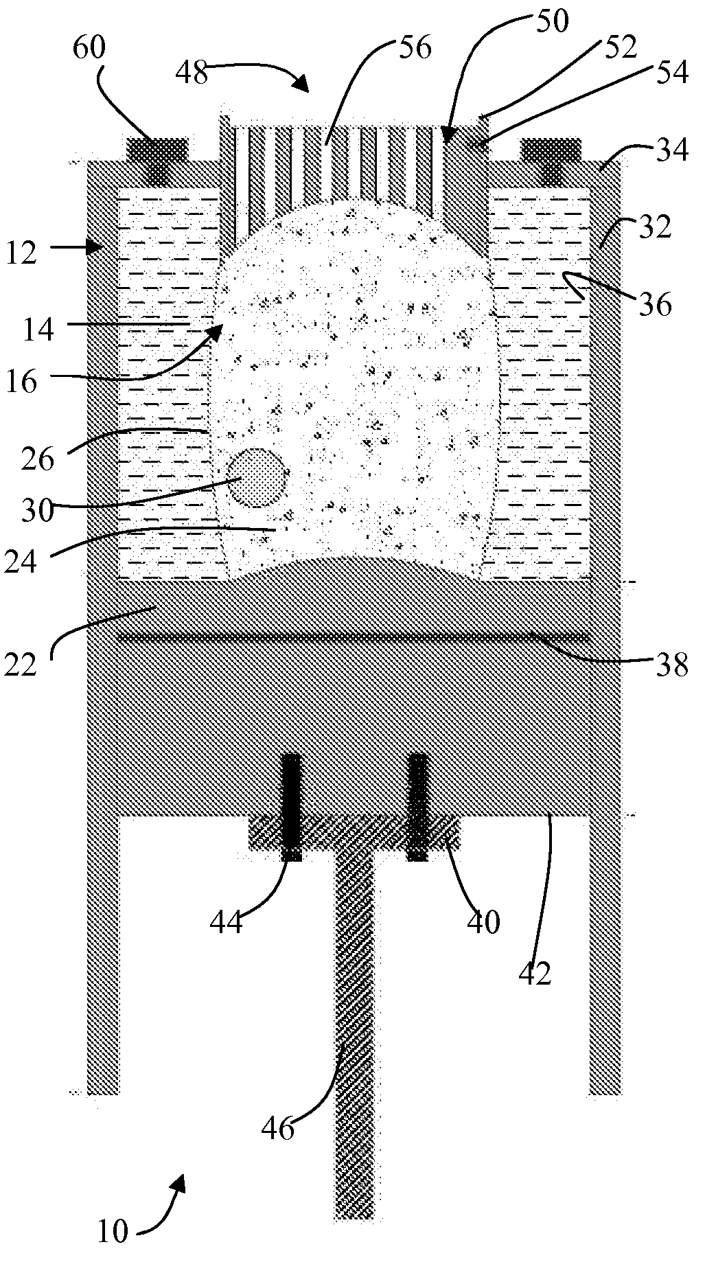
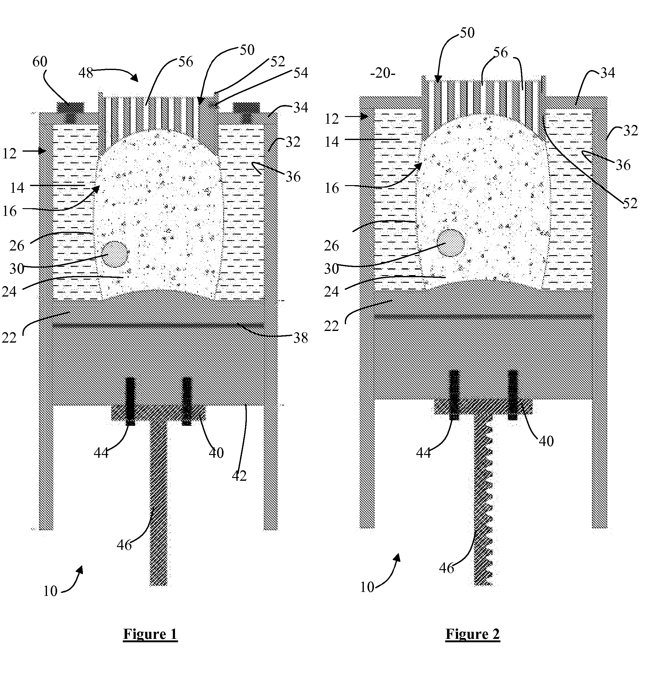
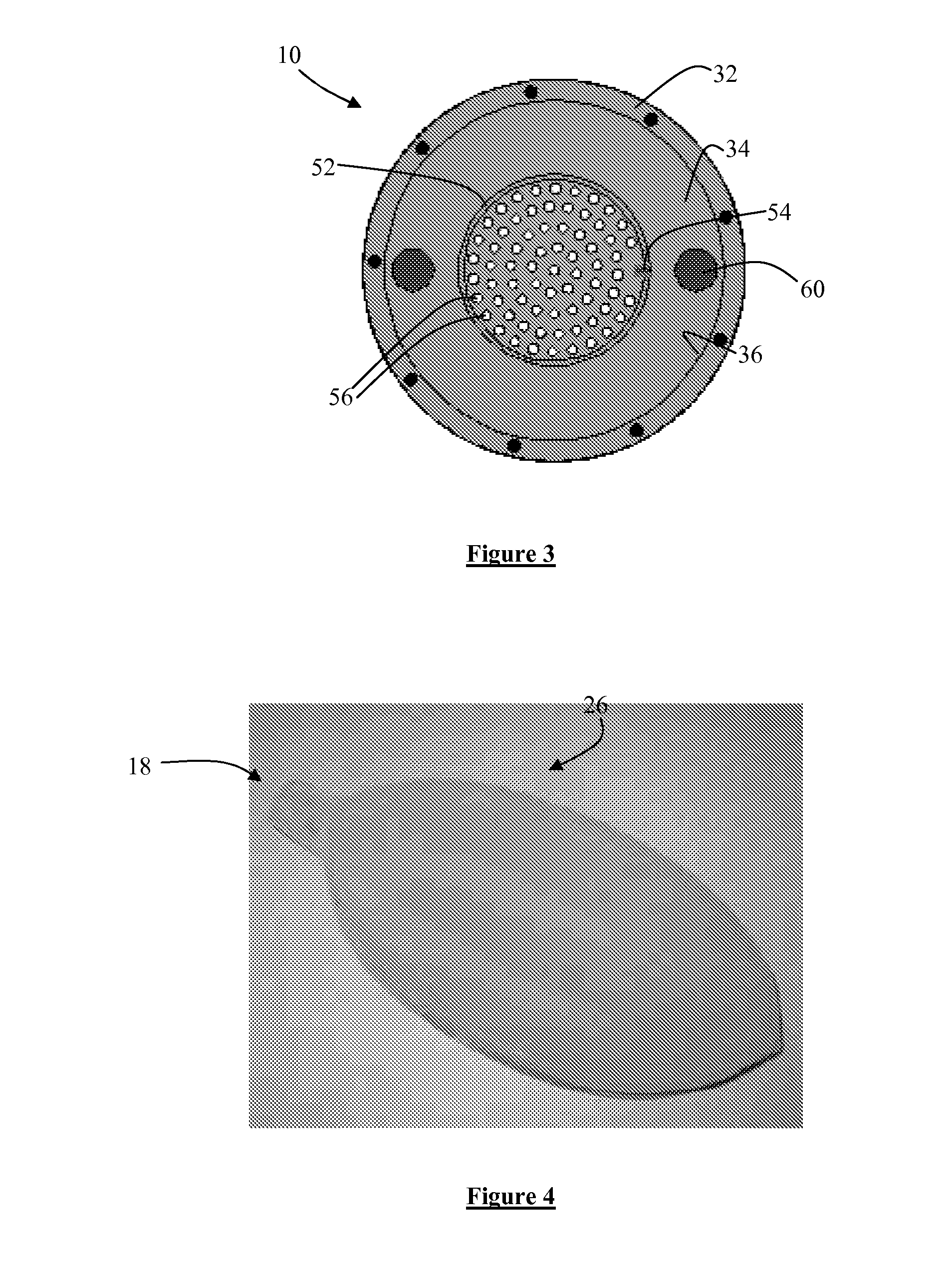
[0057]Referring initially to FIGS. 1 to 3, there is illustrated a deformable phantom apparatus 10 for simulating motion of a patient's anatomy in 3D during breathing. In a broad aspect, the deformable phantom apparatus 10 comprises a chamber 12 fillable with a first fluid 14, a deformable member 16 comprising tissue equivalent material of the anatomy being simulated, the deformable member 16 being positionable within the chamber 12 in the first fluid 14 and having an open end 18 (FIG. 4) in fluid communication with a second fluid 20 outside the chamber 12 in use; and a mechanism for causing the second fluid 20 to flow through the open end 18 to deform the deformable member 16 between a normal state and a deformed (extended or inflated) state to simulate anatomical motion during breathing.

[0058]In a preferred embodiment described below, the first fluid 14 is water and the second fluid 20 is air such that the chamber 12 is filled with water and the deformable member 16 is in fluid com...


PUM


Abstract

A deformable phantom apparatus for simulating motion of a patient's anatomy in 3D during breathing, the apparatus comprising a chamber fillable with a first fluid, a deformable member comprising tissue equivalent material of the anatomy being simulated, the deformable member being positionable within the chamber in the first fluid and having an open end in fluid communication with a second fluid outside the chamber in use; and a mechanism for causing the second fluid to flow through the open end to deform the deformable member between a normal state and a deformed state to simulate motion during breathing.

Description

CROSS REFERENCE TO RELATED APPLICATION[0001]The Applicants hereby claim priority from U.S. patent application No. 60 / 924,697 filed 29 May 2007, the contents of which are hereby incorporated by reference in its entirety.FIELD OF THE INVENTION[0002]The present invention concerns a deformable phantom apparatus for simulating motion of a patient's anatomy, such as a lung, during breathing.BACKGROUND OF THE INVENTION[0003]Radiotherapy uses ionizing radiation to treat or destroy cancerous tumours and lesions. The damage to tumour cells from the radiation is related to the absorbed dose (i.e., energy absorbed from ionizing radiation per unit mass). Therefore increasing the dose to the tumour increases the number of treated or destroyed cancer cells. However, as higher dose levels may also affect healthy tissue and other structures surrounding the tumour, the amount of ionizing radiation used must be controlled to provide as high as possible a dose to the tumour site whilst minimizing damag...

Claims


Application Information

Patent Timeline
Patent Type & Authority Applications(United States)
IPC IPC(8): A61B6/00G09B23/32
CPCA61B6/583A61N5/1037A61N5/1048A61B6/037A61N2005/1076G09B23/288G09B23/32A61N2005/1072
Inventor SERBAN, MONICASTROIAN, GABRIELAHEATH, EMILYSEUNTJENS, JAN
Owner MCGILL UNIV
Why Eureka
Try Eureka